

Endothelial Damage in Acute Respiratory Distress Syndrome
- PMID: 33233715
- PMCID: PMC7699909
- DOI: 10.3390/ijms21228793
Endothelial Damage in Acute Respiratory Distress Syndrome
Abstract
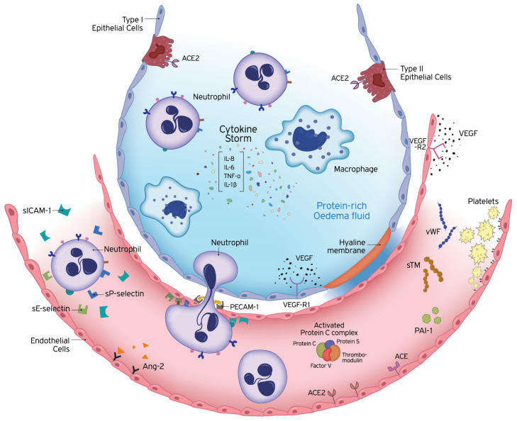
The pulmonary endothelium is a metabolically active continuous monolayer of squamous endothelial cells that internally lines blood vessels and mediates key processes involved in lung homoeostasis. Many of these processes are disrupted in acute respiratory distress syndrome (ARDS), which is marked among others by diffuse endothelial injury, intense activation of the coagulation system and increased capillary permeability. Most commonly occurring in the setting of sepsis, ARDS is a devastating illness, associated with increased morbidity and mortality and no effective pharmacological treatment. Endothelial cell damage has an important role in the pathogenesis of ARDS and several biomarkers of endothelial damage have been tested in determining prognosis. By further understanding the endothelial pathobiology, development of endothelial-specific therapeutics might arise. In this review, we will discuss the underlying pathology of endothelial dysfunction leading to ARDS and emerging therapies. Furthermore, we will present a brief overview demonstrating that endotheliopathy is an important feature of hospitalised patients with coronavirus disease-19 (COVID-19).
Keywords: ARDS; biomarkers; coagulation; dysfunction; inflammation.
Conflict of interest statement
The authors declare no conflict of interest.
Figures
Similar articles
-
[Therapy of coronavirus disease 2019 induced acute respiratory distress syndrome is different from traditional acute respiratory distress syndrome].Zhonghua Shao Shang Za Zhi. 2020 May 20;36(5):330-333. doi: 10.3760/cma.j.cn501120-20200407-00214. Zhonghua Shao Shang Za Zhi. 2020. PMID: 32456368 Chinese.
-
Kallikrein-kinin blockade in patients with COVID-19 to prevent acute respiratory distress syndrome.Elife. 2020 Apr 27;9:e57555. doi: 10.7554/eLife.57555. Elife. 2020. PMID: 32338605 Free PMC article.
-
Platelet functions and activities as potential hematologic parameters related to Coronavirus Disease 2019 (Covid-19).Platelets. 2020 Jul 3;31(5):627-632. doi: 10.1080/09537104.2020.1762852. Epub 2020 May 13. Platelets. 2020. PMID: 32397915 Review.
-
Hemostasis profile in COVID-19 infection.Rev Assoc Med Bras (1992). 2020 May;66(5):571-572. doi: 10.1590/1806-9282.66.5.571. Epub 2020 Jul 3. Rev Assoc Med Bras (1992). 2020. PMID: 32638971 No abstract available.
-
Mesenchymal stem cell therapy for acute respiratory distress syndrome: from basic to clinics.Protein Cell. 2020 Oct;11(10):707-722. doi: 10.1007/s13238-020-00738-2. Epub 2020 Jun 9. Protein Cell. 2020. PMID: 32519302 Free PMC article. Review.
Cited by
-
Molecular hydrogen is a potential protective agent in the management of acute lung injury.Mol Med. 2022 Mar 3;28(1):27. doi: 10.1186/s10020-022-00455-y. Mol Med. 2022. PMID: 35240982 Free PMC article. Review.
-
Initial Tumor Necrosis Factor-Alpha and Endothelial Activation Are Associated with Hemorrhagic Complications during Extracorporeal Membrane Oxygenation.J Clin Med. 2023 Jul 6;12(13):4520. doi: 10.3390/jcm12134520. J Clin Med. 2023. PMID: 37445555 Free PMC article.
-
Reduning alleviates sepsis-induced acute lung injury by reducing apoptosis of pulmonary microvascular endothelial cells.Front Immunol. 2023 Jul 3;14:1196350. doi: 10.3389/fimmu.2023.1196350. eCollection 2023. Front Immunol. 2023. PMID: 37465664 Free PMC article.
-
Histone H4 induces heparan sulfate degradation by activating heparanase in chlorine gas-induced acute respiratory distress syndrome.Respir Res. 2022 Jan 24;23(1):14. doi: 10.1186/s12931-022-01932-y. Respir Res. 2022. PMID: 35073921 Free PMC article.
-
BPC 157 as Potential Treatment for COVID-19.Med Hypotheses. 2021 Nov 9;158:110736. doi: 10.1016/j.mehy.2021.110736. Online ahead of print. Med Hypotheses. 2021. PMID: 34798584 Free PMC article.
References
-
- Bernard G.R., Artigas A., Brigham K.L., Carlet J., Falke K., Hudson L., Lamy M., Legall J.R., Morris A., Spragg R. The American-European Consensus Conference on ARDS. Definitions, mechanisms, relevant outcomes, and clinical trial coordination. Am. J. Respir. Crit. Care Med. 1994;149(Pt 1):818–824. doi: 10.1164/ajrccm.149.3.7509706. - DOI - PubMed
Publication types
MeSH terms
LinkOut - more resources
Full Text Sources
