. 2021 Jan 11;6(1):e144455.
doi: 10.1172/jci.insight.144455.

An immune-based biomarker signature is associated with mortality in COVID-19 patients

Affiliations

An immune-based biomarker signature is associated with mortality in COVID-19 patients

Michael S Abers et al. JCI Insight. .

Abstract

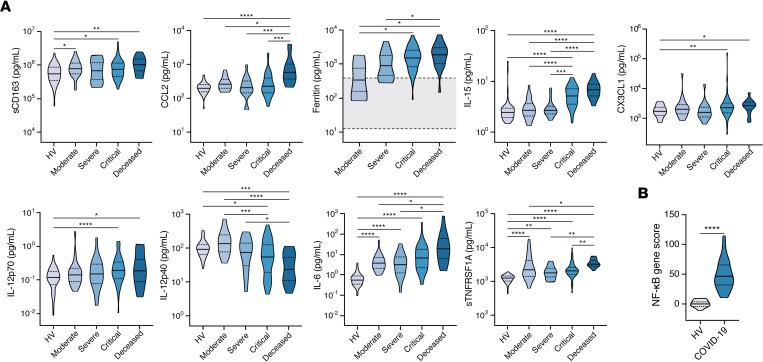
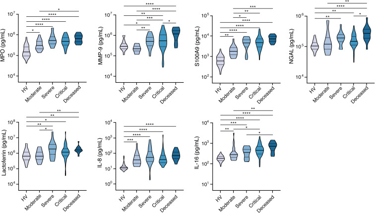
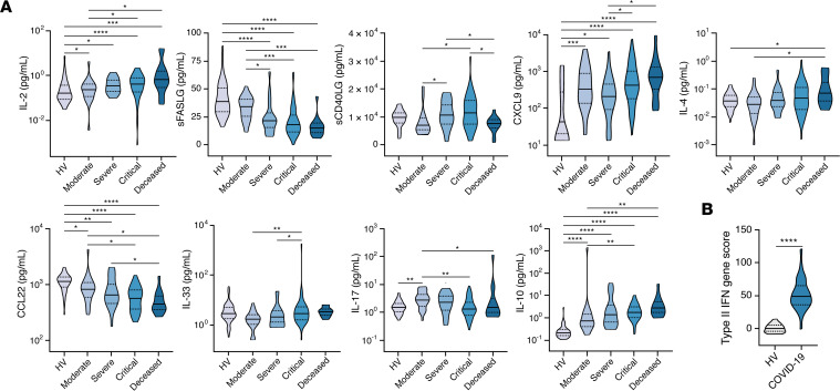
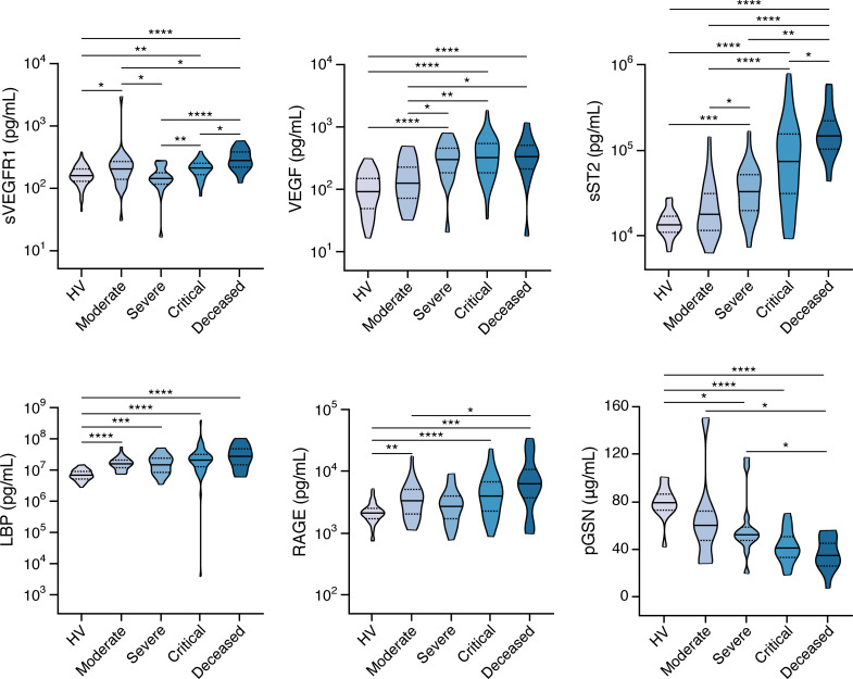
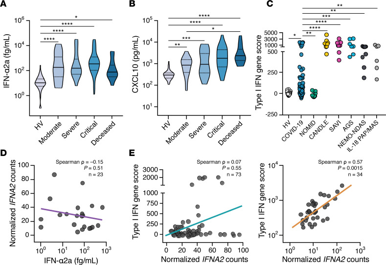
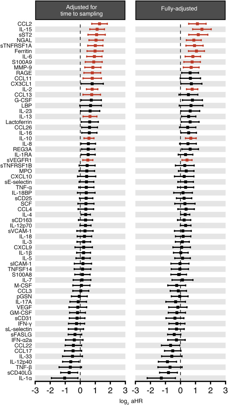
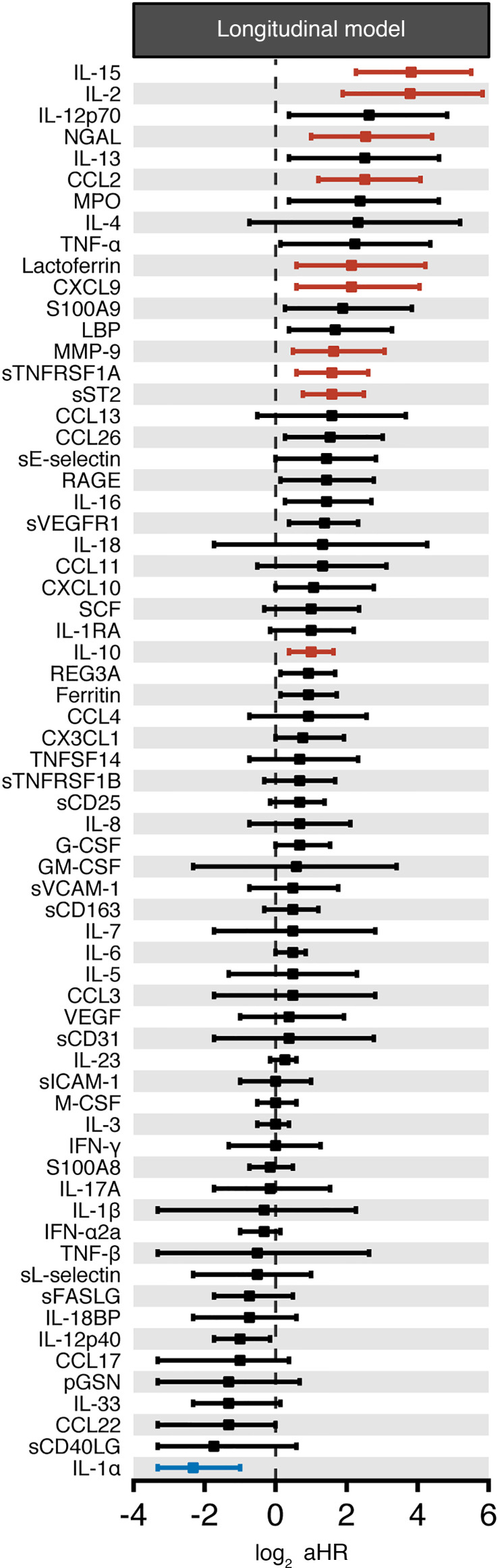
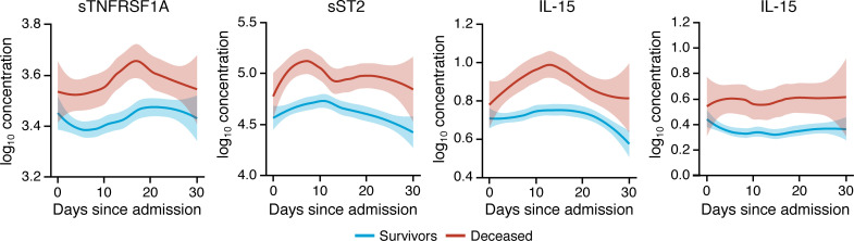
Immune and inflammatory responses to severe acute respiratory syndrome coronavirus 2 (SARS-CoV-2) contribute to disease severity of coronavirus disease 2019 (COVID-19). However, the utility of specific immune-based biomarkers to predict clinical outcome remains elusive. Here, we analyzed levels of 66 soluble biomarkers in 175 Italian patients with COVID-19 ranging from mild/moderate to critical severity and assessed type I IFN-, type II IFN-, and NF-κB-dependent whole-blood transcriptional signatures. A broad inflammatory signature was observed, implicating activation of various immune and nonhematopoietic cell subsets. Discordance between IFN-α2a protein and IFNA2 transcript levels in blood suggests that type I IFNs during COVID-19 may be primarily produced by tissue-resident cells. Multivariable analysis of patients' first samples revealed 12 biomarkers (CCL2, IL-15, soluble ST2 [sST2], NGAL, sTNFRSF1A, ferritin, IL-6, S100A9, MMP-9, IL-2, sVEGFR1, IL-10) that when increased were independently associated with mortality. Multivariate analyses of longitudinal biomarker trajectories identified 8 of the aforementioned biomarkers (IL-15, IL-2, NGAL, CCL2, MMP-9, sTNFRSF1A, sST2, IL-10) and 2 additional biomarkers (lactoferrin, CXCL9) that were substantially associated with mortality when increased, while IL-1α was associated with mortality when decreased. Among these, sST2, sTNFRSF1A, IL-10, and IL-15 were consistently higher throughout the hospitalization in patients who died versus those who recovered, suggesting that these biomarkers may provide an early warning of eventual disease outcome.

Keywords: COVID-19; Chemokines; Cytokines; Immunology.


Conflict of interest statement

Conflict of interest: SLL and MJD are employees of and own stock in BioAegis Therapeutics, Inc, which is developing recombinant human plasma gelsolin for potential clinical use.

Figures

Figure 1
Figure 1. Biomarkers associated with activation of monocytes/macrophages and NF-κB signaling are markedly induced in COVID-19 patients.
(A) Shown are levels of soluble CD163 (sCD163), CCL2, ferritin, IL-15, CX3CL1, IL-12p70, IL-12p40, IL-6, and sTNFRSF1A in peripheral blood of COVID-19 patients with various severity groups (n = 94–119 depending on the biomarker) relative to healthy volunteers (HV; n = 45–60 depending on the biomarker). Ferritin concentrations were determined by clinical assays performed in Italian hospitals. The area shaded in gray reflects the normal range for HVs reported by the clinical laboratory. Groups were compared by Kruskal-Wallis test. When P < 0.05, pairwise comparisons were made using Dunn’s test with Benjamini-Hochberg adjustment for multiple comparisons. (B) Expression of 11 NF-κB–regulated genes was measured by NanoString and expressed as summary z scores in whole blood of COVID-19 patients (n = 29) and HVs (n = 22). Groups were compared by an unpaired Student’s t test. *P < 0.05, **P < 0.01, ***P < 0.001, ****P < 0.0001.
Figure 2
Figure 2. Neutrophil activation–associated biomarkers are increased in COVID-19 patients with more severe disease.
Shown are levels of MPO, MMP-9, S100A9, NGAL, lactoferrin, IL-8, and IL-16 in peripheral blood of COVID-19 patients with various severity groups (n = 80–119 depending on the biomarker) relative to healthy volunteers (HV; n = 12–60 depending on the biomarker). Groups were compared by Kruskal-Wallis test. When P < 0.05, pairwise comparisons were made using Dunn’s test with Benjamini-Hochberg adjustment for multiple comparisons. *P < 0.05, **P < 0.01, ***P < 0.001, ****P < 0.0001.
Figure 3
Figure 3. Th1-type immune response–associated biomarkers are predominantly increased in patients with COVID-19 relative to Th2 and Th17 immune response–associated biomarkers, while sFASLG and sCD40LG are decreased.
(A) Shown are levels of IL-2, sFASLG, sCD40LG, CXCL9, IL-4, CCL22, IL-33, IL-17, and IL-10 in peripheral blood of COVID-19 patients with various severity groups (n = 94–119 depending on the biomarker) relative to healthy volunteers (HV; n = 34–60 depending on the biomarker). Groups were compared by Kruskal-Wallis test. When P < 0.05, pairwise comparisons were made using Dunn’s test with Benjamini-Hochberg adjustment for multiple comparisons. (B) Expression of 15 type II IFN–regulated (IFN-γ–regulated genes was measured by NanoString and expressed as summary z scores in whole blood of COVID-19 patients (n = 29) and HVs (n = 22). Groups were compared by an unpaired Student’s t test. *P < 0.05, **P < 0.01, ***P < 0.001, ****P < 0.0001.
Figure 4
Figure 4. Abnormal levels of biomarkers associated with endothelial integrity and sepsis severity in COVID-19 patients.
Shown are levels of soluble VEGF receptor 1 (sVEGFR1), VEGF, sST2 LPS binding protein (LBP), receptor of advanced glycation end products (RAGE), and plasma gelsolin (pGSN) in peripheral blood of COVID-19 patients with various severity groups (n = 93–119) relative to healthy volunteers (HV; n = 14–60 depending on the biomarker). Groups were compared by Kruskal-Wallis test. When P < 0.05, pairwise comparisons were made using Dunn’s test with Benjamini-Hochberg adjustment for multiple comparisons. *P < 0.05, **P < 0.01, ***P < 0.001, ****P < 0.0001.
Figure 5
Figure 5. Type I IFN mediators are increased in COVID-19 patients, but the transcriptional response of type I IFN genes in circulating immune cells is disproportionally low.
(AB) Shown are (A) IFN-α2a and (B) CXCL10 levels in peripheral blood of COVID-19 patients with various severity groups (n = 94–114 depending on the biomarker) relative to healthy volunteers (HV; n = 45–67 depending on the biomarker). Groups were compared by Kruskal-Wallis test. When P < 0.05, pairwise comparisons were made using Dunn’s test with Benjamini-Hochberg adjustment for multiple comparisons. *P < 0.05, **P < 0.01, ***P < 0.001, ****P < 0.0001. (C) Expression of 28 type I IFN–induced genes was measured by NanoString and expressed as log10-transformed summary z scores. Shown is comparison of HVs (n = 22), COVID-19 patients (n = 84), and patients with the NLRP3 inflammasomopathy NOMID (n = 11); and the type I IFNopathies CANDLE (n = 9), SAVI (n = 9), and AGS (n = 7); the CANDLE mimic NEMO-NDAS (n = 9); and the IL-18opathy IL-18 PAP/MAS (n = 6). NOMID, neonatal onset multisystem inflammatory disease; CANDLE, chronic atypical neutrophilic dermatosis with lipodystrophy and elevated temperature; SAVI, STING-associated vasculopathy with onset in infancy; AGS, Aicardi-Goutières syndrome; NEMO-NDAS, NF-κB essential modulator-deleted exon 5 autoinflammatory syndrome; IL18 PAP/MAS, IL-18–mediated pulmonary alveolar proteinosis and macrophage activation syndrome. (D) Correlation of the transcript levels of IFNA2 in whole blood with blood levels of IFN-α2a in patients with COVID-19 (n = 22). (E) Correlation of the 28 type I IFN–induced gene score with transcript levels of IFNA2 in patients with COVID-19 (left panel) (n = 73) compared with the indicated type I IFNopathies (right panel) (n = 34).
Figure 6
Figure 6. A subset of immune-based biomarkers is associated with mortality in COVID-19 patients in multivariable analyses.
Shown are forest plots and adjusted HRs (aHRs) of all 66 tested biomarkers and their association with mortality during COVID-19 by multivariable analysis, irrespective of when the first sample was collected relative to the hospital admission when adjusting for (left panel) the time of sample collection relative to hospital admission or (right panel) the time of sample collection relative to hospital admission with age, chronic kidney disease, and receipt of immunomodulatory medications. For biomarkers significantly associated with mortality (i.e., q < 0.025), aHR CIs are shown in red.
Figure 7
Figure 7. Association between the longitudinal trajectory of biomarkers and the risk of death after COVID-19.
Shown are forest plots of the immune-based biomarkers (n = 66) whose longitudinal trajectories were significantly associated with increased patient mortality after controlling the FDR irrespective of when the first sample was collected relative to the hospital admission. aHR CIs for biomarkers significantly associated with mortality (i.e., q < 0.025) are shown in red when aHR > 1 and in blue when aHR < 1. aHR CIs for biomarkers with q > 0.025 are shown in black.
Figure 8
Figure 8. sTNFRSF1A, sST2, IL-10, and IL-15 may differentiate between survivors and patients who succumb to COVID-19 throughout the entire hospitalization.
Shown are loess-smoothed means with 95% CIs (shaded intervals) of sTNFRSF1A, sST2, IL-10, and IL-15 concentration throughout the hospitalization in patients with COVID-19 who survived or succumbed to the infection (n = 175). All biomarker concentrations are in pg/mL.

Similar articles

Cited by

References

    1. John Hopkins Coronavirus Resource Center. COVID-19 Dashboard by the Center for Systems Science and Engineering (CSSE) at Johns Hopkins University (JHU). https://coronavirus.jhu.edu/map.html Updated November 30, 2020. Accessed November 30, 2020.
    1. Guan WJ, et al. Clinical characteristics of coronavirus disease 2019 in China. N Engl J Med. 2020;382(18):1708–1720. doi: 10.1056/NEJMoa2002032. - DOI - PMC - PubMed
    1. Chen N, et al. Epidemiological and clinical characteristics of 99 cases of 2019 novel coronavirus pneumonia in Wuhan, China: a descriptive study. Lancet. 2020;395(10223):507–513. doi: 10.1016/S0140-6736(20)30211-7. - DOI - PMC - PubMed
    1. Huang C, et al. Clinical features of patients infected with 2019 novel coronavirus in Wuhan, China. Lancet. 2020;395(10223):497–506. doi: 10.1016/S0140-6736(20)30183-5. - DOI - PMC - PubMed
    1. Richardson S, et al. Presenting characteristics, comorbidities, and outcomes among 5700 patients hospitalized with COVID-19 in the New York City area. JAMA. 2020;323(20):2052–2059. doi: 10.1001/jama.2020.6775. - DOI - PMC - PubMed

Publication types

MeSH terms

Substances