

Aortic "Disease-in-a-Dish": Mechanistic Insights and Drug Development Using iPSC-Based Disease Modeling
- PMID: 33195187
- PMCID: PMC7655792
- DOI: 10.3389/fcell.2020.550504
Aortic "Disease-in-a-Dish": Mechanistic Insights and Drug Development Using iPSC-Based Disease Modeling
Abstract
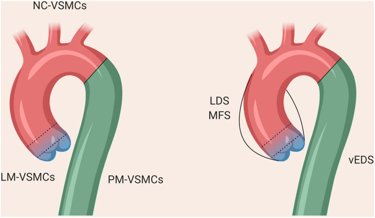
Thoracic aortic diseases, whether sporadic or due to a genetic disorder such as Marfan syndrome, lack effective medical therapies, with limited translation of treatments that are highly successful in mouse models into the clinic. Patient-derived induced pluripotent stem cells (iPSCs) offer the opportunity to establish new human models of aortic diseases. Here we review the power and potential of these systems to identify cellular and molecular mechanisms underlying disease and discuss recent advances, such as gene editing, and smooth muscle cell embryonic lineage. In particular, we discuss the practical aspects of vascular smooth muscle cell derivation and characterization, and provide our personal insights into the challenges and limitations of this approach. Future applications, such as genotype-phenotype association, drug screening, and precision medicine are discussed. We propose that iPSC-derived aortic disease models could guide future clinical trials via "clinical-trials-in-a-dish", thus paving the way for new and improved therapies for patients.
Keywords: Loeys-Dietz; Marfan; aortic aneurysm; disease-in-a-dish; induced pluripotent stem cell; vascular smooth muscle.
Copyright © 2020 Davaapil, Shetty and Sinha.
Figures

Similar articles
-
Modeling aortic diseases using induced pluripotent stem cells.Stem Cells Transl Med. 2021 Feb;10(2):190-197. doi: 10.1002/sctm.20-0322. Epub 2020 Nov 12. Stem Cells Transl Med. 2021. PMID: 33179450 Free PMC article. Review.
-
Differentiation and Application of Induced Pluripotent Stem Cell-Derived Vascular Smooth Muscle Cells.Arterioscler Thromb Vasc Biol. 2017 Nov;37(11):2026-2037. doi: 10.1161/ATVBAHA.117.309196. Epub 2017 Aug 31. Arterioscler Thromb Vasc Biol. 2017. PMID: 28860223 Review.
-
Reverse engineering human neurodegenerative disease using pluripotent stem cell technology.Brain Res. 2016 May 1;1638(Pt A):30-41. doi: 10.1016/j.brainres.2015.09.023. Epub 2015 Sep 28. Brain Res. 2016. PMID: 26423934 Free PMC article. Review.
-
Genetics of thoracic aortic aneurysm: at the crossroad of transforming growth factor-β signaling and vascular smooth muscle cell contractility.Circ Res. 2013 Jul 19;113(3):327-40. doi: 10.1161/CIRCRESAHA.113.300675. Circ Res. 2013. PMID: 23868829 Review.
-
Novel insights into disease modeling using induced pluripotent stem cells.Biol Pharm Bull. 2013;36(2):182-8. doi: 10.1248/bpb.b12-00960. Biol Pharm Bull. 2013. PMID: 23370349 Review.
Cited by
-
Integration of Transformative Platforms for the Discovery of Causative Genes in Cardiovascular Diseases.Cardiovasc Drugs Ther. 2021 Jun;35(3):637-654. doi: 10.1007/s10557-021-07175-1. Epub 2021 Apr 15. Cardiovasc Drugs Ther. 2021. PMID: 33856594 Free PMC article. Review.
-
Using Genomics to Identify Novel Therapeutic Targets for Aortic Disease.Arterioscler Thromb Vasc Biol. 2024 Feb;44(2):334-351. doi: 10.1161/ATVBAHA.123.318771. Epub 2023 Dec 14. Arterioscler Thromb Vasc Biol. 2024. PMID: 38095107 Free PMC article. Review.
-
An in vitro approach reveals molecular mechanisms underlying endocrine disruptor-induced epimutagenesis.Elife. 2024 Oct 3;13:RP93975. doi: 10.7554/eLife.93975. Elife. 2024. PMID: 39361026 Free PMC article.
-
Understanding genomic medicine for thoracic aortic disease through the lens of induced pluripotent stem cells.Front Cardiovasc Med. 2024 Feb 19;11:1349548. doi: 10.3389/fcvm.2024.1349548. eCollection 2024. Front Cardiovasc Med. 2024. PMID: 38440211 Free PMC article. Review.
-
Enigma of aortic aneurysms continues to be enigmatic!Indian J Thorac Cardiovasc Surg. 2022 Apr;38(Suppl 1):1-2. doi: 10.1007/s12055-021-01326-7. Epub 2022 Jan 15. Indian J Thorac Cardiovasc Surg. 2022. PMID: 35463704 Free PMC article. No abstract available.
References
Publication types
Grants and funding
LinkOut - more resources
Full Text Sources