. 2021 Jan 6;109(1):73-90.e7.
doi: 10.1016/j.neuron.2020.10.027. Epub 2020 Nov 11.

Mechanical Allodynia Circuitry in the Dorsal Horn Is Defined by the Nature of the Injury

Affiliations

Mechanical Allodynia Circuitry in the Dorsal Horn Is Defined by the Nature of the Injury

Cedric Peirs et al. Neuron. .

Abstract

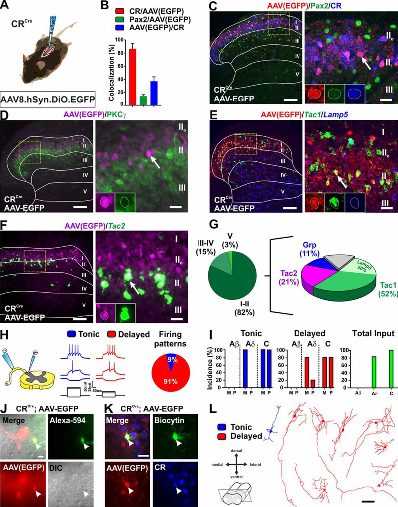
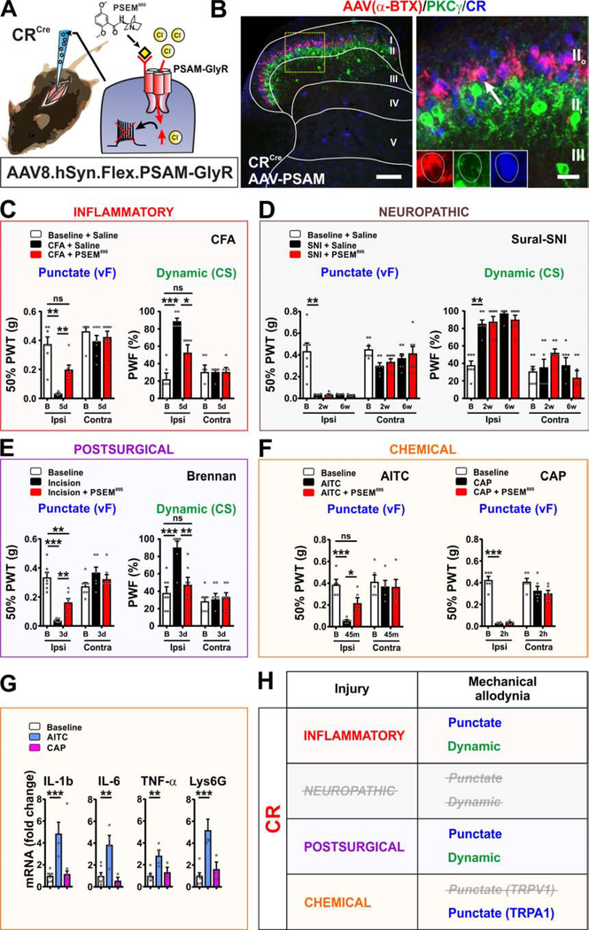
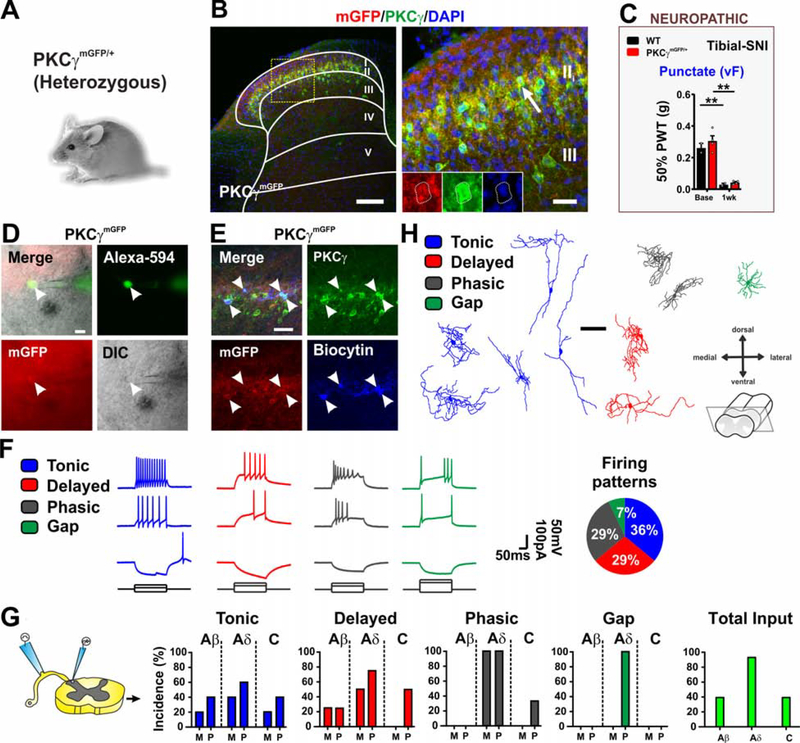
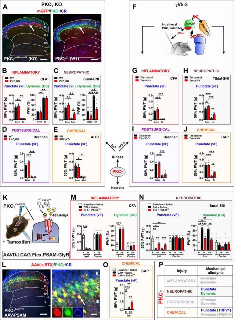
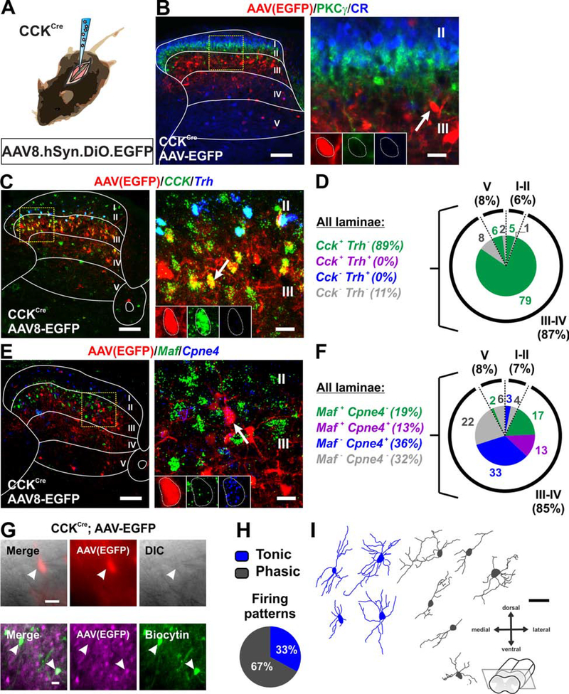
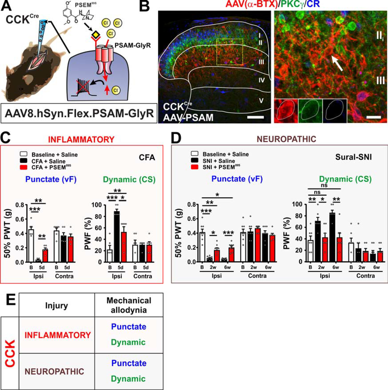
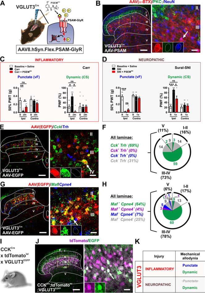
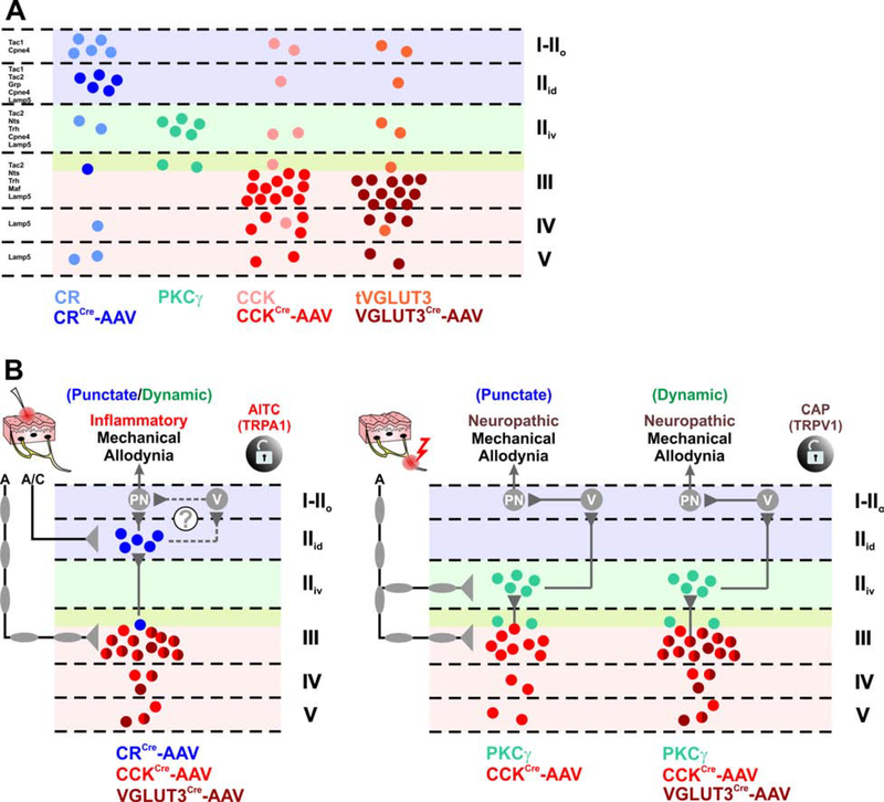
The spinal dorsal horn is a major site for the induction and maintenance of mechanical allodynia, but the circuitry that underlies this clinically important form of pain remains unclear. The studies presented here provide strong evidence that the neural circuits conveying mechanical allodynia in the dorsal horn differ by the nature of the injury. Calretinin (CR) neurons in lamina II inner convey mechanical allodynia induced by inflammatory injuries, while protein kinase C gamma (PKCγ) neurons at the lamina II/III border convey mechanical allodynia induced by neuropathic injuries. Cholecystokinin (CCK) neurons located deeper within the dorsal horn (laminae III-IV) are important for both types of injuries. Interestingly, the Maf+ subset of CCK neurons is composed of transient vesicular glutamate transporter 3 (tVGLUT3) neurons, which convey primarily dynamic allodynia. Identification of an etiology-based circuitry for mechanical allodynia in the dorsal horn has important implications for the mechanistic and clinical understanding of this condition.

Keywords: calretinin; cholecystokinin; dorsal horn; inflammatory pain; mechanical allodynia; neural circuitry; neuropathic pain; pain; protein kinase C gamma; vesicular glutamate transporter 3.


Conflict of interest statement

Declaration of Interests The authors declare no competing interests.

Figures

Figure 1.
Figure 1.. Molecular, electrophysiological and morphological characterization of virally targeted CR neurons.
(A) Schematic of CRCre mouse intraspinally injected with AAV8.hSyn.DIO.EGFP virus. (B-C) Most virally targeted EGFP+ neurons (red) express CR (blue) and a small number of those also express Pax2 (green). Yellow box shows location of insert. Arrow shows CR and EGFP colocalized cell. Scale bars = 50 μm and 10 μm. (D) EGFP+ neurons (magenta) do not overlap (arrow) with PKCγ (green). Yellow box shows location of insert. Arrow shows example of EGFP+ cell. Scale bars = 50 μm and 10 μm. (E-G) Viral expression of EGFP (red) is largely contained within dorsal horn lamina II. Infected neurons overlap with Tac1 (with and without Lamp5) (52%), Tac2 (21%) and Grp (11%). Yellow box shows location of insert. Arrow shows example of colocalized cell. Scale bars = 50 μm and 10 μm. (H-I) Patch clamp recording in transverse lumbar spinal cord slices with dorsal root and DRG still attached. Nearly all EGFP+ neurons show delayed firing pattern in response to current injection. One shows tonic firing. Neurons receive monosynaptic Aδ and mono- and poly-synaptic C-fiber input with no Aβ input. (n=11 cells, N= 4 mice). M= monosynaptic, P= polysynaptic. (J-K) Recorded cells are filled with Alexa-594 and biocytin for post-hoc reconstruction and to confirm EGFP and CR expression. Scale bars = 20 μm. (L) Morphology of recorded cells in (H). Scale bar = 50 μm.
Figure 2.
Figure 2.. Targeted CR neurons in lamina II are important for conveying mechanical allodynia induced by inflammatory, but not neuropathic pain models.
(A) Schematic of CRCre mice injected intraspinally with AAV8.hSyn.FLEX.PSAM-GlyR virus. PSEM89S inhibits PSAM-GlyR expressing cells. (B) Dorsal horn shows α-BTX-Alexa647 binding (red) to PSAM-GlyR is confined to lamina II. Receptor overlaps with CR (blue) but not PKCγ (green) neurons. Yellow box shows location of insert. Arrow shows colocalized cell. Scale bars = 100 μm and 20 μm. (C) Mechanical allodynia induced by the CFA model is completely reversed by PSEM89S (p=0.0031, punctate; p=0.0185, dynamic). N=7 and 6 mice respectively. Paw withdrawal threshold (PWT) and paw withdrawal frequency (PWF). (D) PSEM89S injection has no effect on mechanical allodynia at 2 and 6 weeks after sural-SNI. N=7 mice. (E) In the postsurgical pain model, PSEM89S injection partially reverses punctate allodynia, and completely reverses dynamic allodynia (p=0.0082, punctate; p=0.0050, dynamic). N=7 mice. (F) Punctate mechanical allodynia after mustard oil (AITC) injection is reversed by PSEM89S (p=0.0485). N=5 mice. Punctate allodynia induced by capsaicin (CAP) injection is unaffected by PSEM89S. N=6 mice. (G) Levels of mRNAs for inflammatory mediators in the skin are increased after AITC, but not capsaicin (CAP) injection compared to vehicle injection. Skin was harvested immediately after allodynia measurements. N=7 baseline, 4 AITC and 3 CAP mice (H) Summary of the role of targeted CR neurons in mechanical allodynia induced by persistent pain models. See also Figure S1 and S2.
Figure 3.
Figure 3.. Electrophysiological and morphological characterization of PKCγ interneurons.
(A-B) Dorsal horn of adult mice heterozygous for myristoylated GFP knockin allele (PKCγmGFP/+) immunostained for mGFP (red), PKCγ (green) and DAPI (blue). GFP and PKCγ are co-expressed in lamina II (arrow). Yellow box shows location of insert. Scale bars = 50 μm and 10 μm. (C) PKCγmGFP/+ mice develop punctate mechanical allodynia similar to WT littermates after tibial-SNI. N= 2 WT and 4 PKCγmGFP/+ mice. (D-E) Patch clamp recording of mGFP+ cells in lamina II and II/III border in transverse spinal cord slices with dorsal root and DRG still attached. Recorded cells are filled with Alexa-594 and biocytin for post-hoc reconstruction and to confirm mGFP (red) and PKCγ (green) expression. Scale bars = 20 μm. (F-G) Firing patterns in response to current injection are tonic, delayed or phasic with a few gap. Tonic and delayed cells receive all input types, except delay neurons do not show monosynaptic C-fiber input. Phasic firing cells receive largely Aδ and some C-input but no Aβ input. Gap firing cells receive only polysynaptic Aδ input. (n=14 cells, N=6 mice). M= monosynaptic, P= polysynaptic. (H) Morphology of the recorded cells in (F). Scale bar = 50 μm.
Figure 4.
Figure 4.. PKCγ is important for conveying mechanical allodynia induced by neuropathic, but not inflammatory pain models.
(A) Mice homozygous for the mGFP knockin allele (PKCγmGFP/mGFP) express EGFP (red) but not the kinase (green) (PKCγ KO mice). Scale bar = 50 μm. (B) Normal punctate and dynamic allodynia after CFA in PKCγ KO mice. N=3–4 mice per genotype. (C) Attenuation of punctate (p=0.0138) and dynamic (p=0.0001) mechanical allodynia induced by sural-SNI in PKCγ KO (red bars) mice compared to WT littermates (black bars). N=4–9 mice per genotype. (D) Punctate allodynia induced by the postsurgical pain model does not differ between PKCγ KO and WT littermates. N=4 mice per genotype. (E) PKCγ KOs and WT littermates show similar punctate allodynia after AITC injection. N=3–4 mice per genotype. (F) Schematic of i.t. delivery of the PKCγ inhibitor, tat-γV5–3. The inhibitor prevents PKCγ from binding to the scaffolding protein RACK and its subsequent phosphorylation of substrates. (G) Compared to the tat-control peptide (100 pmoles) (black bars), injection of tat-γV5–3 (100 pmoles) (red bars) has no effect on punctate mechanical allodynia induced by CFA. N= 9 tat-γV5–3, N=6 tat control mice. (H) i.t. injection of tat-γV5–3 (100 pmoles) (red bars) partially reverses punctate mechanical allodynia (p=0.008) induced by tibial-SNI (black bars). A small effect of tat-γV5–3 versus tat-control peptide at baseline is noted (p=0.05). N= 10 tat-γV5–3, N=8 tat control mice. (I) i.t. injection of tat-γV5–3 (100 pmoles) (red bars) has no effect on punctate mechanical allodynia (black bars) in the postsurgical pain model. N= 10 tat-γV5–3, N=6 tat control mice. (J) Injection of tat-γV5–3 (100 pmoles) (red bars), but not the tat-control peptide (100 pmoles) (black bars) partially reverses punctate allodynia (p=0.0347) induced by CAP. N= 13 tat-γV5–3, N=9 tat control mice. (K) Schematic showing intraspinal injection of AAVDJ.CAG.FLEX.PSAM-GlyR virus into the PKCγCreERT2 mice. (L) Dorsal horn of PKCγCreERT2 mice expressing PSAM-GlyR shows colocalization of α-BTX-Alexa647 (red) with PKCγ (green) but not CR (blue). (M) Injection of PSEM89S has no effect on punctate or dynamic mechanical allodynia induced by CFA. N=6 mice. (N) Injection of PSEM89S at 2 and 6 weeks after sural-SNI partially reverses punctate (p=0.0036, 2wk; p=0.0002; 6wk) and fully reverses dynamic (p=0.0009, 2wk; p=0.0033, 6wk) allodynia. N=7 mice. (O) Punctate allodynia induced by CAP injected into the ankle is also partially reversed (p=0.0051) by PSEM89S. N=6 mice. (P) Summary of the role of PKCγ neurons in conveying mechanical allodynia induced by persistent pain models. See also Figure S3.
Figure 5.
Figure 5.. Molecular, electrophysiological and morphological characterization of virally targeted CCK neurons.
(A-B) Schematic of CCKCre mice injected with AAV8.hSyn.DIO.EGFP virus. In the dorsal horn, EGFP+ neurons (red) are predominantly in lamina III with scatter cells in laminae II, IV and V. Cells do not colocalize with PKCγ (green) or CR (blue). Yellow box shows location of insert. Arrow shows example of EGFP+ cell. Scale bars = 100 μm and 20 μm. (C-D) Nearly all EGFP+ neurons (red) express Cck (green). Consistent with no PKCγ colocalization, none express Trh (blue). Values are percentage of total EGFP+ neurons. Yellow box shows location of insert. Arrow shows example of colocalized cell. Scale bars = 100 μm and 20 μm. (E-F) Most EGFP+ neurons (red) overlap with Maf (green) and/or Cpne4 (blue). Values are percentage of total EGFP+ neurons. Yellow box shows location of insert. Arrow shows example of colocalized cell. Scale bars = 100 μm and 20 μm. (G) Patch clamp recordings (EGFP+, red or magenta) in transverse slices of lumbar dorsal horn under DIC and filled with Biocytin (green). Scale bars = 20 μm. (H) Firing patterns in response to current injection were tonic (67%) and phasic (33%). n=20 cells, N=6 mice. (I) Morphology of neurons recorded in (H). Scale bar = 50 μm.
Figure 6.
Figure 6.. Targeted CCK neurons convey mechanical allodynia induced by both inflammatory and neuropathic pain models.
(A-B) Schematic of CCKCre mice injected with AAV8.hSyn.FLEX.PSAM-GlyR virus. Dorsal horn shows α-BTX-Alexa647 binding (red) in lamina III with scattered cells in laminae II, IV and V. No overlap with PKCγ (green) or CR (blue) in laminae II or III. Yellow box shows location of insert. Arrow shows example of non-colocalized cell. Scale bars = 100 μm and 20 μm. (C) PSEM89S injection in CFA model partially reverses punctate (p=0.0051) and dynamic (p=0.0185) allodynia. N= 6 mice. (D) PSEM89S injection at 2 and 6 weeks after sural-SNI partially reverses punctate (p=0.0264, 2wk; p=0.0003, 6wk) and completely reverses dynamic (p=0.0155, 2wk; p=0.0022, 6wk) allodynia. N=7 mice. (E) Summary of the role of targeted CCK neurons in conveying mechanical allodynia in persistent pain models. See also Figure S4.
Figure 7.
Figure 7.
Role of targeted VGLUT3 neurons in conveying mechanical allodynia induced by inflammatory and neuropathic pain models. (A-B) Schematic of AAV8.hSyn.FLEX.PSAM-GlyR virus injected into VGLUT3Cre mice. Staining with α-BTX-Alexa647 (red) shows expression in lamina III with scattered cells in laminae II, IV and V. The α-BTX-Alexa647 (red) does not overlap with PKCγ (green). Yellow box shows location of insert. Arrow shows example of α-BTX-Alexa647 (PSAM-GlyR) positive cell. Scale bars = 100 μm and 20 μm. (C) PSEM89S injection reverses punctate (p=0.0109) and dynamic (p=0.0126) allodynia in the carrageenan model. N=7 mice (D) PSEM89S does not reverse punctate allodynia, but markedly reverses dynamic allodynia (p=0.0188) in the sural-SNI model. N= 6 mice. (E-F) Dorsal horns from VGLUT3Cre mice injected with AAV8.hSyn.DIO.EGFP show EGFP+ neurons (red) primarily in dorsal horn laminae III-IV, where most overlap with Cck (green). Fewer EGFP cells express Cck in laminae II and V. No EGFP+ neurons overlap with Trh (blue). Values are percentage of total EGFP+ neurons. Yellow box shows location of insert. Arrow shows example of colocalized cell. Scale bars = 100 μm and 20 μm. (G-H) EGFP+ neurons (red) overlap with only Maf (red) (64%). Few express only Cpne4 (blue) (7%) or both Maf and Cpne4 (4%). Yellow box shows location of insert. Arrow shows example of a colocalized cell. Scale bars = 100 μm and 20 μm. (I-J) VGLUT3EGFP BAC transgenic crossed to CCKCre;Ai14-tdTomato mice show both reporters reside in lamina III with scattered cells in laminae II, IV and V. Most EGFP+ neurons (84%) express tdTomato. Yellow box shows location of insert. Arrow shows example of colocalized cell. Scale bars = 100 μm and 20 μm. (K) Summary of the role of targeted tVGLUT3 neurons in conveying mechanical allodynia induced by persistent pain models. See also Figure S5.
Figure 8.
Figure 8.. Dorsal horn circuitry underlying mechanical allodynia depends on the injury type.
(A) Schematic of the laminar distribution of virally targeted (AAV) and lineage-derived populations discussed in this study. Virally targeted CR neurons (dark blue) are mostly restricted to the dorsal-inner part of lamina II (IIid) and largely exclude other CR-lineage neurons (light blue) in lamina I, the deep layers, and those expressing PKCγ. Nearly all PKCγ neurons were targeted by the virus (light green). Virally targeted CCK neurons (dark red) are highly concentrated in lamina III with scattered cells in laminae IV-V and generally exclude CCK-lineage neurons in the superficial laminae including those expressing PKCγ (light red). Virally targeted tVGLUT3 neurons (dark brown) are highly concentrated in lamina III with scattered cells in laminae IV-V and exclude tVGLUT3-lineage neurons in superficial laminae including those expressing PKCγ (light brown). (B) Schematic diagram of the mechanical allodynia circuitry in the context of injury. Targeted CR neurons receive A- and C-fiber input whereas targeted CCK/tVGLUT3 neurons receive predominantly A-fiber input. Left circuit: After inflammation, punctate or dynamic mechanical allodynia engage neurons expressing CCK/tVGLUT3 in lamina III, which activate targeted CR neurons in lamina IIid. These CR neurons ultimately activate projection neurons (PN) directly, or potentially indirectly through vertical cells, to elicit mechanical allodynia. A similar circuit is observed after activating TRPA1 through peripheral injection of mustard oil (AITC). Right circuit: After nerve injury, punctate mechanical allodynia engages CCK, but not tVGLUT3 neurons in lamina III. These neurons then activate PKCγ neurons in lamina IIid and ultimately projection neurons (PN) indirectly through vertical cells. A similar circuit is observed for dynamic mechanical allodynia, but also includes targeted CCK/tVGLUT3 neurons in lamina III. Mechanical allodynia induced by peripheral injection of the TRPV1 agonist capsaicin (CAP) also relies on PKCγ and CCK neurons.

Comment in

Similar articles

Cited by

References

    1. Abraira VE, Kuehn ED, Chirila AM, Springel MW, Toliver AA, Zimmerman AL, Orefice LL, Boyle KA, Bai L, Song BJ, et al. (2017). The Cellular and Synaptic Architecture of the Mechanosensory Dorsal Horn. Cell 168, 295–310 e219. - PMC - PubMed
    1. Alba-Delgado C, El Khoueiry C, Peirs C, Dallel R, Artola A, and Antri M (2015). Subpopulations of PKCgamma interneurons within the medullary dorsal horn revealed by electrophysiologic and morphologic approach. Pain 156, 1714–1728. - PubMed
    1. Alba-Delgado C, Mountadem S, Mermet-Joret N, Monconduit L, Dallel R, Artola A, and Antri M (2018). 5-HT2A Receptor-Induced Morphological Reorganization of PKCgamma-Expressing Interneurons Gates Inflammatory Mechanical Allodynia in Rat. J Neurosci 38, 10489–10504. - PMC - PubMed
    1. Alexander GM, Rogan SC, Abbas AI, Armbruster BN, Pei Y, Allen JA, Nonneman RJ, Hartmann J, Moy SS, Nicolelis MA, et al. (2009). Remote control of neuronal activity in transgenic mice expressing evolved G protein-coupled receptors. Neuron 63, 27–39. - PMC - PubMed
    1. Artola A, Voisin D, and Dallel R (2020). PKCgamma interneurons, a gateway to pathological pain in the dorsal horn. J Neural Transm (Vienna) 127, 527–540. - PubMed

Publication types

Substances