. 2020 Nov 12;16(11):e1008959.
doi: 10.1371/journal.pgen.1008959. eCollection 2020 Nov.

Meiosis reveals the early steps in the evolution of a neo-XY sex chromosome pair in the African pygmy mouse Mus minutoides

Affiliations

Meiosis reveals the early steps in the evolution of a neo-XY sex chromosome pair in the African pygmy mouse Mus minutoides

Ana Gil-Fernández et al. PLoS Genet. .

Abstract

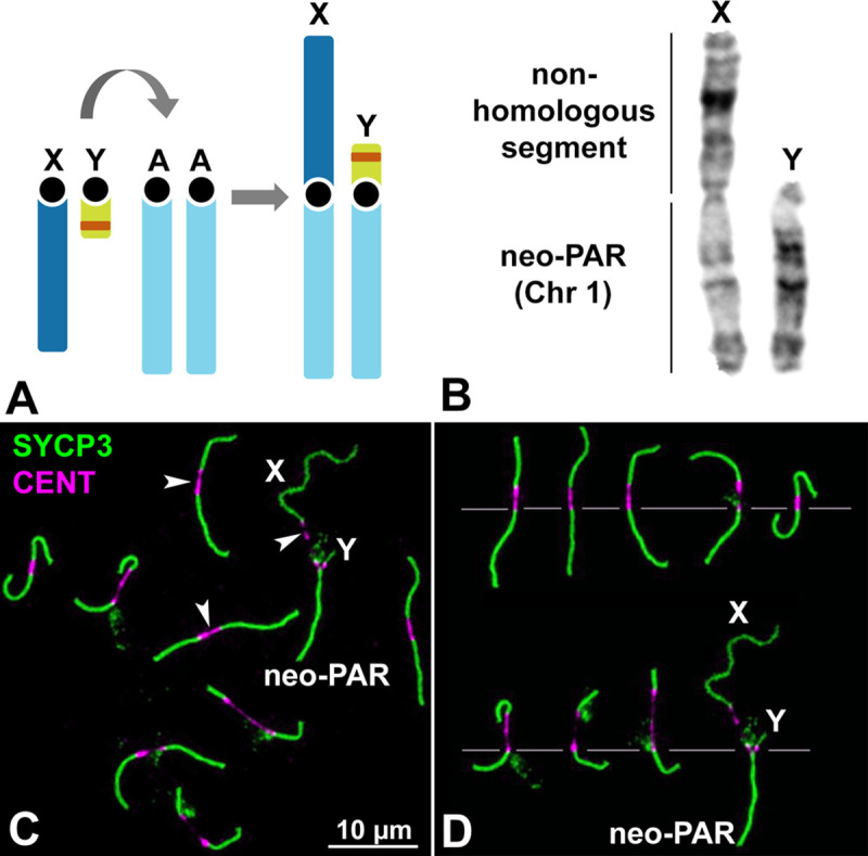
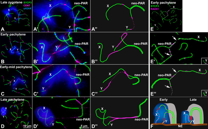
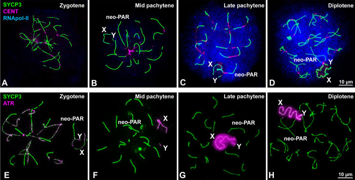
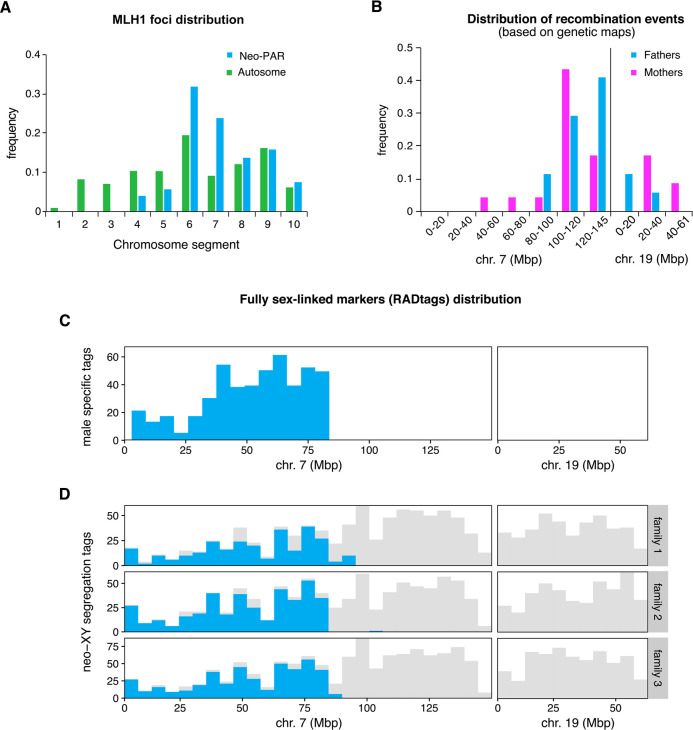
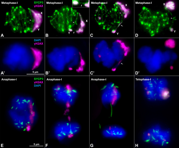
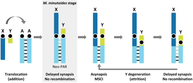
Sex chromosomes of eutherian mammals are highly different in size and gene content, and share only a small region of homology (pseudoautosomal region, PAR). They are thought to have evolved through an addition-attrition cycle involving the addition of autosomal segments to sex chromosomes and their subsequent differentiation. The events that drive this process are difficult to investigate because sex chromosomes in almost all mammals are at a very advanced stage of differentiation. Here, we have taken advantage of a recent translocation of an autosome to both sex chromosomes in the African pygmy mouse Mus minutoides, which has restored a large segment of homology (neo-PAR). By studying meiotic sex chromosome behavior and identifying fully sex-linked genetic markers in the neo-PAR, we demonstrate that this region shows unequivocal signs of early sex-differentiation. First, synapsis and resolution of DNA damage intermediates are delayed in the neo-PAR during meiosis. Second, recombination is suppressed or largely reduced in a large portion of the neo-PAR. However, the inactivation process that characterizes sex chromosomes during meiosis does not extend to this region. Finally, the sex chromosomes show a dual mechanism of association at metaphase-I that involves the formation of a chiasma in the neo-PAR and the preservation of an ancestral achiasmate mode of association in the non-homologous segments. We show that the study of meiosis is crucial to apprehend the onset of sex chromosome differentiation, as it introduces structural and functional constrains to sex chromosome evolution. Synapsis and DNA repair dynamics are the first processes affected in the incipient differentiation of X and Y chromosomes, and they may be involved in accelerating their evolution. This provides one of the very first reports of early steps in neo-sex chromosome differentiation in mammals, and for the first time a cellular framework for the addition-attrition model of sex chromosome evolution.


Conflict of interest statement

The authors have declared that no competing interests exist.

Figures

Fig 1
Fig 1. Sex chromosomes in M. minutoides.
A. Schematic representation of the Rb translocation between the sex chromosomes (X and Y) and autosomal chromosome 1 (A). The ancestral sex chromosomes are completely differentiated and, thus, achiasmate. The translocation results in a large neo-PAR constituted by chromosome 1. The orange band in the Y chromosome represents the sex-determining locus. B. G-banding of the sex chromosomes indicating the neo-PAR and the non-homologous segment. C. Spread spermatocyte at pachytene labelled with antibodies against SYCP3 (green) and centromeres (magenta). The trajectories of SCs are interrupted at the centromeres, which appear largely stretched (arrowheads). Non-homologous segments of the sex chromosomes (X, Y) and the neo-PAR are indicated. D. Meiotic karyotype of M. minutoides. Bivalents are arranged according to the length of their largest arm, as previously described [50, 72]. The sex bivalent is largely heteromorphic in the non-homologous segments.
Fig 2
Fig 2. Synapsis and DNA repair in the sex chromosomes.
A-D”. Spread spermatocytes in prophase-I labelled with antibodies against SYCP3 (green), centromeres (magenta) and γH2AX (blue). Non-homologous segments of the sex chromosomes (X, Y) and the neo-PAR are indicated. A-A”. Zygotene. Most autosomes have completed synapsis. Enlarged view of the sex chromosomes (A’) and their schematic representation (A”). Both the non-homologous segments and a large portion of the neo-PAR are unsynapsed and labelled with γH2AX. In some cases, centromeres are so elongated that the signal is very weak (represented as dashed lines). B-B”. Early pachytene. Autosomes have fully synapsed but sex chromosomes still have not completed synapsis in the neo-PAR. The centromeres appear greatly stretched. C-C”. Mid-pachytene. Synapsis in the sex chromosomes has extended but still is not complete. D-D”. Late pachytene. The neo-PAR has completely synapsed. Although the non-homologous segments of the sex chromosomes remain unsynapsed, they are in close proximity, forming a single chromatin mass that is strongly labelled with γH2AX. E-E”. Details and schematic representation of a sex bivalent from an early pachytene spermatocyte labelled with antibodies against SYCP3 (green) and RAD51 (magenta). The proximal regions of the neo-PAR are unsynapsed and accumulate RAD51 foci. The non-homologous segments also accumulate RAD51. Arrows and dashed lines (in E”) indicate putative centromere positions. F. Schematic representation of synapsis and DNA repair in the sex chromosomes. neo-PAR AEs/LEs are represented in green, AEs of X and Y differential segments are represented in blue and yellow respectively, centromeres in red, TFs in yellow, RAD51 in pink, chromatin in grey, γH2AX in blue and nuclear envelope (NE) in orange. At early pachytene, synapsis proceeds from the distal end of the neo-PAR; however, compared with autosomes, it is delayed. Unsynapsed regions accumulate DNA repair proteins (pink foci). As synapsis proceeds, the X chromosome bends to allow the complete synapsis of the neo-PAR. Repair proteins are also removed from the neo-PAR but remain on the unsynapsed non-homologous segments.
Fig 3
Fig 3. Inactivation of sex chromosomes.
A-D. Spread spermatocytes at different stages of prophase-I labelled with antibodies against SYCP3 (green), centromeres (magenta) and RNA polymerase-II (blue). Non-homologous segments of the sex chromosomes (X, Y) and the neo-PAR are indicated. A. RNA pol-II is absent from the nucleus during zygotene. B. At mid pachytene, a weak RNA pol-II signal is detected in several areas of the nucleus. C. By late pachytene, the nuclear RNA pol-II signal has greatly increased and is maintained through diplotene (D). In contrast to the neo-PAR and autosomes, the non-homologous segments of the sex chromosomes (X, Y) are mostly or completely devoid of RNA pol-II. E-H. Spread spermatocytes at different stages of prophase-I labelled with antibodies against SYCP3 (green) and ATR (magenta). E. At zygotene, ATR foci are observed along the unsynapsed regions of the autosomes and sex chromosomes. F. At mid pachytene, ATR is only seen on the unsynapsed regions of the sex chromosomes. At late pachytene (G) and diplotene (H), the ATR signal has extended to the chromatin of the non-homologous segments of the sex chromosomes. The neo-PAR is not labelled by ATR.
Fig 4
Fig 4. Recombination and position of fully sex-linked genetic markers on the neo-sex chromosomes.
A. Distribution of MLH1 foci along the neo-PAR in M. minutoides (blue) and the longest autosomal arm (green) in 91 spermatocytes from three males. The chromosomes have been divided into 10 equal segments from the centromere (1) to the distal telomere (10). B. Distribution of recombination events across the neo-sex chromosomes in the parents of families 1 and 3 (magenta: mothers, blue: fathers) based on the recombination maps build independently for the four parents. C. Distribution of male-specific RADtags across the neo-sex chromosomes, according to their mapping position to M. musculus domesticus chromosomes 7 and 19. D. Distribution of tags carrying SNPs with a fully sex-linked segregation pattern along the neo-sex chromosomes, according to their mapping position to M. musculus domesticus chromosomes 7 and 19 (shown in blue). Tags that carry at least one SNP, regardless of their transmission pattern, are shown in grey.
Fig 5
Fig 5. Sex chromosome segregation.
Squashed spermatocytes labelled with antibodies against SYCP3 (green) and γH2AX (magenta) and counterstained with DAPI (blue). Non-homologous segments of the sex chromosomes (X, Y) are indicated. A-D’. Metaphase-I. Different configurations of the sex bivalent can be observed. The neo-PAR always shows a chiasma (arrowheads), however, the non-homologous segments can be associated with each other by forming a common chromatin mass (A-A’), or through a SYCP3-positive filament (arrow) (see enlarged detail on the top) (B-B’) or a γH2AX-positive filament (arrow) (C-C’). Alternatively, these segments can appear completely separated (D-D’). E-G. Anaphase-I. γH2AX connections (E) and/or SYCP3 (F, G) filaments are observed between segregating chromosomes during early anaphase, resembling the associations observed at metaphase-I. At telophase-I (H), no lagging or mis-segregated chromosomes are observed.
Fig 6
Fig 6. Schematic representation of a possible scenario for the evolution of the neo-sex chromosomes in the addition-attrition model for Mus minutoides.
Before sex-autosome translocation, the autosomal pair shows standard (chromosome-wide) synapsis and recombination (black bars). Following translocation, due to structural constraints imposed by the attachment of chromosomes to the nuclear envelop and the difference in size between the X and Y, synapsis is delayed and recombination halted in the centromeric region of the neo-PAR (grey bars). These processes could consequently lead to genetic differentiation of the neo-X and neo-Y, and once meiotically essential genes have been transposed/translocated to autosomes, the region will be free to undergo large rearrangements triggering asynapsis and MSCI. This is the last nail in the coffin, causing the attrition of the Y. At this point, a new round of synapsis and recombination impairment could start over, promoting the recurrent attrition of the Y chromosome.

Similar articles

Cited by

References

    1. Ohno S. Sex chromosomes and sex linked genes. Berlin: Springer; 1967.
    1. Bachtrog D. Y-chromosome evolution: emerging insights into processes of Y-chromosome degeneration. Nat Rev Genet. 2013;14(2):113–24. Epub 2013/01/19. 10.1038/nrg3366 - DOI - PMC - PubMed
    1. Charlesworth D, Charlesworth B, Marais G. Steps in the evolution of heteromorphic sex chromosomes. Heredity. 2005;95(2):118–28. 10.1038/sj.hdy.6800697 . - DOI - PubMed
    1. Rice WR. Evolution of the Y Sex Chromosome in Animals: Y chromosomes evolve through the degeneration of autosomes. BioScience. 1996;46(5):331–43. 10.2307/1312947 - DOI
    1. Bachtrog D. A dynamic view of sex chromosome evolution. Curr Opin Genet Dev. 2006;16(6):578–85. Epub 2006/10/24. 10.1016/j.gde.2006.10.007 . - DOI - PubMed

Publication types

Grants and funding

This work was supported by Ministerio de Economía y Competitividad (Spain) (Grant number CGL2014-53106-P to JP), French National Research Agency (ANR grant SEXYMUS 10-JCJC-1605 and and ANR grant SEXREV 18-CE02-0018-01 to FV), Del Duca Foundation from the Institut de France (“subvention scientifique”), and the Swiss National Science Foundation (grant 31003A 166323 to NP). A.G-F. was supported by a predoctoral fellowship from the Ministerio de Economía y Competitividad (Spain) and the European Social Fund (European Commission). P.A.S was supported by a postdoctoral fellowship from MUSE program “investissements d’avenir” (ANR-16-IDEX-0006). Computations were performed at Vital-IT (www.vital-it.ch), a center for high-performance computing of the SIB Swiss Institute of Bioinformatics, and the Montpellier Bioinformatics Biodiversity (MBB) platform of ISEM, supported by the LabEx CeMEB. Some cytogenetic works were also performed at the CytoEvol platform of ISEM supported by the Labex CeMEB. The funders had no role in study design, data collection and analysis, decision to publish, or preparation of the manuscript.

LinkOut - more resources