

Innate Immune Effectors Play Essential Roles in Acute Respiratory Infection Caused by Klebsiella pneumoniae
- PMID: 33163539
- PMCID: PMC7607282
- DOI: 10.1155/2020/5291714
Innate Immune Effectors Play Essential Roles in Acute Respiratory Infection Caused by Klebsiella pneumoniae
Abstract
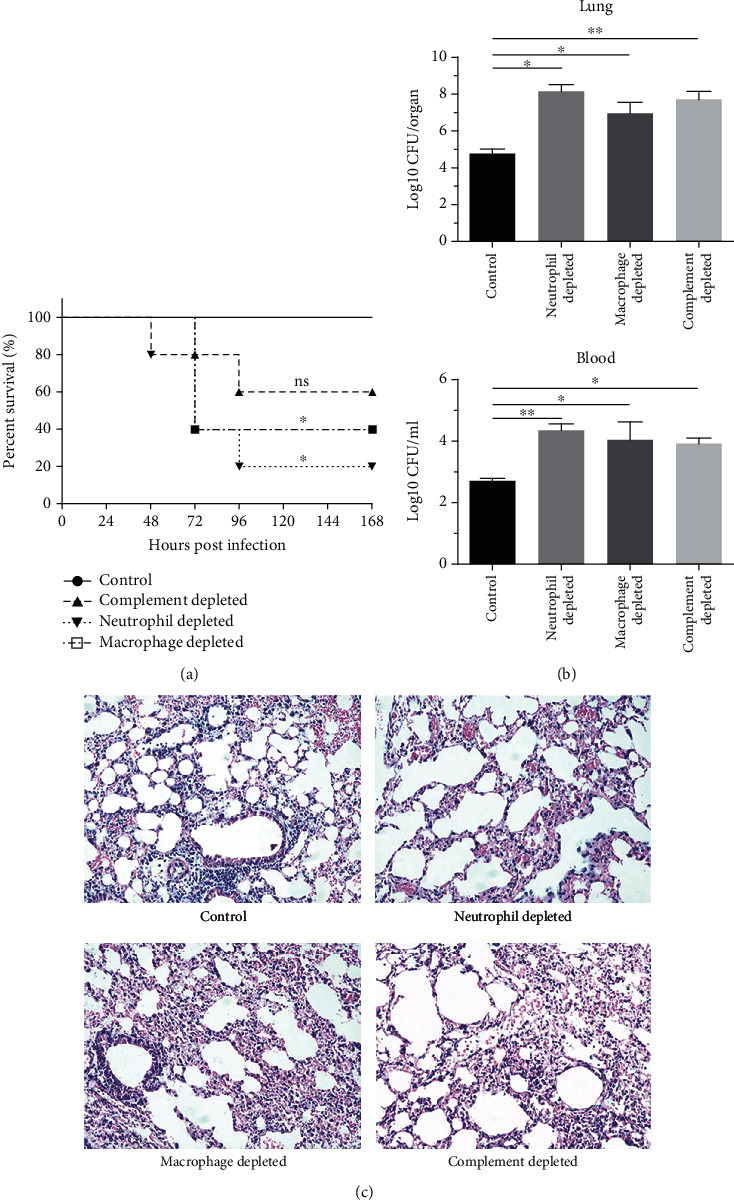
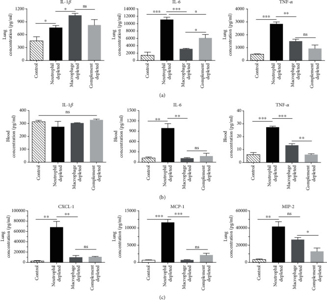
Innate immune effectors constitute the first line of host defense against pathogens. However, the roles of these effectors are not clearly defined during Klebsiella pneumoniae (K. pneumoniae) respiratory infection. In the current study, we established an acute pneumonia model of K. pneumoniae respiratory infection in mice and confirmed that the injury was most severe 48 h post infection. Flow cytometric assay demonstrated that alveolar macrophages were the predominant cells in BALF before infection, and neutrophils were quickly recruited after infection, and this was in consistent with the kinetics of chemokine expression. Further, we depleted neutrophils, macrophages, and complement pathways in vivo and challenged these mice with a sublethal dose of K. pneumonia, the result showed that 80%, 60%, and 40% of mice were died in these groups, respectively, while no deaths occurred in the control group. Besides, innate immune effector depleted mice showed higher bacterial burdens in lungs and blood, companied with more severe lung damage and increased levels of cytokine/chemokine expression. These results demonstrated that the innate immune effectors are critical in the early controlling of K. pneumoniae infection, and neutrophils are the most important. Thus, alternative strategies targeting these innate immune effectors may be effective in controlling of K. pneumoniae respiratory infection.
Copyright © 2020 Dong Liu et al.
Conflict of interest statement
The authors declare that the research was conducted in the absence of any commercial or financial relationships that could be construed as a potential conflict of interest.
Figures
Similar articles
-
SKAP2 is required for defense against K. pneumoniae infection and neutrophil respiratory burst.Elife. 2020 Apr 30;9:e56656. doi: 10.7554/eLife.56656. Elife. 2020. PMID: 32352382 Free PMC article.
-
Cyclic di-GMP stimulates protective innate immunity in bacterial pneumonia.Infect Immun. 2007 Oct;75(10):4942-50. doi: 10.1128/IAI.01762-06. Epub 2007 Jul 23. Infect Immun. 2007. PMID: 17646358 Free PMC article.
-
Nitric oxide is required for effective innate immunity against Klebsiella pneumoniae.Infect Immun. 1997 May;65(5):1870-5. doi: 10.1128/iai.65.5.1870-1875.1997. Infect Immun. 1997. PMID: 9125574 Free PMC article.
-
Modulation of pulmonary innate immunity during bacterial infection: animal studies.Arch Immunol Ther Exp (Warsz). 2002;50(3):159-67. Arch Immunol Ther Exp (Warsz). 2002. PMID: 12098931 Review.
-
Innate immunity, cytokines, and pulmonary host defense.Infect Dis Clin North Am. 1998 Sep;12(3):555-67, vii. doi: 10.1016/s0891-5520(05)70198-7. Infect Dis Clin North Am. 1998. PMID: 9779378 Review.
Cited by
-
Genomic and Immunological Characterization of Hypermucoviscous Carbapenem-Resistant Klebsiella pneumoniae ST25 Isolates from Northwest Argentina.Int J Mol Sci. 2022 Jul 1;23(13):7361. doi: 10.3390/ijms23137361. Int J Mol Sci. 2022. PMID: 35806365 Free PMC article.
-
Inhibiting lipid droplet biogenesis enhances host protection against hypervirulent Klebsiella pneumoniae infections.Med Microbiol Immunol. 2024 Nov 14;213(1):26. doi: 10.1007/s00430-024-00807-x. Med Microbiol Immunol. 2024. PMID: 39541006 Free PMC article.
-
Host Immune Response to Clinical Hypervirulent Klebsiella pneumoniae Pulmonary Infections via Transcriptome Analysis.J Immunol Res. 2022 Sep 20;2022:5336931. doi: 10.1155/2022/5336931. eCollection 2022. J Immunol Res. 2022. PMID: 36249423 Free PMC article.
-
Respiratory Commensal Bacteria Increase Protection against Hypermucoviscous Carbapenem-Resistant Klebsiella pneumoniae ST25 Infection.Pathogens. 2022 Sep 19;11(9):1063. doi: 10.3390/pathogens11091063. Pathogens. 2022. PMID: 36145495 Free PMC article.
-
The Therapeutic Treatment with the GAG-Binding Chemokine Fragment CXCL9(74-103) Attenuates Neutrophilic Inflammation and Lung Dysfunction during Klebsiella pneumoniae Infection in Mice.Int J Mol Sci. 2022 Jun 2;23(11):6246. doi: 10.3390/ijms23116246. Int J Mol Sci. 2022. PMID: 35682923 Free PMC article.
References
-
- Candevir Ulu A., Kurtaran B., Inal A. S., et al. Risk factors of carbapenem-resistant Klebsiella pneumoniae infection: a serious threat in ICUs. Medical science monitor: international medical journal of experimental and clinical research. 2015;21:219–224. doi: 10.12659/MSM.892516. - DOI - PMC - PubMed
MeSH terms
Substances
LinkOut - more resources
Full Text Sources
