Review
. 2021 Apr:67:101-109.
doi: 10.1016/j.sbi.2020.10.007. Epub 2020 Nov 5.

NEDD8 and ubiquitin ligation by cullin-RING E3 ligases

Affiliations
Review

NEDD8 and ubiquitin ligation by cullin-RING E3 ligases

Kheewoong Baek et al. Curr Opin Struct Biol. 2021 Apr.

Abstract

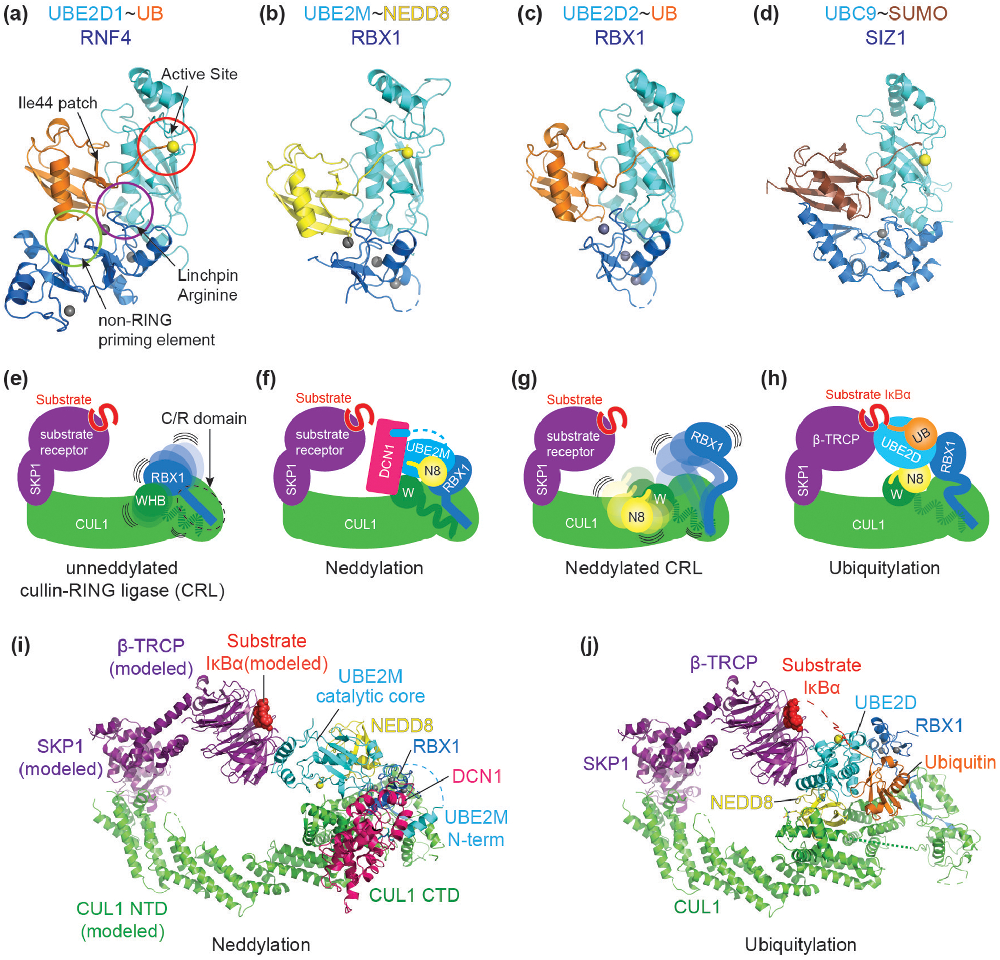
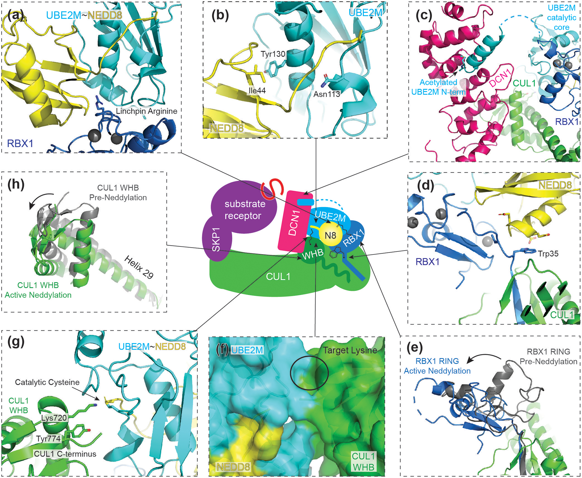
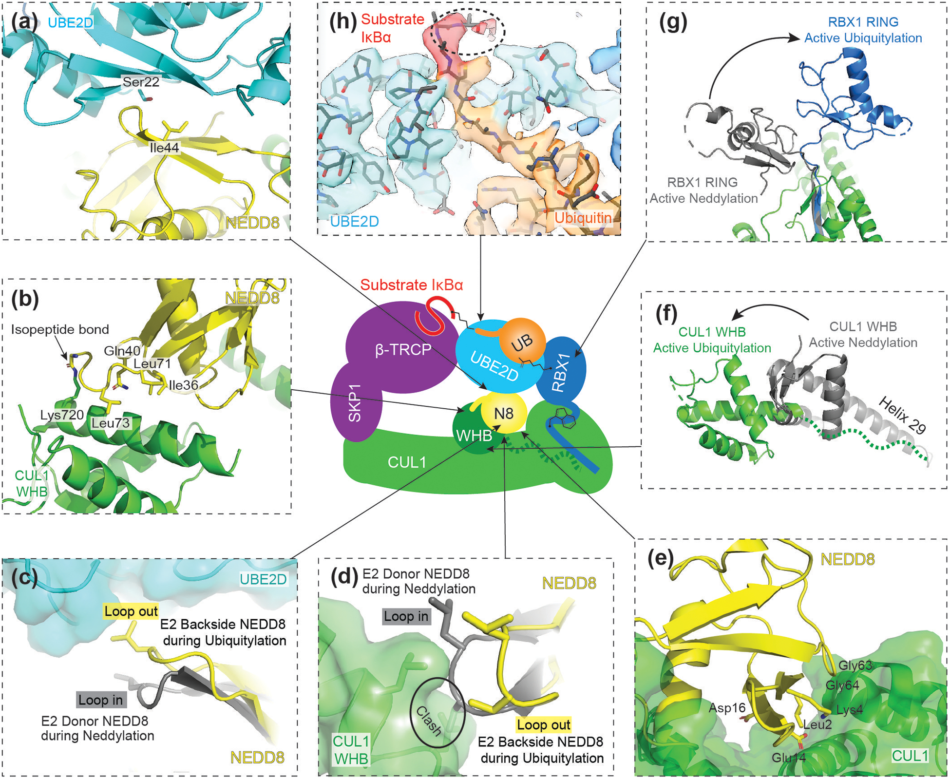
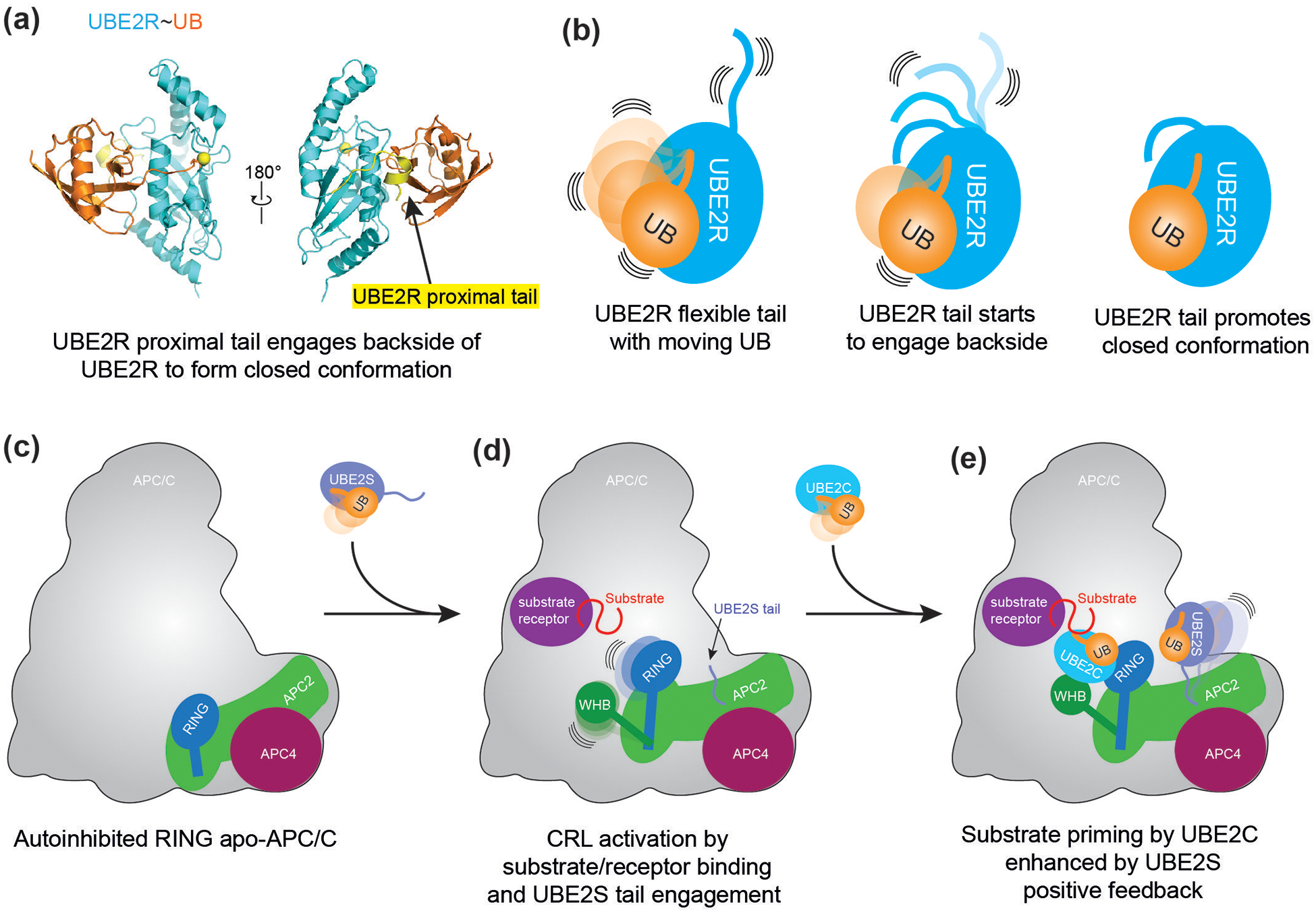
RING E3s comprise the largest family of ubiquitin (UB) and ubiquitin-like protein (UBL) ligases. RING E3s typically promote UB or UBL transfer from the active site of an associated E2 enzyme to a distally-recruited substrate. Many RING E3s - including the cullin-RING ligase family - are multifunctional, interacting with various E2s (or other E3s) to target distinct proteins, transfer different UBLs, or to initially modify substrates with UB or subsequently elongate UB chains. Here we consider recent structures of cullin-RING ligases, and their partner E2 enzymes, representing ligation reactions. The studies collectively reveal multimodal mechanisms - interactions between ancillary E2 or E3 domains, post-translational modifications, or auxiliary binding partners - directing cullin-RING E3-E2 enzyme active sites to modify their specific targets.


Conflict of interest statement

Conflict of interest

The authors declare no competing interest.

Figures

Figure 1
Figure 1. Context determines specificity of structurally similar RING-E2~UB/UBL modules.
(a) A common closed conformation across RING E3-E2~UB/UBL assemblies (“~” refers to thioester linkage between E2 catalytic cysteine and UB/UBL C-terminus). In the closed conformation, ubiquitin’s Ile44 hydrophobic interacts with the E2. This orientation is reinforced through interaction with a RING E3 linchpin (often an arginine, purple circle) and non-RING priming element (light green circle). The closed conformation stabilizes the position of the UB/UBL C-terminal tail, thereby activating the E2~ubiquitin active site (red circle) for catalysis. Highlighted in yellow sphere is the catalytic residue of E2 throughout Figure 1. Representative example shown is RNF4-UBE2D1~UB (PDB ID: 4AP4). (b) Cullin associated RBX1’s RING domain activates multiple E2~UBL conjugates. RBX1 promotes closed conformation of UBE2M~NEDD8 (PDB ID: 4P5O). (c) RBX1’s RING domain interacts with and promotes the closed conformation of UBE2D~UB (PDB ID: 6TTU). (d) SIZ1 promotes UBC9~SUMO closed conformation (PDB ID: 5JNE). (e) Schematic of a unneddylated cullin-RING ligase (CRL) bound to a SKP1-Fbox-substrate complex with its N-terminus on the left side (EMDB ID: 10582). On the right side, RBX1’s catalytic RING domain is flexibly tethered to RBX1’s N-terminal strand, embedded within the cullin forming a “C/R” domain (dotted circle). CUL1’s WHB domain is flexibly tethered to its connecting helix-29. (f) Schematic of a cullin-RING ligase neddylation intermediate (PDB ID: 4P5O). RBX1 RING facilitates closed conformation of UBE2M~NEDD8, and with the participation of a co-E3 ligase DCN1, promotes transfer of NEDD8 to CUL1’s Lys720 located within the WHB domain. (g) Schematic of a neddylated cullin-RING ligase (EMDB ID: 10583). Once NEDD8 is isopeptide linked to CUL1’s WHB domain, RBX1’s RING domain is further liberated. NEDD8, the linked WHB domain, and RBX1’s RING domain sample multiple orientations with enhanced flexibility. (h) Schematic of a cullin-RING ligase intermediate during ubiquitylation (PDB ID: 6TTU). RBX1 promotes closed conformation of UBE2D~UB. (i) Crystal structure of CUL1-RBX1-DCN1-UBE2M-NEDD8 representing an intermediate for neddylation (PDB ID: 4P5O. CUL1’s N-terminal domain (NTD) and SKP1-β-TRCP-IκBα are modeled (from PDB ID: 6TTU). (j) Cryo-EM structure of CUL1-RBX1-SKP1-β-TRCP-IκBα-UBE2D-Ubiquitin representing an intermediate for ubiquitylation (PDB ID: 6TTU).
Figure 2
Figure 2. Structure of CUL1-RBX1 intermediate during neddylation.
Central cartoon represents the CUL1 neddylation intermediate structure (PDB ID: 4P5O), with arrows indicating locations of structural details. (a) RBX1 displays a linchpin arginine in a non-conical location, which promotes closing of the UBE2M~NEDD8 conjugate. (b) When activated, NEDD8’s Ile44 hydrophobic patch interacts with UBE2M’s central helix. The C-terminal tail of NEDD8 is locked and primed for catalysis by Asn113 of UBE2M. (c) UBE2M~NEDD8 recruitment to CRL is facilitated by both RING E3 RBX1 and co-E3 DCN1. UBE2M’s acetylated N-terminus engages a hydrophobic pocket of DCN1 while its catalytic domain interacts with the RBX1 RING domain. (d) Unique residues of NEDD8 orient RBX1. NEDD8 steers UBE2M~NEDD8 by aligning two charged residues unique to NEDD8 to RBX1’s Trp35. Other residues in between RBX1’s cullin-binding strand and catalytic RING domain also function as non-RING priming elements. (e) Comparison of RBX1’s RING position before (grey) and during active neddylation (blue). During neddylation, RBX1 is reoriented, positioning UBE2M~NEDD8 for its active site to align with CUL1’s target lysine. (f) CUL1’s WHB domain and UBE2M present complementary surfaces to buttress the active conformation. (g) CUL1’s acceptor Lys720 is guided by neighboring residues of CUL1, including Tyr774 and CUL1’s C-terminus. The acceptor lysine itself intrinsically activates UBE2M~NEDD8. (h) Comparison of CUL1’s WHB domain before (grey) and during active neddylation (green). CUL1’s WHB domain and its connecting helix 29 translocate during neddylation.
Figure 3
Figure 3. Structure of neddylated CRL1 intermediate during ubiquitylation.
Central cartoon represents neddylated CRL1β-TRCP ubiquitylation intermediate structure, where arrows indicate locations of structural details. (a) NEDD8’s Ile44 hydrophobic patch interacts with UBE2D’s backside, opposite of the active site, during ubiquitylation. This interaction encompasses several residues of UBE2D’s backside including Ser22. (b) During ubiquitylation, NEDD8 and its isopeptide-linked CUL1 WHB interact via an interface that involves NEDD8’s Ile36 hydrophobic patch. (c) To bind the backside of UBE2D, NEDD8 adopts a “loop-out” (shown in yellow) conformation. The “loop-in” (shown in grey) conformation of NEDD8, which is typically observed in the E2 active site bound donor position, is incompatible with UBE2D backside binding. (d) The “loop-out” (yellow) conformation of NEDD8 is also required to interact with the CUL1 WHB domain. (e) Residues unique to NEDD8 make direct contacts with CUL1. (f) Comparison of CUL1’s WHB domain position during neddylation (grey) and ubiquitylation (green). CUL1’s WHB domain translocates to a position beyond the length of helix requiring remodeling to accommodate the location of the WHB linked NEDD8. (g) Comparison of RBX1’s RING domain during active neddylation (grey) and active ubiquitylation (blue). During ubiquitylation, the RING domain adopts a unique orientation accommodating the position of UBE2D~UB. (h) Cryo-EM density of UBE2D~UB active site. The density for the substrate IκBα is shown in red. Highlighted in dotted circle is the density of backbone atoms of IκBα, showing how a target lysine could potentially be guided by neighboring residues of the substrate.
Figure 4
Figure 4
(a) Crystal structure of UBE2R~UB. Highlighted in yellow are residues of UBE2R’s C-terminal extension comprising the proximal tail. CC0651, an inhibitor of UBE2R, is not depicted for simplification. Yellow sphere connected to Ubiquitin’s (orange) C-terminus indicates catalytic residue of UBE2R. (b) Schematic of the role of UBE2R C-terminal extension in stabilizing the closed conformation of the UBE2R~UB intermediated. (c) Schematic of APC11 RING domain in autoinhibited, apo-APC/C. (d) Upon substrate receptor/substrate recruitment to APC/C, the CRL unit of APC/C (APC2-APC11) shifts upward, and the RING domain is relieved from autoinhibition. The WHB domain of APC2 is liberated to engage UBE2C, while the C-terminal tail of UBE2S can interact with a separate region of APC2 and APC4. (e) APC/C substrates are primed by UBE2C. The priming reaction can be allosterically activated by the C-terminal tail of the chain elongating E2, UBE2S. Acceleration of priming by UBE2S enhances the formation of UBE2S’s target substrate for polyubiquitylation.

Similar articles

Cited by

References

    1. Cappadocia L, Lima CD: Ubiquitin-like Protein Conjugation: Structures, Chemistry, and Mechanism. Chem Rev 2018, 118:889–918. - PMC - PubMed
    1. Pan ZQ, Kentsis A, Dias DC, Yamoah K, Wu K: Nedd8 on cullin: building an expressway to protein destruction. Oncogene 2004, 23:1985–1997. - PubMed
    1. Komander D, Rape M: The ubiquitin code. Annu Rev Biochem 2012, 81:203–229. - PubMed
    1. Pruneda JN, Littlefield PJ, Soss SE, Nordquist KA, Chazin WJ, Brzovic PS, Klevit RE: Structure of an E3:E2~Ub complex reveals an allosteric mechanism shared among RING/U-box ligases. Mol Cell 2012, 47:933–942. - PMC - PubMed
    2. A NMR-based study and structural model defining a key hydrogen bond between a conserved side chain within E3s and carbonyl of E2s for stabilizing the closed E2~UB conformation.

    1. Hamilton KS, Ellison MJ, Barber KR, Williams RS, Huzil JT, McKenna S, Ptak C, Glover M, Shaw GS: Structure of a conjugating enzyme-ubiquitin thiolester intermediate reveals a novel role for the ubiquitin tail. Structure 2001, 9:897–904. - PubMed

Publication types

LinkOut - more resources