. 2020 Dec 9;40(50):9602-9616.
doi: 10.1523/JNEUROSCI.1736-20.2020. Epub 2020 Nov 6.

Macrophage-Derived Vascular Endothelial Growth Factor-A Is Integral to Neuromuscular Junction Reinnervation after Nerve Injury

Affiliations

Macrophage-Derived Vascular Endothelial Growth Factor-A Is Integral to Neuromuscular Junction Reinnervation after Nerve Injury

Chuieng-Yi Lu et al. J Neurosci. .

Abstract

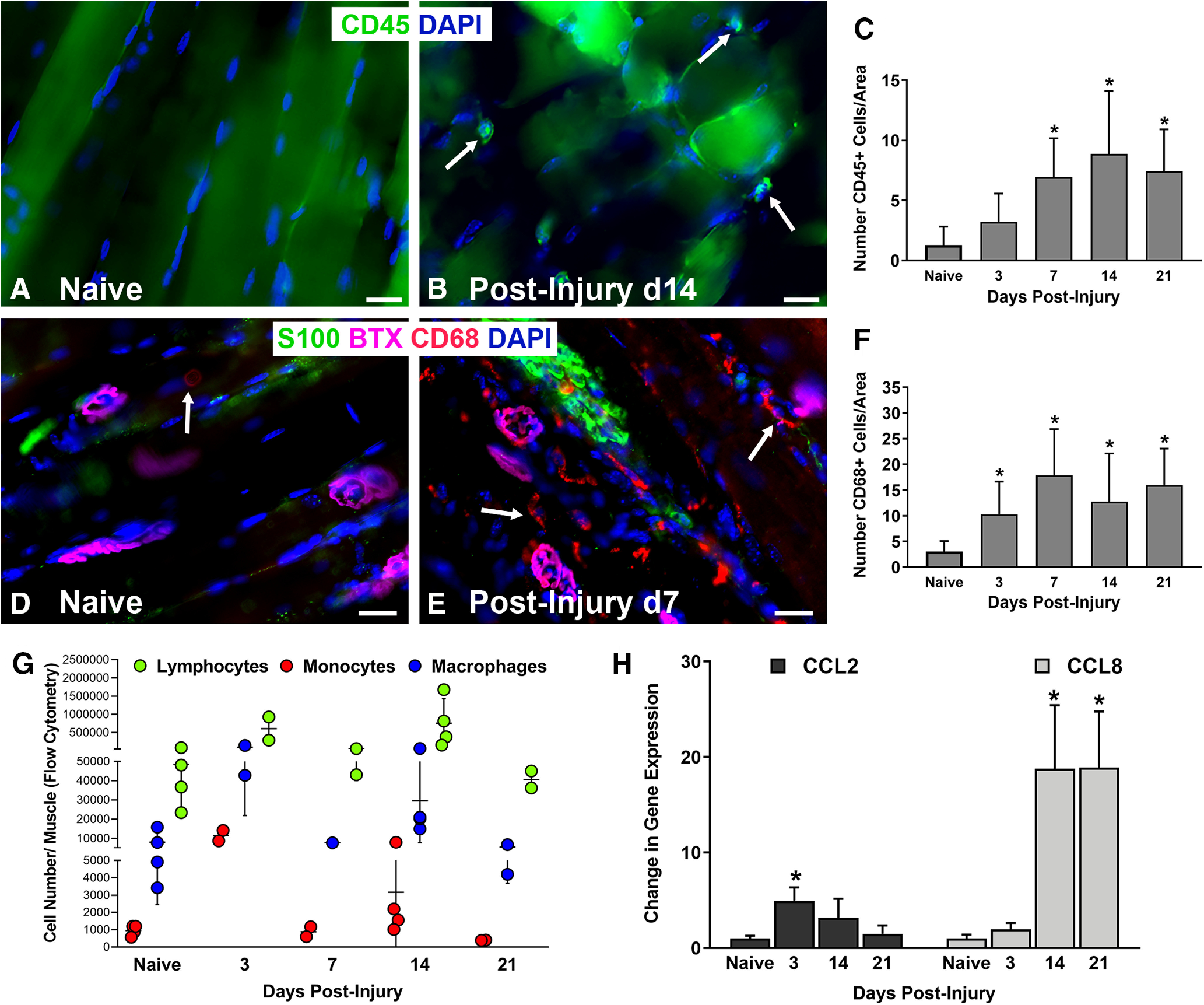
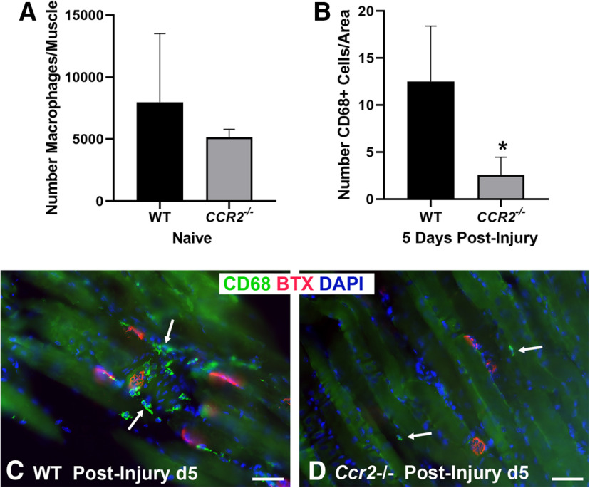
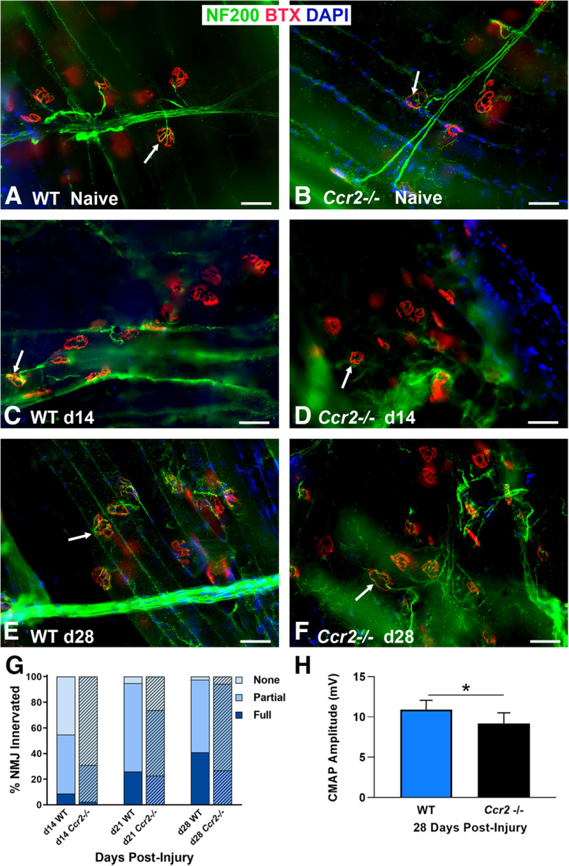
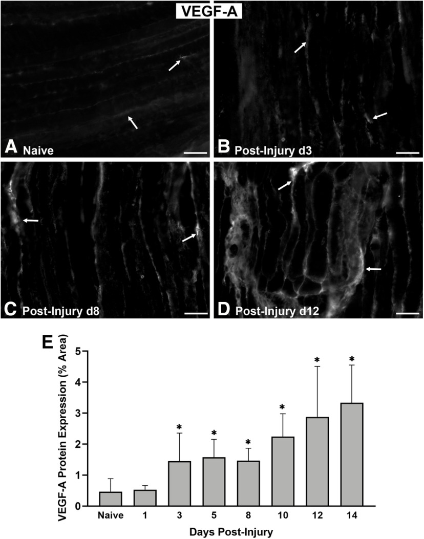
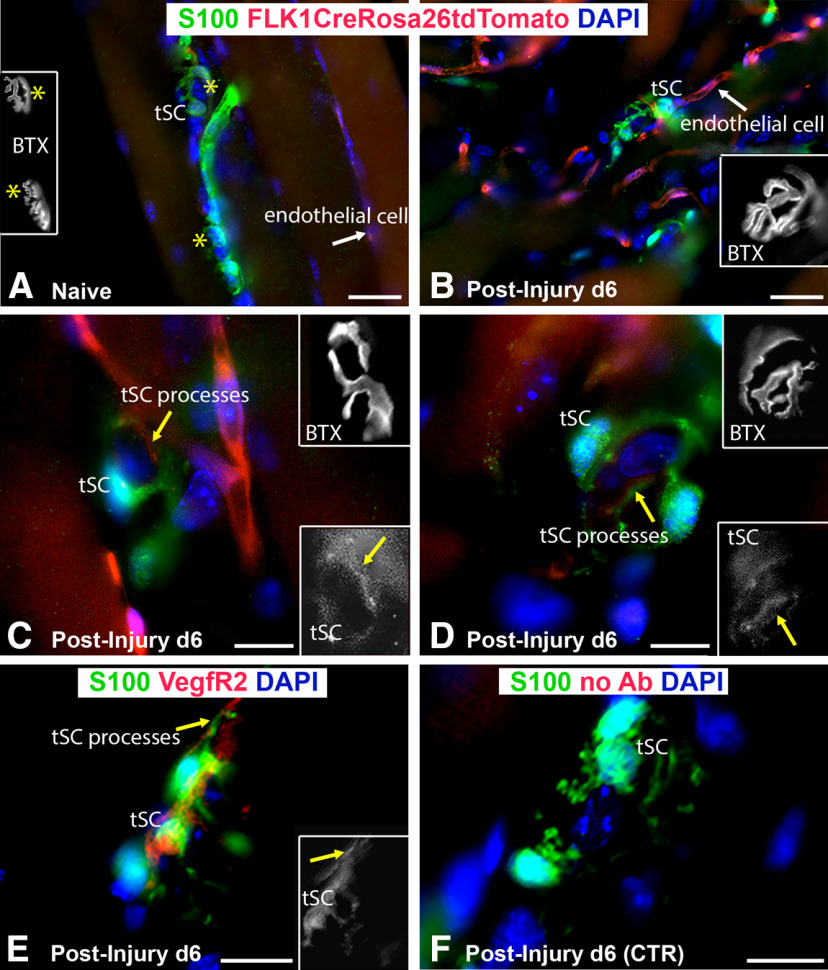
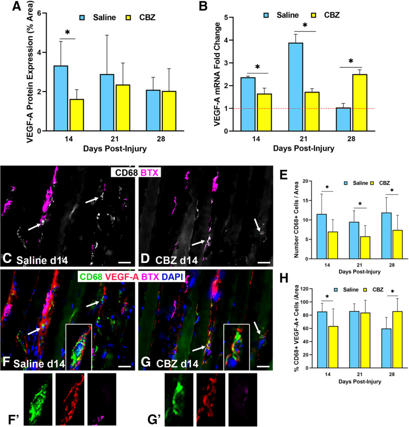
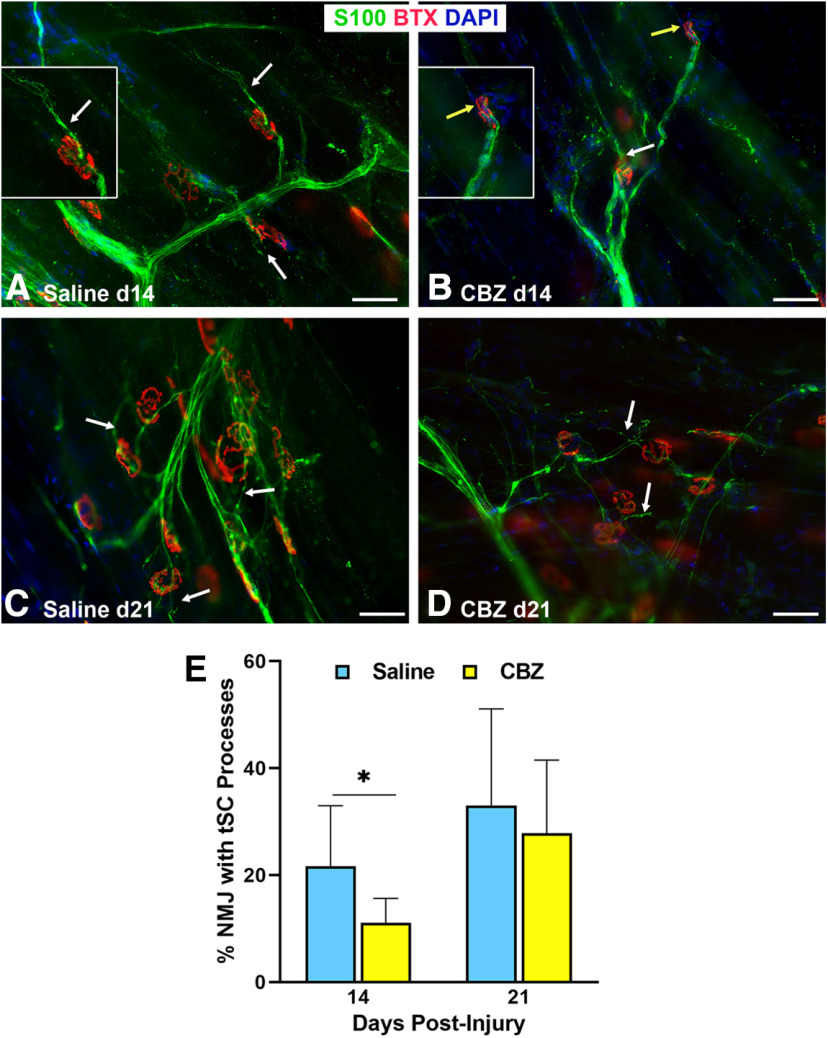
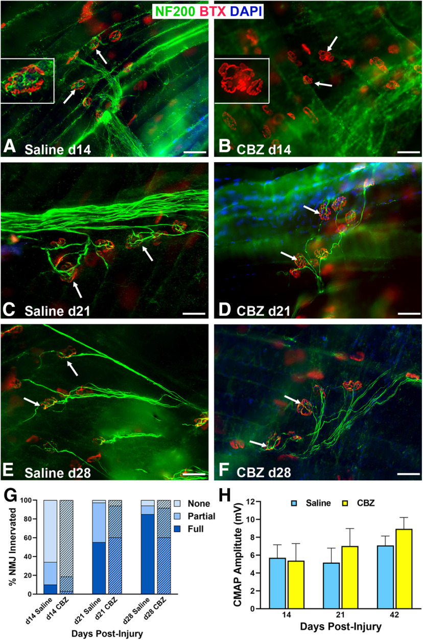
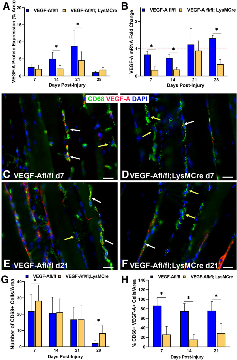
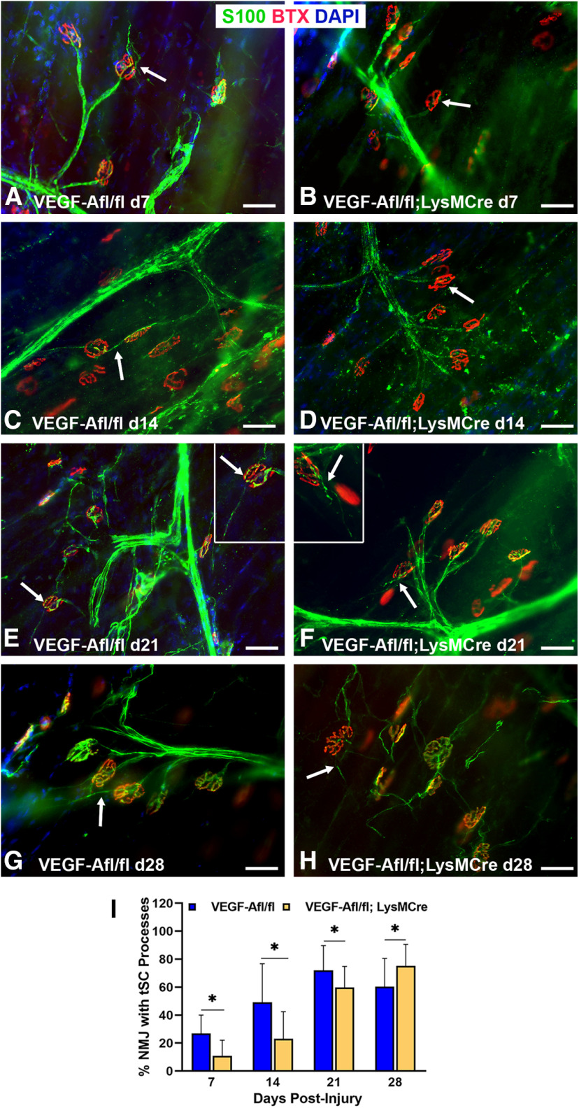
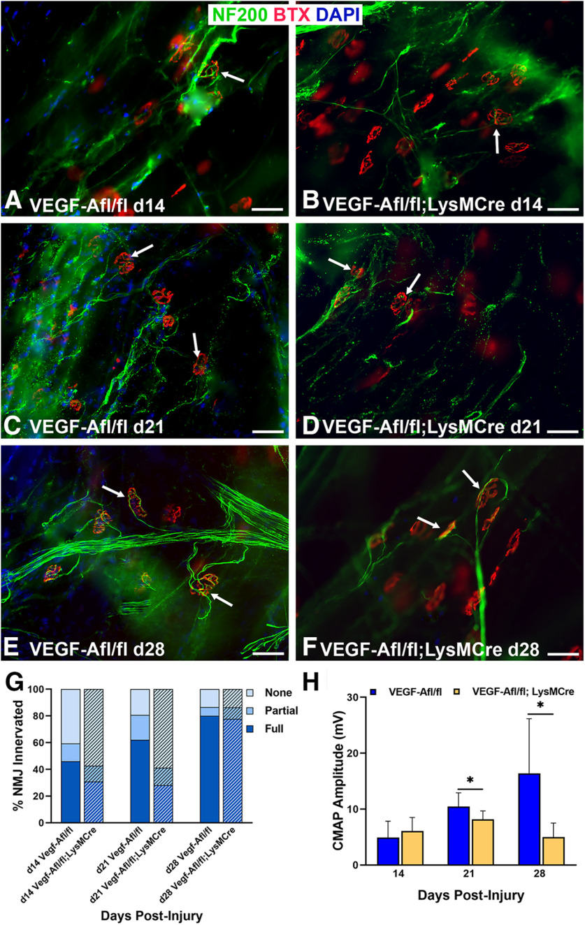
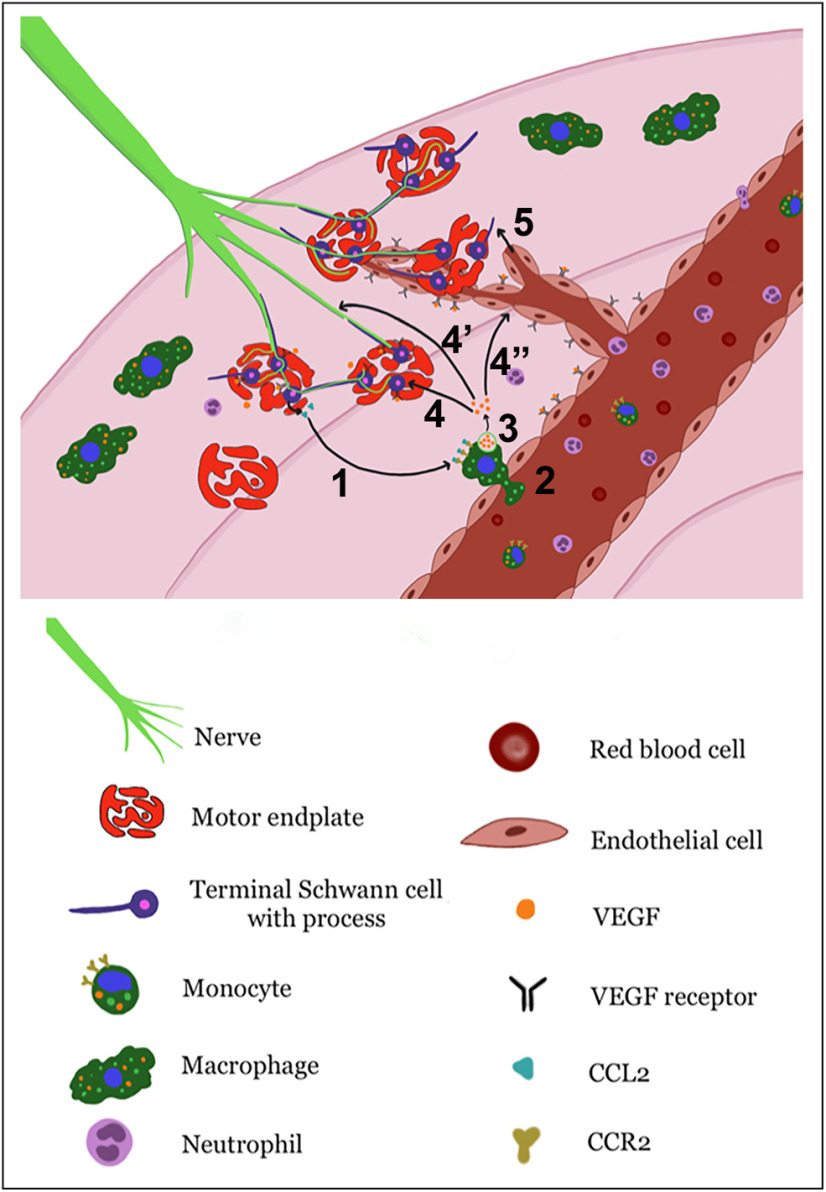
Functional recovery in the end target muscle is a determinant of outcome after peripheral nerve injury. The neuromuscular junction (NMJ) provides the interface between nerve and muscle and includes non-myelinating terminal Schwann cells (tSCs). After nerve injury, tSCs extend cytoplasmic processes between NMJs to guide axon growth and NMJ reinnervation. The mechanisms related to NMJ reinnervation are not known. We used multiple mouse models to investigate the mechanisms of NMJ reinnervation in both sexes, specifically whether macrophage-derived vascular endothelial growth factor-A (Vegf-A) is crucial to establishing NMJ reinnervation at the end target muscle. Both macrophage number and Vegf-A expression increased in end target muscles after nerve injury and repair. In mice with impaired recruitment of macrophages and monocytes (Ccr2-/- mice), the absence of CD68+ cells (macrophages) in the muscle resulted in diminished muscle function. Using a Vegf-receptor 2 (VegfR2) inhibitor (cabozantinib; CBZ) via oral gavage in wild-type (WT) mice resulted in reduced tSC cytoplasmic process extension and decreased NMJ reinnervation compared with saline controls. Mice with Vegf-A conditionally knocked out in macrophages (Vegf-Afl/fl; LysMCre mice) demonstrated a more prolonged detrimental effect on NMJ reinnervation and worse functional muscle recovery. Together, these results show that contributions of the immune system are integral for NMJ reinnervation and functional muscle recovery after nerve injury.SIGNIFICANCE STATEMENT This work demonstrates beneficial contributions of a macrophage-mediated response for neuromuscular junction (NMJ) reinnervation following nerve injury and repair. Macrophage recruitment occurred at the NMJ, distant from the nerve injury site, to support functional recovery at the muscle. We have shown hindered terminal Schwann cell (tSC) injury response and NMJ recovery with inhibition of: (1) macrophage recruitment after injury; (2) vascular endothelial growth factor receptor 2 (VegfR2) signaling; and (3) Vegf secretion from macrophages. We conclude that macrophage-derived Vegf is a key component of NMJ recovery after injury. Determining the mechanisms active at the end target muscle after motor nerve injury reveals new therapeutic targets that may translate to improve motor recovery following nerve injury.

Keywords: muscle recovery; nerve injury; neuromuscular junction; reinnervation; terminal Schwann cell; vascular endothelial growth factor.


Figures

Figure 1.
Figure 1.
Increased leukocytes are present in the end target muscle following peripheral nerve injury in WT mice. A–C, Lymphocyte (CD45+, arrows) cell numbers significantly increased after nerve injury compared with naive, uninjured muscles and peaked at day 14. D–F, Macrophage (CD68+, arrows) cell numbers were significantly higher than in naive muscles at all time points from days 3 to 21 postinjury. G, Flow cytometry revealed peak cell numbers in lymphocytes and macrophages at days 3 and 14 after nerve injury. H, Gene expressions of the Ccr2 ligands, chemokine-ligand-2 (Ccl2) and chemokine-ligand-8 (Ccl8), were significantly higher at days 3 and 14, respectively, compared with naive muscles. Ccl8 remained elevated at day 21. A, B, CD45 Ab = lymphocytes (green). D, E, CD68 Ab = macrophages (red), S100 Ab = glial cells (green), BTX = α‐bungarotoxin (endplates, purple), DAPI = nuclear staining (blue). Scale bars: 20 μm (A, B) and 50 μm (D, E). Data: mean ± SD; N = 3–4 mice/time point; *p < 0.05.
Figure 2.
Figure 2.
Ccr2−/− mice have impaired macrophage recruitment to the NMJ. A, Flow cytometry showed a comparable number of macrophages/muscle in naive, uninjured WT, and Ccr2−/− mice. B–D, WT mice showed increased CD68+ macrophages/image area (green, arrows) around the NMJ 5 d after nerve injury, while Ccr2−/− mice had nearly no macrophage presence at the NMJ 5 d after nerve injury. CD68 Ab = macrophages (green), BTX = α‐bungarotoxin (endplates, red), DAPI = nuclear staining (blue). Scale bar: 50 μm (C, D). Data: mean ± SD; N = 3–4 mice/genotype/time point; *p < 0.05.
Figure 3.
Figure 3.
Ccr2−/− mice exhibit decreased NMJ reinnervation and diminished muscle function following nerve injury and repair. A, B, WT and Ccr2−/− mice have normal NMJ morphology and innervation (arrows) in naive, uninjured muscles. C, D, NMJ reinnervation, represented by colocalization of neurofilament and motor endplate staining (arrows), was significantly decreased in Ccr2−/− mice compared with WT mice at 14 d after nerve injury and repair. A significant difference in innervation remained at 21 d (images not shown), but was mitigated by 28 d (E, F). G, Bar graph summarizes the quantitative findings according to full, partial, or no NMJ innervation (none). H, Evoked CMAPs of the TA muscle were significantly lower in Ccr2−/− mice compared with WT mice at 28 d after nerve injury and repair, despite no difference in reinnervation at that time point. NF200 Ab = axons (green), BTX = α‐bungarotoxin (endplates, red), DAPI = nuclear staining (blue). Scale bar: 50 μm. Data: mean ± SD; N = 3–6 mice/genotype/time point; *p < 0.05.
Figure 4.
Figure 4.
Vegf-A expression increases in the end target muscle following nerve injury in WT mice. A, Immunostaining of Vegf-A protein (pseudocolored in gray, arrows) was nearly undetectable in naive, uninjured muscles. B–D, From the 3rd to the 12th day postinjury, Vegf-A expression steadily increased. E, The increasing Vegf-A protein expression/image area under 20× magnification was quantitatively summarized for all time points. Vegf-A Ab (gray). Scale bar: 50 μm. Data: mean ± SD; N= 3 mice/time point; *p < 0.05.
Figure 5.
Figure 5.
VegfR2 is expressed in tSC cytoplasm and processes connecting cell bodies after nerve injury. In order to determine VegfR2 localization, we used Flk-1Cre; Rosa26tdTomato mice, where Rosa26TdTomato staining (red) is present at locations of VegfR2 (Flk-1) expression. A, In naive muscles without previous nerve injury, VegfR2 was located on ECs (white arrows) but not in tSCs or around NMJs. B, At day 6 after nerve injury and repair, the NMJ with tSCs was surrounded by VegfR2-expressing ECs (white arrow). C, D, At higher magnifications, VegfR2 localized within tSC cell bodies and cytoplasmic processes (yellow arrows) in addition to ECs. The single channel insets, pseudocolored in gray, highlight the positive staining of VegfR2 within the tSCs and tSC processes (yellow arrows). E, F, Colocalization of S100 staining (glia) with anti-Flk1 is seen at tSCs at 6 d postinjury. The inset (E) represents a single red channel pseudocolored in gray showing the positive staining of VegfR2 in tSCs. Immunostaining for BTX (endplates, yellow stars) in panels A–D are shown as insets pseudocolored in gray. These single-channel images represent BTX staining of the area shown in color. Flk-1 = VegfR2 (red), S100 Ab = glial cells (green), DAPI = nuclear staining (blue). N = 3 mice/time point. Scale bars: 50 μm (A, B) and 20 μm (C, D).
Figure 6.
Figure 6.
VegfR2 inhibition decreases Vegf-A expression and macrophage cell numbers after nerve injury. A, CBZ, a VegfR2 inhibitor, treatment (daily from days 6 to 13 following nerve injury) decreased Vegf-A protein expression at 14 d after nerve injury compared with saline-gavaged control mice. B, In control mice, Vegf-A mRNA expression was elevated by 14 d postinjury, peaked at 21 d, and returned to baseline by 28 d. CBZ inhibited Vegf-A mRNA expression at days 14 and 21 postinjury, but the effect was not sustained by day 28. C–E, CBZ resulted in significantly decreased macrophage numbers at all time points. Arrows point to CD68+ cells (pseudocolored in gray in C, D). α‐Bungarotoxin staining of (BTX purple) demonstrates relationship of macrophages near NMJs. F–H, The percent of macrophages (green) colocalizing (arrows) with Vegf-A (red) was significantly less in the CBZ-treated group than saline-treated controls at day 14. The magnified images of single cells in F', G' highlight the colocalization of Vegf-A with a macrophages via isolated color channels. Vegf-A Ab (red), CD68 Ab = macrophages (green), BTX = α‐bungarotoxin (endplates, purple), DAPI = nuclear staining (blue). Scale bar: 20 μm. Data: mean ± SD; N = 3–5 mice/treatment/time point; *p < 0.05.
Figure 7.
Figure 7.
VegfR2/Vegf-A inhibition reduces tSC process extension beyond NMJ area after nerve injury. A, B, tSC cytoplasmic processes in the CBZ-treated mice (yellow arrows) were absent, short, or fragmented, and extended from fewer NMJs at day 14 compared with long tSC processes in saline-treated control mice (white arrows). The insets highlight long tSC processes (white arrow) in saline-treated mice (A) and lack of processes (yellow arrow) in CBZ-treated mice (B). C, D, This effect did not persist on day 21 postinjury and repair (8 d after the last dose of CBZ), as there was no difference between CBZ-treated mice and controls relative to percent NMJs with tSC processes. E, Quantitatively, the percentage of NMJs with tSC processes in CBZ-treated mice was half that of controls at 14 d and not significantly different from controls at 21 d. S100Ab = glial cells (green), BTX = α‐bungarotoxin (endplates, red), DAPI = nuclear staining (blue). Scale bar: 50 μm. Data: mean ± SD; N = 3–5 mice/treatment/time point; *p < 0.05.
Figure 8.
Figure 8.
VegfR2/Vegf-A inhibition reduces NMJ reinnervation after nerve injury. A, B, NMJ reinnervation, defined as overlap of axonal staining (NF200 Ab, green) with α‐bungarotoxin (BTX, endplates, red) staining (arrows), was significantly less in CBZ-treated mice compared with controls at day 14, but not at days 21 (C, D) or 28 (E, F) after nerve injury and repair. The insets highlight innervated endplate (A) and denervated endplate (B) in saline-treated and CBZ-treated mice, respectively. G, Quantification analyses showed both full and partial NMJ reinnervation in CBZ-treated mice were approximately half the values of controls at 14 d postinjury. The percentage of fully reinnervated NMJs did not change from 21 to 28 d postinjury in the CBZ group. H, There were no differences in CMAP amplitudes between CBZ-treated mice and saline-treated controls at all time points postinjury. DAPI = nuclear staining (blue). Scale bar: 50 μm. Data: mean ± SD; N = 3–5 mice/treatment/time point.
Figure 9.
Figure 9.
Vegf-A expression within macrophages is decreased in Vegf-Afl/fl; LysMCre mice. A, Vegf-A protein expression was significantly decreased in Vegf-Afl/fl; LysMCre mice at days 14 and 21 following nerve injury and repair. Vegf-A levels peaked in both conditional knock-out mice and controls (Vegf-Afl/fl) at 21 d postinjury. B, Vegf-A mRNA expression was significantly decreased in Vegf-Afl/fl; LysMCre mice compared with the control mice. Vegf-A mRNA expression peaked at day 21 in the Vegf-Afl/fl; LysMCre mice before significantly decreasing again at 28 d after nerve injury. C–H, Macrophage (green) number was higher in the Vegf-Afl/fl; LysMCre mice compared with controls at days 7 and 28 after nerve injury but did not differ between Vegf-Afl/fl; LysMCre mice and controls at days 14 and 21. Both groups had fewer macrophages present in the muscle at day 28. G, The percentage of CD68+ cells (green) colocalizing with Vegf-A (red, white arrows in C–F), however, was significantly decreased in Vegf-Afl/fl; LysMCre mice compared with controls at all time points. Macrophages not colocalized with Vegf-A are indicated by yellow arrows. Vegf-A Ab (red), CD68 Ab = macrophages (green), DAPI = nuclear staining (blue). Scale bar: 20 μm. Data: mean ± SD; N = 3 mice/genotype/time point; *p < 0.05.
Figure 10.
Figure 10.
tSC process extension is diminished after nerve injury in Vegf-Afl/fl; LysMCre mice. A, At day 7 after nerve injury, tSCs extended their processes (arrow) in control mice. In contrast, tSC process extension was absent (arrow) at the NMJs in Vegf-Afl/fl; LysMCre mice (B). Significant differences in tSC process extension persisted at days 14 (C, D) and 21 (E, F) postinjury. The inset in panel E shows an NMJ with a long tSC process (arrow) in control mice. The inset in panel F shows two endplates in Vegf-Afl/fl; LysMCre mice but only one (arrow) with a very short tSC process. By day 28, however, thinner tSC processes were present in the NMJs of Vegf-Afl/fl; LysMCre mice (H) than in controls (G). I, Significant differences in the percent of NMJs with tSC process extension was most evident from days 7 to 21. S100 Ab = glial cells (green), BTX = α‐bungarotoxin (endplates, red), DAPI = nuclear staining (blue). Scale bar: 50 μm. Data: mean ± SD; N = 3 mice/genotype/time point; *p < 0.05.
Figure 11.
Figure 11.
Innervation and functional NMJ recovery in Vegf-Afl/fl; LysMCre mice is persistently worse than controls. NMJ reinnervation, as seen by the colocalization of the neurofilaments (green) with motor endplates (red, arrows), is shown at days 14 (A, B), 21 (C, D), and 28 (E, F) postinjury and nerve repair. G, NMJ reinnervation was significantly worse in the Vegf-Afl/fl; LysMCre mice, achieving only half of the reinnervation rate of controls, at day 21. Vegf-Afl/fl; LysMCre mice showed a significantly lower percentage of fully innervated NMJs and higher percentage of denervated (none) NMJs compared with controls. By day 28, both groups of mice were able to reach nearly 80% of motor endplates fully innervated. H, Vegf-Afl/fl; LysMCre mice demonstrated significantly poorer CMAP performance at 21 and 28 d postinjury compared with control mice, indicating a functional impact extending beyond morphologic differences. NF200 Ab = axons (green), BTX = α‐bungarotoxin (endplates, red), DAPI = nuclear staining (blue). Scale bar: 50 μm. Data: mean ± SD; N = 3–5 mice/genotype/time point; *p < 0.05.
Figure 12.
Figure 12.
Illustration of proposed mechanism of Vegf-A effects on NMJ reinnervation. Ccl2–Ccr2 interactions (1) recruit circulating monocytes/macrophages into muscle (2) following nerve injury. Macrophages then secrete Vegf-A (3), which may have three targets: VegfRs located on tSCs, which induce tSC process extension to guide axons to the NMJs (4), VegfRs on nerve terminals and myelinating SCs to promote axonal regeneration (4'), and VegfRs on ECs, resulting in increased vessel permeability and angiogenesis to allow delivery of additional circulating inflammatory cells to the NMJ for establishing reinnervation (4”). Angiogenesis may then guide tSC process extensions (5) to aid NMJ reinnervation.

Similar articles

Cited by

References

    1. Alexander KA, Chang MK, Maylin ER, Kohler T, Müller R, Wu AC, Van Rooijen N, Sweet MJ, Hume DA, Raggatt LJ, Pettit AR (2011) Osteal macrophages promote in vivo intramembranous bone healing in a mouse tibial injury model. J Bone Miner Res 26:1517–1532. 10.1002/jbmr.354 - DOI - PubMed
    1. Arnold L, Henry A, Poron F, Baba-Am Y, van Rooijen N, Plonquet A, Gherardi RK, Chazaud B (2007) Inflammatory monocytes recruited after skeletal muscle injury switch into antiinflammatory macrophages to support myogenesis. J Exp Med 204:1057–1069. 10.1084/jem.20070075 - DOI - PMC - PubMed
    1. Barik A, Li L, Sathyamurthy A, Xiong W-C, Mei L (2016) Schwann cells in neuromuscular junction formation and maintenance. J Neurosci 36:9770–9781. 10.1523/JNEUROSCI.0174-16.2016 - DOI - PMC - PubMed
    1. Barrette B, Hébert M-A, Filali M, Lafortune K, Vallières N, Gowing G, Julien J-P, Lacroix S (2008) Requirement of myeloid cells for axon regeneration. J Neurosci 28:9363–9376. 10.1523/JNEUROSCI.1447-08.2008 - DOI - PMC - PubMed
    1. Bogdanik LP, Osborne MA, Davis C, Martin WP, Austin A, Rigo F, Bennett CF, Lutz CM (2015) Systemic, postsymptomatic antisense oligonucleotide rescues motor unit maturation delay in a new mouse model for type II/III spinal muscular atrophy. Proc Natl Acad Sci USA 112:E5863–E5872. 10.1073/pnas.1509758112 - DOI - PMC - PubMed

Publication types

Substances