. 2021 Jan;99(1):60-77.
doi: 10.1124/molpharm.120.000072. Epub 2020 Nov 5.

Protective Effects of Flavonoids in Acute Models of Light-Induced Retinal Degeneration

Affiliations

Protective Effects of Flavonoids in Acute Models of Light-Induced Retinal Degeneration

Joseph T Ortega et al. Mol Pharmacol. 2021 Jan.

Abstract

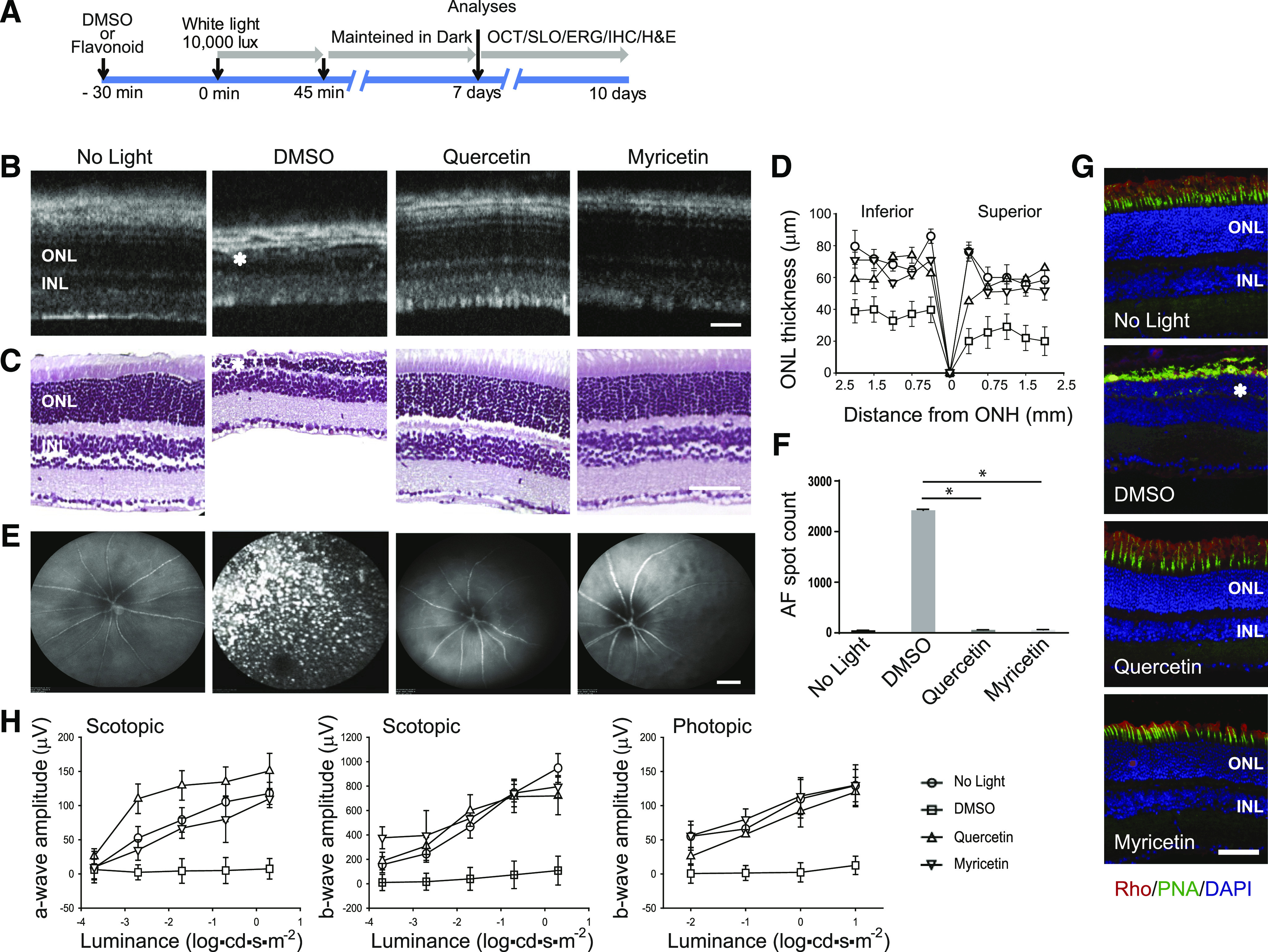
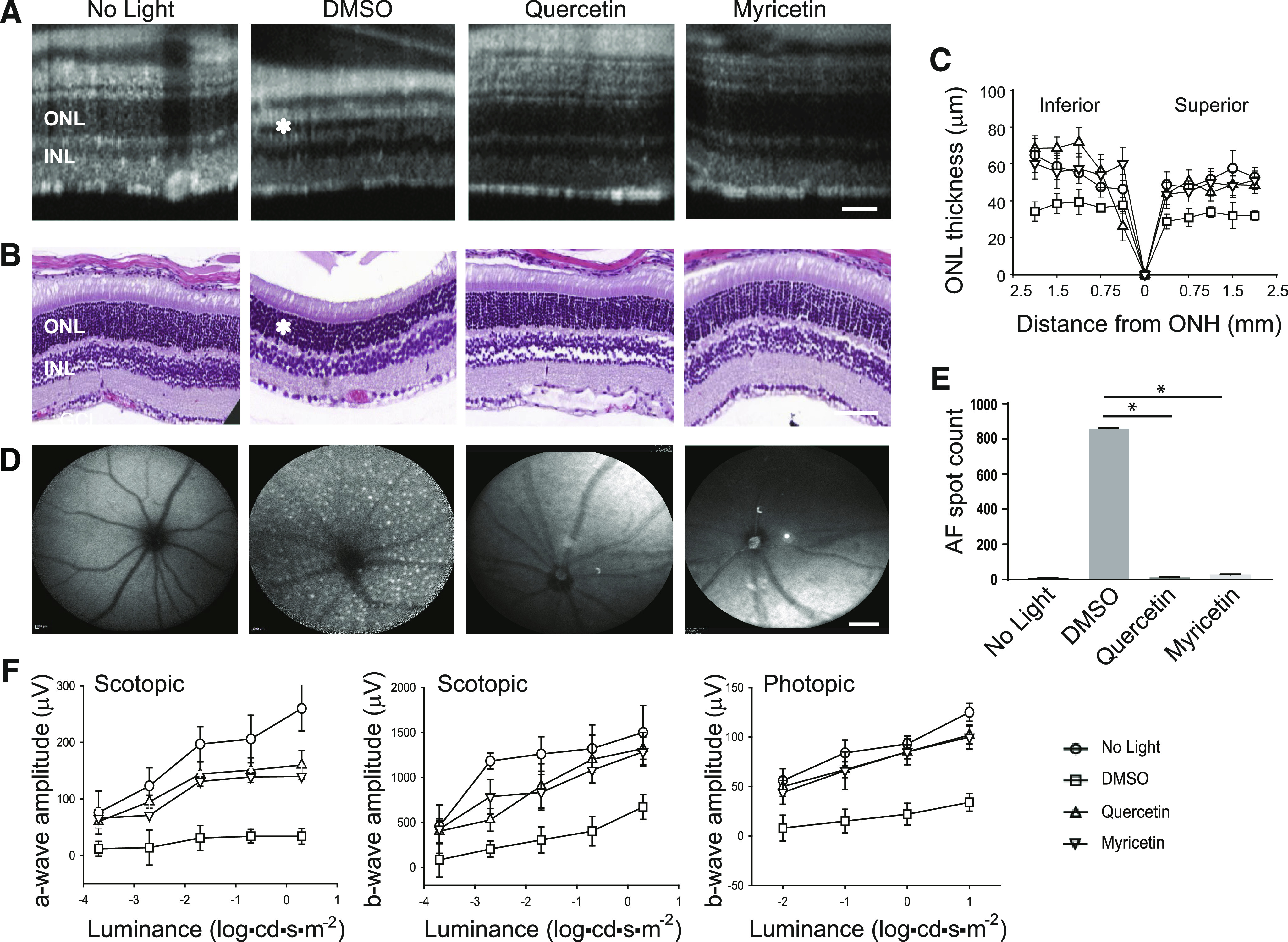
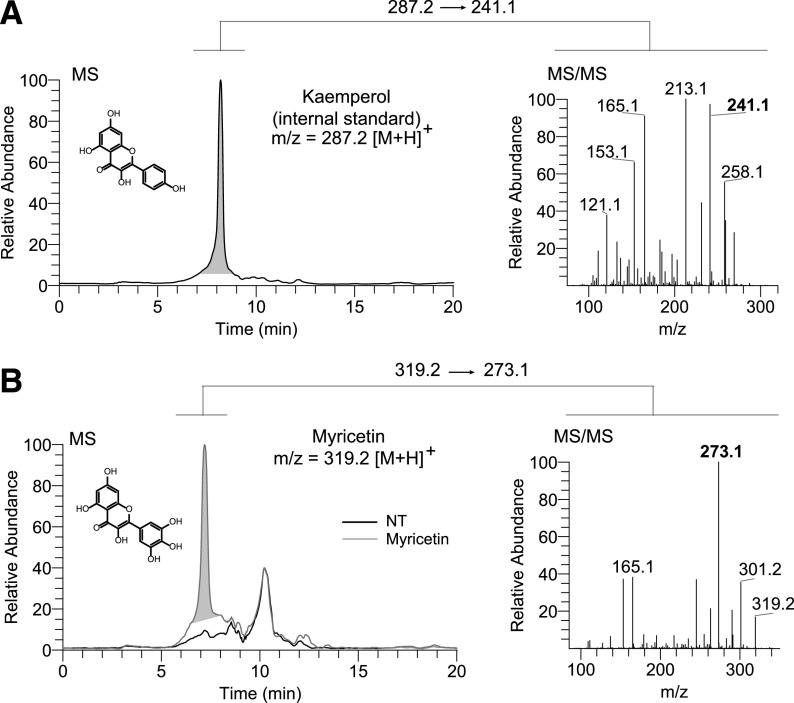
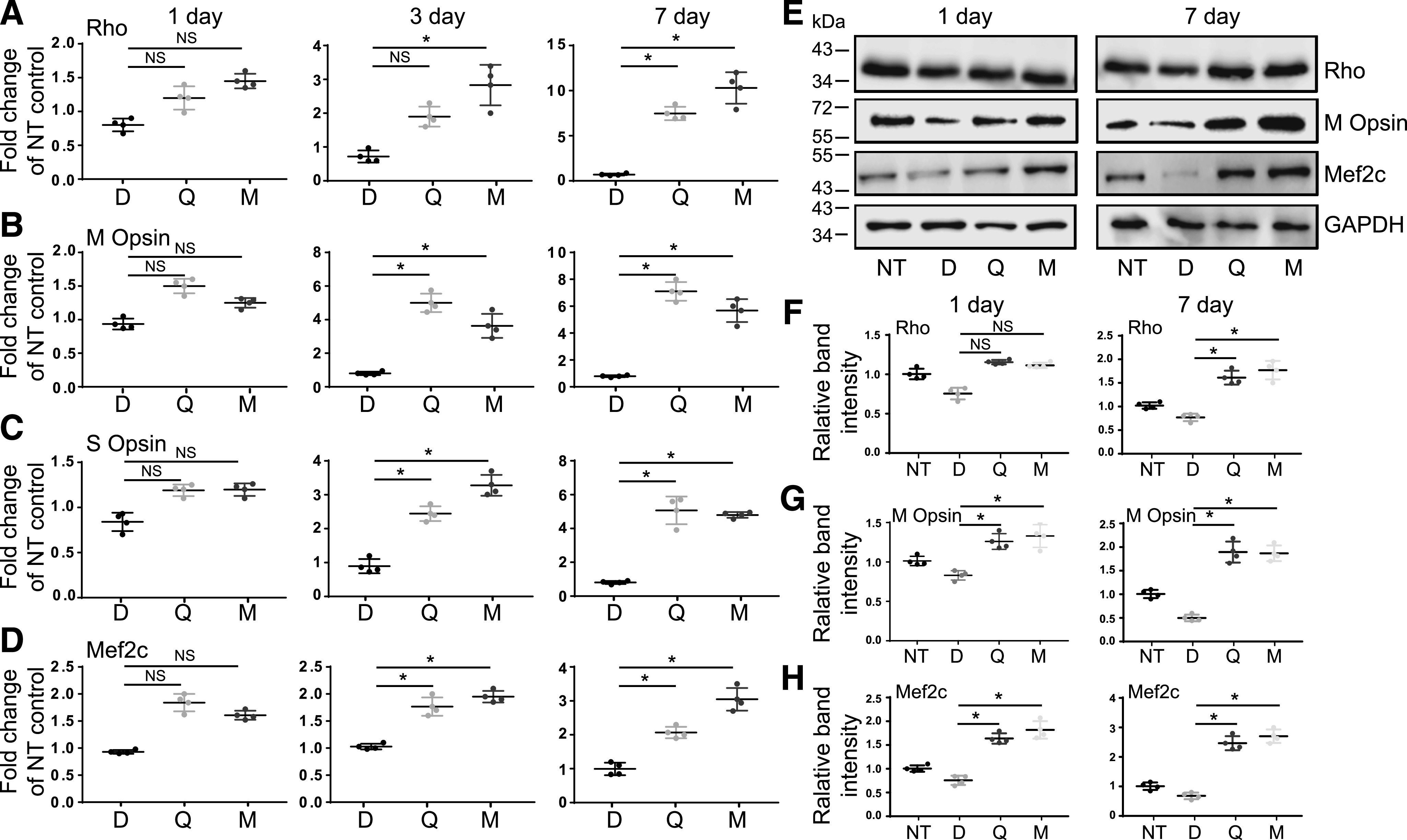
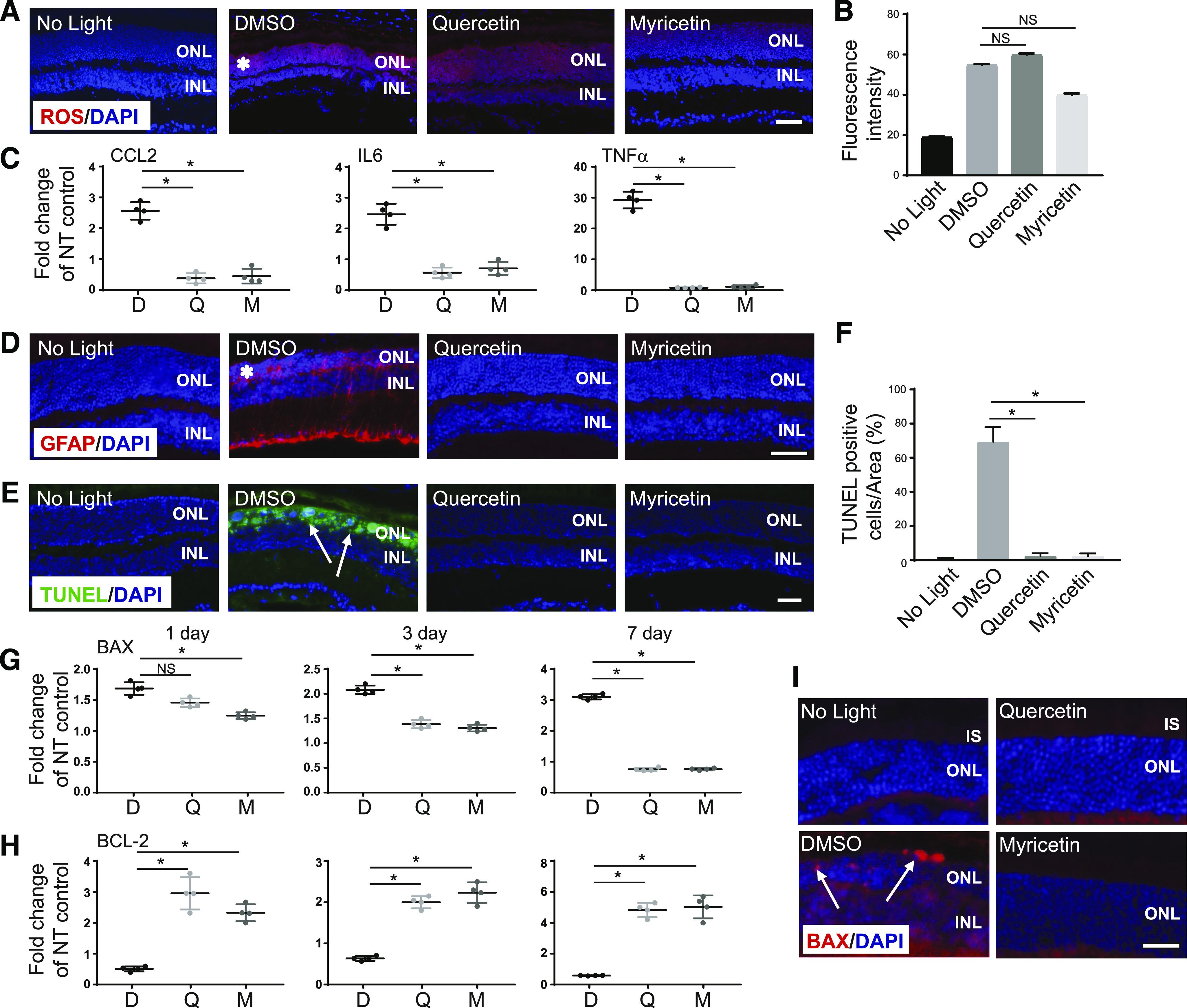
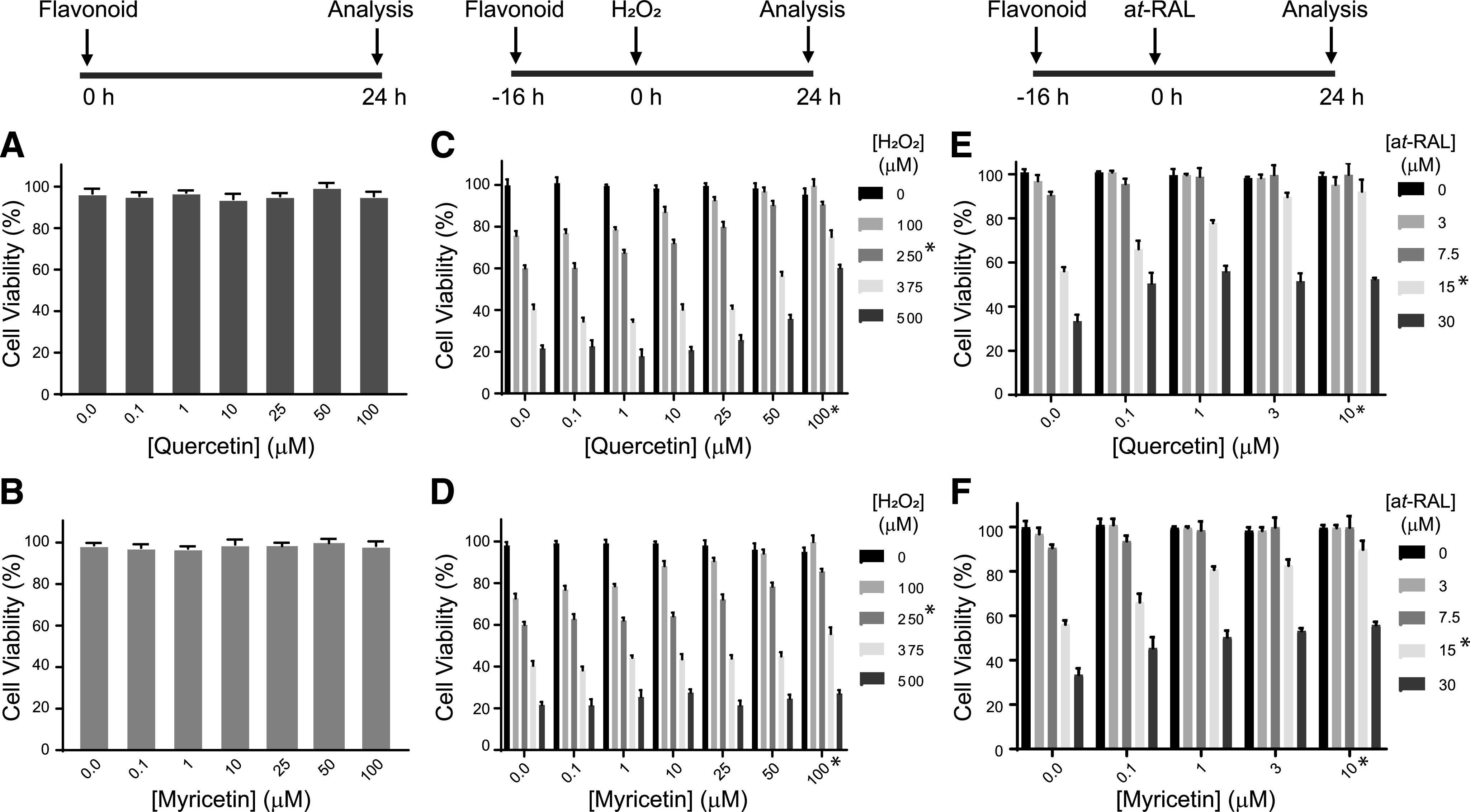
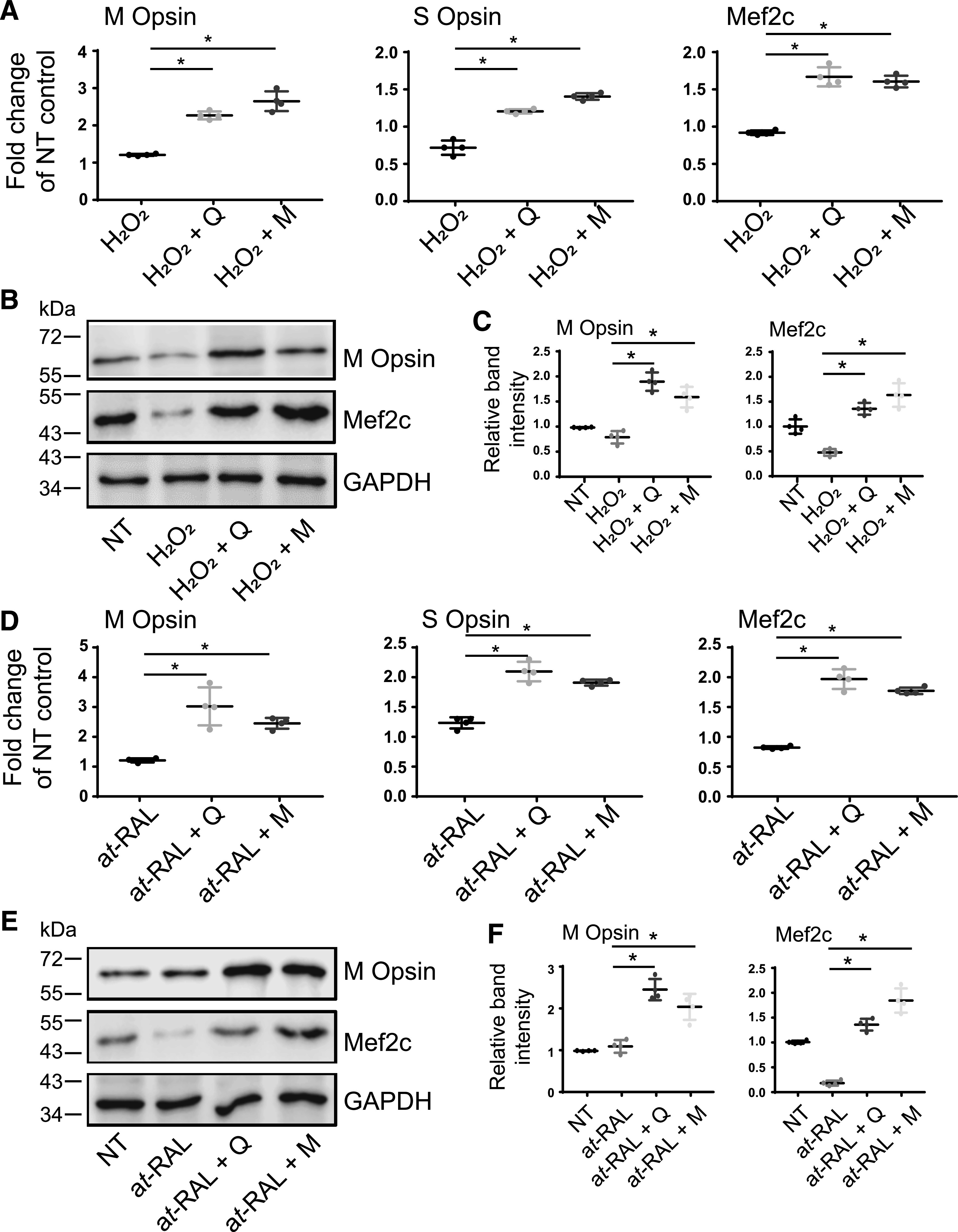
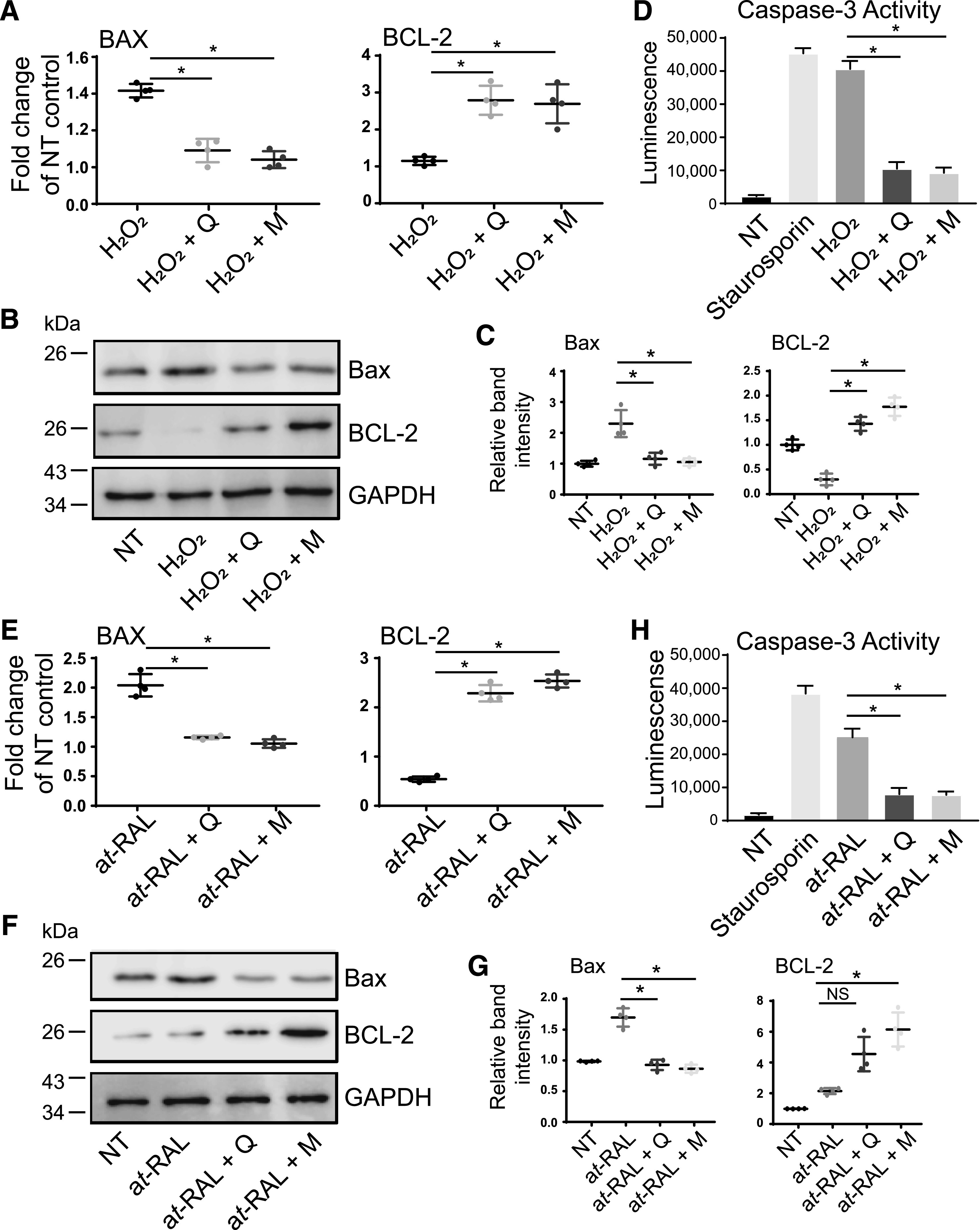
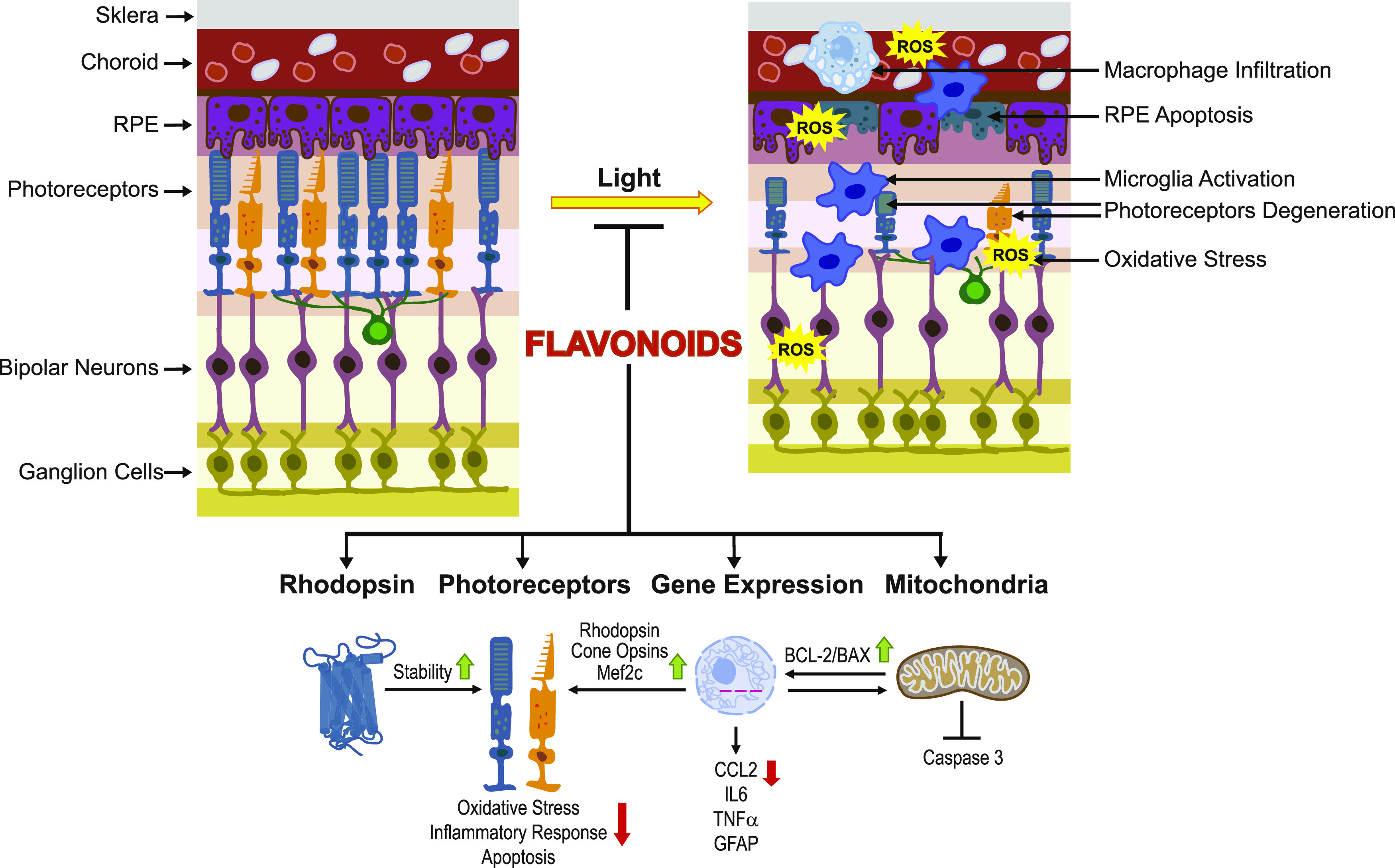
Degeneration of photoreceptors caused by excessive illumination, inherited mutations, or aging is the principal pathology of blinding diseases. Pharmacological compounds that stabilize the visual receptor rhodopsin and modulate the cellular pathways triggering death of photoreceptors could avert this pathology. Interestingly, flavonoids can modulate the cellular processes, such as oxidative stress, inflammatory responses, and apoptosis, that are activated during retinal degeneration. As we found previously, flavonoids also bind directly to unliganded rod opsin, enhancing its folding, stability, and regeneration. In addition, flavonoids stimulate rhodopsin gene expression. Thus, we evaluated the effect of two main dietary flavonoids, quercetin and myricetin, in ATP-binding cassette subfamily A member 4 -/- /retinol dehydrogenase 8 -/- and wild-type BALB/c mice susceptible to light-induced photoreceptor degeneration. Using in vivo imaging, such as optical coherence tomography, scanning laser ophthalmoscopy, and histologic assessment of retinal morphology, we found that treatment with these flavonoids prior to light insult remarkably protected retina from deterioration and preserved its function. Using high-performance liquid chromatography-mass spectrometry analysis, we detected these flavonoids in the eye upon their intraperitoneal administration. The molecular events associated with the protective effect of quercetin and myricetin were related to the elevated expression of photoreceptor-specific proteins, rhodopsin and cone opsins, decreased expression of the specific inflammatory markers, and the shift of the equilibrium between cell death regulators BCL2-associated X protein (BAX) and B-cell lymphoma 2 toward an antiapoptotic profile. These results were confirmed in photoreceptor-derived 661W cells treated with either H2O2 or all-trans-retinal stressors implicated in the mechanism of retinal degeneration. Altogether, flavonoids could have significant prophylactic value for retinal degenerative diseases. SIGNIFICANCE STATEMENT: Flavonoids commonly present in food exhibit advantageous effects in blinding diseases. They bind to and stabilize unliganded rod opsin, which in excess accelerates degenerative processes in the retina. Additionally, flavonoids enhance the expression of the visual receptors, rod and cone opsins; inhibit the inflammatory reactions; and induce the expression of antiapoptotic markers in the retina, preventing the degeneration in vivo. Thus, flavonoids could have a prophylactic value for retinal degenerative diseases.


Conflict of interest statement

Conflicts of Interest The authors declare that they have no conflicts of interest with the contents of this article.

Figures

Fig. 1.
Fig. 1.
Protective effect of flavonoids against retinal degeneration in Abca4−/−Rdh8−/− mice induced by bright light. (A) Experimental design of mouse treatment. Flavonoids (20 mg/kg b.wt.) or DMSO vehicle were administered to 4–6-week-old Abca4−/−Rdh8−/− mice by intraperitoneal injection 30 minutes before exposure to 10,000-lux light for 45 minutes. After illumination, mice were kept in the dark for 7–10 days before their examination. The health of the retina was inspected by OCT and SLO in vivo imaging, H&E staining, and immunohistochemistry. Retinal function was examined by ERG. (B) Representative OCT images of mouse eye obtained on the 7th day after the indicated treatment and exposure to bright light. INL, inner nuclear layer. Asterisk indicates a disorganized photoreceptor layer in DMSO-treated control mice. Scale bar, 50 µm. (C) Retinal sections prepared from eyes collected from mice either unexposed to light or exposed to bright light after the indicated treatment and stained with H&E. Asterisk indicates disorganized photoreceptor structures in DMSO-treated control mice. Scale bar, 50 µm. (D) The ONL thickness was measured at inferior and superior sites at 250, 500, 750, 1000, 1500, and 2000 μm from the ONH. Measurements were performed in 20 mice, with five mice per treatment group. Error bars indicate S.D. Changes in the ONL thickness observed between dark-adapted group and DMSO-treated, exposed-to-light group were statistically different. Changes in the ONL thickness observed after treatment with flavonoids compared with the DMSO-treated group were statistically different. No significant difference in the ONL thickness was observed between mice kept in the dark and those treated with flavonoids. (E) Representative SLO images. AF spots were detected only in the retina of DMSO-treated and exposed-to-bright-light mice. AF spots were not detected in mice kept in the dark or mice treated with either quercetin or myricetin before illumination. Scale bar, 50 µm. (F) Quantification of AF spots performed in 20 mice, with five mice per treatment group. Error bars indicate S.D. Changes in the number of AF spots after treatment with flavonoids compared with DMSO-treated mice were statistically different and are indicated with asterisk (*). No significant difference was observed between mice kept in the dark and those exposed to light after treatment with flavonoids. (G) Immunohistochemistry in cryo-sections prepared from eyes collected either from mice unexposed to light or exposed to bright light after the indicated treatment. Sections were stained with an anti-rhodopsin (Rho) C-terminus–specific antibody (red) showing the structural organization of rod photoreceptors, PNA staining (green) showing the health of cone photoreceptors, and DAPI staining of nuclei (blue). Asterisk shows a severely disrupted photoreceptor layer in DMSO-treated control mice. Scale bar, 50 µm. (H) Retinal function examined by ERG responses. Retinal function was significantly protected in mice treated with flavonoid before exposure to bright light insult as compared with DMSO-treated mice in both scotopic a- and b-waves and in photopic b-waves. ERG measurements were carried out in 20 mice, with five mice per treatment group. Changes in the ERG responses compared between dark-adapted mice and DMSO-treated, exposed-to-light mice were statistically different. Changes in the ERG responses after treatment with flavonoids compared with DMSO-treated mice were statistically different. No statistical difference was observed between the dark-adapted group and mice treated with flavonoids. Statistical analysis was performed with the one-way ANOVA and post hoc Dunnett’s tests.
Fig. 2.
Fig. 2.
The effect of flavonoids on bright light–induced retinal degeneration in WT mice. Flavonoids were intraperitoneally injected into BALB/c mice (20 mg/kg b.wt.) 30 minutes before exposure to light at 12,000 lux for 2 hours. Then mice were kept in the dark for 7–10 days before examination of retinal morphology and function. (A) Representative OCT images. The ONL was protected in mice treated with either quercetin or myricetin as compared with DMSO-treated control mice. Asterisk indicates a shorter photoreceptor layer in DMSO-treated control mice. Scale bar, 50 µm. (B) Examination of retinal morphology after H&E staining of paraffin sections of eyes collected from mice either kept in the dark or exposed to light after indicated treatment. Asterisk indicates a shorter photoreceptor layer in DMSO-treated control mice. Scale bar, 50 µm. (C) The ONL thickness measured at inferior and superior sites at 250, 500, 750, 1000, 1500, and 2000 μm from the ONH in mice kept in the dark and mice treated with DMSO vehicle or flavonoid before exposure to bright light. Measurements were performed in 20 mice, with five mice per treatment group. Error bars indicate S.D. Changes in the ONL thickness observed between dark-adapted group and DMSO-treated, exposed-to-light group were statistically different. Changes in the ONL thickness observed after treatment with flavonoids compared with the DMSO-treated group were statistically different. No significant difference in the ONL thickness was observed between mice kept in the dark and those treated with flavonoids. (D) Representative SLO images. AF spots were detected in the retina of mice treated with DMSO before exposure to bright light. Only a few AF spots were found in the retina of mice treated with flavonoid before illumination and in mice kept in the dark. Scale bar, 50 µm. (E) Quantification of AF spots was performed in 20 mice, with five mice per treatment group. Error bars indicate S.D. Changes in the number of AF spots compared between dark-adapted group and DMSO-treated, exposed-to-light group were statistically different. Changes in the number of AF spots after treatment with either quercetin or myricetin compared with the DMSO-treated group were statistically different and are indicated with (*). No significant difference was observed between mice unexposed to light and those treated with flavonoids. (F) Retinal function examined by ERG responses. Retinal function was significantly protected in mice treated with flavonoid before exposure to bright-light insult as compared with DMSO-treated mice in both scotopic a- and b-waves and in photopic b-waves. ERG measurements were carried out in 20 mice, with five mice per treatment group. Changes in the ERG responses compared between dark-adapted mice and DMSO-treated, exposed-to-light mice were statistically different. Changes in the ERG responses after treatment with flavonoids compared with DMSO-treated mice were statistically different. No statistical difference was observed between the dark-adapted group and mice treated with flavonoids. Statistical analysis was performed with the one-way ANOVA and post hoc Dunnett’s tests.
Fig. 3.
Fig. 3.
Detection of flavonoids in mouse eyes. (A) MS analysis of kaemperol. Shown is the elution profile for kaemperol used as an internal standard. The chromatogram represents ion intensity for m/z = 287.2 [M + H]+. Fragmentation pattern of kaemperol is shown on the right (MS/MS). (B) MS analysis of flavonoids extracted from eyes of Abca4−/−Rdh8−/− administered with myricetin (20 mg/kg b.wt.). The MS spectrum of elution peak between 7 and 9 minutes (green line) indicates ion corresponding to myricetin m/z = 319.2 [M + H]+. Fragmentation pattern of myricetin is shown on the right (MS/MS). The elution peak detected at ∼10 minutes does not correspond with flavonoid and was also detected in the sample extracted from the eyes of nontreated control mice (NT, black line). The experiment was performed twice.
Fig. 4.
Fig. 4.
The effect of flavonoids on levels of photoreceptor-specific markers in eyes of Abca4−/−Rdh8−/− mice exposed to bright light. Total RNA was isolated from the eyes of mice unexposed to light and mice treated with either DMSO, quercetin, or myricetin prior to illumination. Four or five mice were used for each treatment group. Eyes were collected on days 1, 3, and 7 post-treatment. RT-qPCR was performed to determine the mRNA expression levels of photoreceptor’s specific genes, such as rhodopsin (Rho) (A), M cone opsin (B), S cone opsin (C), and Mef2c transcription factor regulating the expression of Rho and cone opsins (D). Relative fold change of these genes’ expression was normalized to the expression of GAPDH. The mean of data from three independent experiments is shown as a fold change of NT control. The expression of these genes was slightly downregulated in DMSO-treated and illuminated mice, whereas it was significantly upregulated upon treatment with flavonoids. The statistically different changes in the expression of the specific gene compared on different days after treatment between DMSO-treated and flavonoid-treated mice are indicated with asterisk (*). The nonstatistically different changes are indicated as NS. Error bars indicate S.D. Statistical analysis was performed for each gene separately, combining data for all days and using the multivariate two-way ANOVA analysis and Turkey’s post hoc tests. Immunoblot analysis examining the changes in the protein expression of rhodopsin, cone opsins, and Mef2c in mouse eyes on day 1 and day 7 post-treatment. Twenty mice were used with at least four mice for each treatment group. Representative immunoblots are shown (E). Quantification of rhodopsin (F), M cone opsin (G), and Mef2c (H) protein expression levels. Protein bands detected in (E) were quantified by densitometry analysis with ImageJ software. Band intensities were normalized to the intensity of GAPDH. The mean of data from three independent experiments is shown. The statistically different changes in the expression of the specific protein compared between DMSO-treated control and flavonoid-treated mice on different days are indicated with asterisk (*). The nonstatistically different changes are indicated as NS. Error bars indicate S.D. Statistical analysis was performed for each gene separately, including data for all days together, using the multivariate two-way ANOVA analysis and Turkey’s post hoc tests. D, treated with DMSO vehicle; M, treated with myricetin; NT, nontreated; Q, treated with quercetin.
Fig. 5.
Fig. 5.
The effect of flavonoids on ROS generation and levels of inflammatory and apoptotic markers in eyes of Abca4−/−Rdh8−/− mice exposed to bright light. (A) Mice treated either with DMSO vehicle or flavonoid prior to exposure to 10,000-lux light or unexposed mice were injected intraperitoneally with a DHE probe 1 day after illumination. Eyes were collected 1 hour later, fixed, and processed by cryo-sectioning. The red fluorescent signal indicates ROS accumulated in the retina. Staining with DAPI (blue) was used to visualize nuclei. Scale bar, 50 µm. (B) Quantification of fluorescence intensity obtained from different regions of photoreceptor cell layers is presented as mean and S.D. The changes in the fluorescence intensity detected in the retina of mice treated with either quercetin or myricetin as compared with DMSO-treated and illuminated mice were not statistically different, which is indicated as NS. No changes in the fluorescence intensity were detected in the retina of mice treated with quercetin. Statistical analysis was performed with the one-way ANOVA and Dunnett’s post hoc tests. (C) RT-qPCR was performed to determine the mRNA expression levels of genes implicated in inflammation. Eyes were collected from unilluminated mice and mice treated either with DMSO vehicle or flavonoid prior to illumination on day 7 post-treatment. Twenty mice were used, with at least four mice for each treatment group. Total RNA was isolated from collected mouse eyes. RT-qPCR detecting the expression levels of CCL2, IL6, and TNFα was performed with specific primers. Relative fold change of these genes’ expression was normalized to the expression of GAPDH. The mean of data from three independent experiments is shown as the fold change of NT control. The expression of inflammatory markers was upregulated in DMSO-treated and exposed-to-light mice, whereas upon treatment with flavonoids it was significantly downregulated. The statistically different changes in the expression of the specific gene (CCL2, IL6, or TNFα) compared between DMSO-treated control and flavonoid-treated mice are indicated with asterisk (*). Error bars indicate S.D. Statistical analysis was performed with the one-way ANOVA and Dunnett’s post hoc tests. (D) Eye cryo-sections (8 μm–thick) were stained with an antibody against GFAP, a microglia activation marker (red). Nuclei were stained with DAPI (blue). Scale bar, 50 µm. GFAP positive staining was increased in DMSO-treated and light-exposed mice as compared with unilluminated mice. Treatment with flavonoids significantly reduced microglia activation induced by bright-light insult. (E) TUNEL staining performed in eye cryo-sections showed dying photoreceptors (green). Nuclei were stained with DAPI (blue). Scale bar, 50 µm. (F) Quantification of TUNEL-positive photoreceptor cells. The number of TUNEL-positive cells increased in DMSO-treated and illuminated mice, but it was greatly reduced in flavonoid-treated mice. The statistically different changes are indicated with asterisk (*). Error bars indicate S.D. Statistical analysis was performed with the one-way ANOVA and Dunnett’s post hoc tests. (G and H) RT-qPCR performed on days 1, 3, and 7 after treatment to determine time-related changes of the mRNA expression levels of proapoptotic (BAX) and prosurvival (BCL-2) genes in mouse eyes. Twenty mice were used, with at least four mice for each treatment group. Relative fold change of these genes’ expression was normalized to the expression of GAPDH. The mean of data from three independent experiments is shown as a fold change of NT control. The expression of BAX was upregulated in DMSO-treated and exposed-to-bright-light mice but was significantly downregulated upon treatment with flavonoids. The expression of BCL-2 was slightly downregulated in DMSO-treated and exposed-to-bright-light mice but was significantly upregulated upon treatment with flavonoids. The statistically significant changes in the expression of BAX and BCL-2 compared between DMSO-treated and flavonoid-treated mice on different days are indicated with (*). The nonstatistically different changes are indicated as NS. Error bars indicate S.D. Statistical analysis was performed for each gene separately, combining data for all days, using the multivariate two-way ANOVA analysis and Turkey’s post hoc tests. (I) Eye cryo-sections were stained with an antibody against BAX protein. White arrows indicate the expression of BAX signal in the ONL and inner segments (ISs) (red). BAX positive staining was increased in mice treated with DMSO vehicle and exposed to light as compared with unilluminated mice. Treatment with flavonoids mitigated the expression of BAX. Nuclei were stained with DAPI (blue). Scale bar, 50 µm. D, treated with DMSO vehicle; M, treated with myricetin; NT, nontreated; Q, treated with quercetin.
Fig. 6.
Fig. 6.
The effect of flavonoids on H2O2 or all-trans-retinal–induced damage of 661W photoreceptor-derived cells. Schemes of cell treatments with flavonoids alone, flavonoids in combination with H2O2, and flavonoids in combination with all-trans-retinal (at-RAL) are shown above (A, C, and E), respectively. (A and B) The effect of quercetin and myricetin, respectively, on cell viability. 661W cells were treated with increasing concentrations (0–100 µM) of flavonoid, and 24 hours later cell viability was evaluated with an MTT assay. No toxicity was detected. (C and D) The effect of quercetin and myricetin, respectively, on H2O2-induced cell damage. 661W cells were treated with flavonoid at 0–100-µM concentration range 16 hours before applying H2O2 at increasing concentrations (100–500 µM). Cell viability was evaluated 24 hours later with an MTT assay. Concentration-dependent protective effect of flavonoids was noticed in cells stressed with up to 250 µM H2O2. (E and F) The effect of quercetin and myricetin, respectively, on all-trans-retinal–induced cell damage. 661W cells were treated with flavonoid at 0–10-µM concentration range 24 hours before applying all-trans-retinal at increasing concentrations (0–30 µM). Cell viability was evaluated 24 hours after applied stress with an MTT assay. Concentration-dependent protective effect of flavonoids was noticed in cells stressed with up to 15 µM all-trans-retinal. Asterisks indicate concentrations of the stressor and flavonoids used in further experiments.
Fig. 7.
Fig. 7.
The effect of flavonoids on levels of cone photoreceptor’s specific markers in 661W cells exposed to oxidative stress or all-trans-retinal (at-RAL). 661W cells pretreated with 100 µM quercetin or myricetin for 16 hours were then treated either with H2O2 (250 µM) (A–C) or all-trans-retinal (15 µM) (D–F) for 24 hours. (A and D) RT-qPCR was performed to determine the mRNA expression levels of cone-specific genes, such as M and S cone opsins, and Mef2c, their expression regulator. Relative fold change of these genes’ expression was normalized to the expression of GAPDH. The mean of data from three independent experiments is shown. The expressions of cone opsins and Mef2c were upregulated in cells treated with flavonoids. The statistically significant changes in the expression of the specific gene (M and S cone opsins or Mef2c) compared with stressor (H2O2 or at-RAL)-treated cells are indicated with (*). Error bars indicate S.D. Statistical analysis was performed with the one-way ANOVA and Dunnett’s post hoc tests. (B and E) Immunoblot analysis examining changes in the protein expression of M cone opsin and Mef2c in response to stress and flavonoid treatment. (C and F) Quantification of M cone opsin and Mef2c expression levels. Protein bands were quantified using densitometry analysis with ImageJ software. The mean of data from three independent experiments is shown. The statistically different changes in the expression level of the specific protein (M cone opsin or Mef2c) compared between stressor (H2O2 or at-RAL)-treated cells and flavonoid-treated stressed cells are indicated with asterisk (*). Error bars indicate S.D. Statistical analysis was performed with the one-way ANOVA and Dunnett’s post hoc tests. D, treated with DMSO vehicle; M, treated with myricetin; NT, nontreated; Q, treated with quercetin.
Fig. 8.
Fig. 8.
The effect of flavonoids on levels of apoptotic markers in 661W cells exposed to oxidative stress or all-trans-retinal (at-RAL). 661W cells pretreated with 100 µM quercetin or myricetin for 16 hours were then treated either with H2O2 (250 µM) (A–D) or all-trans-retinal (15 µM) (E–H) for 24 hours. (A and E) RT-qPCR was performed to determine the mRNA expression levels of genes implicated in apoptosis, such as BAX and BCL-2. Relative fold change of marker genes expression was normalized to the expression of GAPDH. The mean of data from three independent experiments is shown. The expression of BAX was downregulated, and the expression of BCL-2 was upregulated in cells treated with flavonoid. The statistically significant changes in the expression of the specific gene (BAX or BCL-2) compared with stressor (H2O2 or at-RAL)-treated cells are indicated with asterisk (*). Error bars indicate S.D. Statistical significance was calculated with the one-way ANOVA and Dunnett’s post hoc tests. (B and F) Immunoblot analysis examining changes in the protein expression of BAX and BCL-2 in response to stress and flavonoid treatment. Representative immunoblots are shown. (C and G) Quantification of BAX and BCL-2 protein expression. Protein bands were quantified using densitometry analysis with ImageJ software. The mean of data from three independent experiments is shown. Band intensities were normalized to the intensity of GAPDH. The statistically different changes in the expression level of the specific protein (BAX or BCL-2) between stress-treated cells and flavonoid-treated stressed cells are indicated with asterisk (*). The nonstatistically different changes are indicated as NS. Error bars indicate S.D. Statistical analysis was performed with the one-way ANOVA and Dunnett’s post hoc tests. (D and H) Caspase-3 activity assay. 661W cells were seeded into 96-well plates. After the indicated treatment, media was removed, and 50 μl of caspase reagent was added to each well. Shown is the luciferase activity recorded after 60 minutes incubation in the dark at room temperature. The activity of caspase-3 was induced with staurosporin (positive control), H2O2, or all-trans-retinal, and it was significantly attenuated by treatment with flavonoid (*). Error bars indicate S.D. Statistical analysis comparing stress (H2O2 or at-RAL)-treated cells with flavonoid-treated cells was performed with the one-way ANOVA and Dunnett’s post hoc tests. D, treated with DMSO vehicle; M, treated with myricetin; NT, nontreated; Q, treated with quercetin.
Fig. 9.
Fig. 9.
The mechanism of flavonoids’ protective effect on retinal health injured with bright light. The scheme of the healthy retina is shown on the left. Prolonged exposure to bright light and unfavorable genetic background lead to the degeneration of photoreceptors and RPE cells. These cells are critical to sustaining vision. Bright light triggers an overproduction of ROS leading to oxidative stress in the retina, which activates the inflammatory response in the retina orchestrated by microglia and macrophages to clear dying photoreceptors. Under persisted insult, these immune cells exacerbate the release of inflammatory mediators and exaggerate retinal damage. The scheme of the degenerated retina caused by exposure to excessive light is shown on the right. Treatment with flavonoids before the exposure to bright light prevents retinal degeneration. The mechanism of retinal health protection by flavonoids is related to a synergy of their multiple effects, such as 1) binding and stabilizing rhodopsin (Ortega et al., 2019); 2) enhancing the expression of rhodopsin and cone opsins as well as Mef2c, which regulates their expression; 3) decreasing the expression of inflammatory response mediators, such as CCL2, IL6, TNFα, and GFAP; and 4) shifting the ratio of mitochondrial apoptotic mediators, such as BCL-2/Bax, toward the prosurvival BCL-2 and thus inhibiting the activation of the executive cell apoptosis protease, caspase-3.

Similar articles

Cited by

References

    1. Barzegar A. (2016) Antioxidant activity of polyphenolic myricetin in vitro cell- free and cell-based systems. Mol Biol Res Commun 5:87–95. - PMC - PubMed
    1. Bian M, Zhang Y, Du X, Xu J, Cui J, Gu J, Zhu W, Zhang T, Chen Y. (2017) Apigenin-7-diglucuronide protects retinas against bright light-induced photoreceptor degeneration through the inhibition of retinal oxidative stress and inflammation. Brain Res 1663:141–150. - PubMed
    1. Bungau S, Abdel-Daim MM, Tit DM, Ghanem E, Sato S, Maruyama-Inoue M, Yamane S, Kadonosono K. (2019) Health benefits of polyphenols and carotenoids in age-related eye diseases. Oxid Med Cell Longev 2019:9783429. - PMC - PubMed
    1. Cao X, Liu M, Tuo J, Shen D, Chan CC. (2010) The effects of quercetin in cultured human RPE cells under oxidative stress and in Ccl2/Cx3cr1 double deficient mice. Exp Eye Res 91:15–25. - PMC - PubMed
    1. Chang ML, Wu CH, Jiang-Shieh YF, Shieh JY, Wen CY. (2007) Reactive changes of retinal astrocytes and Müller glial cells in kainate-induced neuroexcitotoxicity. J Anat 210:54–65. - PMC - PubMed

Publication types