

PI3K-Akt Signaling in the Basolateral Amygdala Facilitates Traumatic Stress Enhancements in Fear Memory
- PMID: 33151288
- PMCID: PMC7968623
- DOI: 10.1093/ijnp/pyaa083
PI3K-Akt Signaling in the Basolateral Amygdala Facilitates Traumatic Stress Enhancements in Fear Memory
Abstract
Background: A core symptom of posttraumatic stress disorder is persistent fear memory, which can be defined as fear memory that is resistant to updating, inhibition, or extinction. posttraumatic stress disorder emerges after traumatic stress exposure, but neurobiological mechanisms via which traumatic stress leads to persistent fear memory are not well defined. Akt signaling within the amygdala (Amy) is enhanced with traumatic stress, and phosphatidylinositol kinase 3 (PI3K) activation of Akt within the basolateral Amy (BLA) has been implicated as critical to fear memory formation. These findings raise the possibility that traumatic stress enhances PI3K→Akt signaling in the BLA, which leads to persistent fear memory.
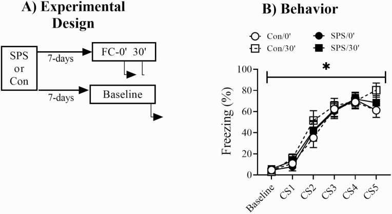
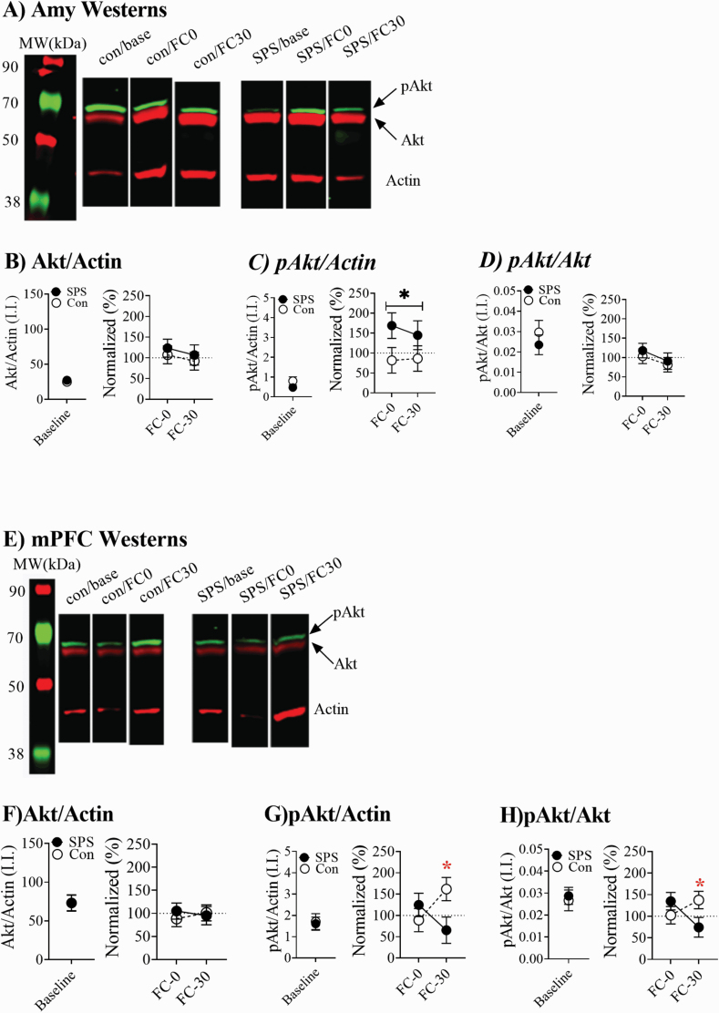
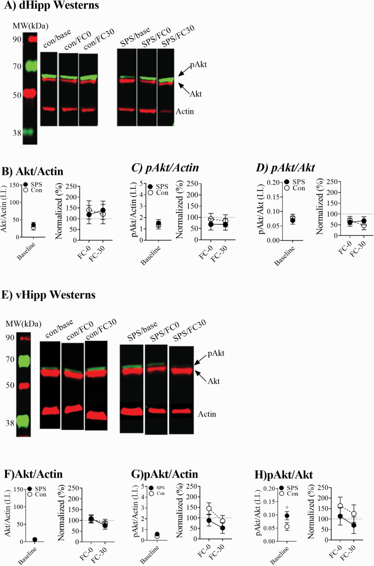
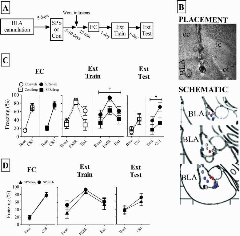
Methods: To test this hypothesis, rats were exposed to traumatic stress using the single prolonged stress model, and changes in Akt phosphorylation were assayed in the Amy at 0 and 30 minutes after fear conditioning (FC). In a separate experiment, we inhibited PI3K→Akt signaling in the BLA prior to FC and observed the effect this had on acquisition, expression, and extinction of FC in stressed and control rats.
Results: Enhanced Akt phosphorylation in the Amy at both time points was observed in stressed rats, but not in control rats. PI3K→Akt inhibition in the BLA had no effect on freezing in control rats but decreased freezing during extinction training and testing in stressed rats.
Conclusion: These findings suggest that PI3K→Akt signaling in the BLA could be a mechanism via which traumatic stress leads to fear memory that is resistant to extinction.
Keywords: Basolateral amygdala; PTSD; extinction; fear memory; single prolonged stress.
© The Author(s) 2020. Published by Oxford University Press on behalf of CINP.
Figures
Similar articles
-
PI3-kinase cascade has a differential role in acquisition and extinction of conditioned fear memory in juvenile and adult rats.Learn Mem. 2016 Nov 15;23(12):723-731. doi: 10.1101/lm.041806.116. Print 2016 Dec. Learn Mem. 2016. PMID: 27918278 Free PMC article.
-
Basolateral amygdala calpain is required for extinction of contextual fear-memory.Neurobiol Learn Mem. 2018 Nov;155:180-188. doi: 10.1016/j.nlm.2018.08.004. Epub 2018 Aug 4. Neurobiol Learn Mem. 2018. PMID: 30086394
-
Inhibition of the PI3 kinase cascade in corticolimbic circuit: temporal and differential effects on contextual fear and extinction.Int J Neuropsychopharmacol. 2013 May;16(4):825-33. doi: 10.1017/S1461145712000636. Epub 2012 Jun 18. Int J Neuropsychopharmacol. 2013. PMID: 22704253
-
Single prolonged stress: toward an animal model of posttraumatic stress disorder.Depress Anxiety. 2009;26(12):1110-7. doi: 10.1002/da.20629. Depress Anxiety. 2009. PMID: 19918929 Review.
-
The role of the basolateral amygdala and infralimbic cortex in (re)learning extinction.Psychopharmacology (Berl). 2019 Jan;236(1):303-312. doi: 10.1007/s00213-018-4957-x. Epub 2018 Jun 30. Psychopharmacology (Berl). 2019. PMID: 29959461 Review.
Cited by
-
Basal forebrain cholinergic systems as circuits through which traumatic stress disrupts emotional memory regulation.Neurosci Biobehav Rev. 2024 Apr;159:105569. doi: 10.1016/j.neubiorev.2024.105569. Epub 2024 Feb 1. Neurosci Biobehav Rev. 2024. PMID: 38309497 Review.
-
Immune-metabolic mechanisms of post-traumatic stress disorder and atherosclerosis.Front Physiol. 2023 Feb 8;14:1123692. doi: 10.3389/fphys.2023.1123692. eCollection 2023. Front Physiol. 2023. PMID: 36846337 Free PMC article. Review.
-
Duration of exposure to epidural anesthesia at delivery, DNA methylation in umbilical cord blood and their association with offspring asthma in Non-Hispanic Black women.Environ Epigenet. 2022 Dec 13;9(1):dvac026. doi: 10.1093/eep/dvac026. eCollection 2023. Environ Epigenet. 2022. PMID: 36694712 Free PMC article.
-
Deciphering the Mysterious Relationship between the Cross-Pathogenetic Mechanisms of Neurodegenerative and Oncological Diseases.Int J Mol Sci. 2023 Sep 29;24(19):14766. doi: 10.3390/ijms241914766. Int J Mol Sci. 2023. PMID: 37834214 Free PMC article. Review.
-
The role of estrogen receptor manipulation during traumatic stress on changes in emotional memory induced by traumatic stress.Psychopharmacology (Berl). 2023 May;240(5):1049-1061. doi: 10.1007/s00213-023-06342-6. Epub 2023 Mar 6. Psychopharmacology (Berl). 2023. PMID: 36879072
References
-
- American Psychiatric Association, Force DSMT (2013) Diagnostic and statistical manual of mental disorders: DSM-5. Arlington, VA: American Psychiatric Association.
-
- Armario A, Escorihuela RM, Nadal R (2008) Long-term neuroendocrine and behavioural effects of a single exposure to stress in adult animals. Neurosci Biobehav Rev 32:1121–1135. - PubMed
-
- Baker KD, Gray AR, Richardson R (2017) The development of perineuronal nets around parvalbumin gabaergic neurons in the medial prefrontal cortex and basolateral amygdala of rats. Behav Neurosci 131:289–303. - PubMed
-
- Beeman CL, Bauer PS, Pierson JL, Quinn JJ (2013) Hippocampus and medial prefrontal cortex contributions to trace and contextual fear memory expression over time. Learn Mem 20:336–343. - PubMed
-
- Bergstrom HC (2016) The neurocircuitry of remote cued fear memory. Neurosci Biobehav Rev 71:409–417. - PubMed
Publication types
MeSH terms
Substances
Grants and funding
LinkOut - more resources
Full Text Sources
Medical
