. 2021 Jan 13;95(3):e01369-20.
doi: 10.1128/JVI.01369-20. Print 2021 Jan 13.

Dual Pathways of Human Immunodeficiency Virus Type 1 Envelope Glycoprotein Trafficking Modulate the Selective Exclusion of Uncleaved Oligomers from Virions

Affiliations

Dual Pathways of Human Immunodeficiency Virus Type 1 Envelope Glycoprotein Trafficking Modulate the Selective Exclusion of Uncleaved Oligomers from Virions

Shijian Zhang et al. J Virol. .

Abstract

The human immunodeficiency virus type 1 (HIV-1) envelope glycoprotein (Env) trimer is transported through the secretory pathway to the infected cell surface and onto virion particles. In the Golgi, the gp160 Env precursor is modified by complex sugars and proteolytically cleaved to produce the mature functional Env trimer, which resists antibody neutralization. We observed mostly uncleaved gp160 and smaller amounts of cleaved gp120 and gp41 Envs on the surface of HIV-1-infected or Env-expressing cells; however, cleaved Envs were relatively enriched in virions and virus-like particles (VLPs). This relative enrichment of cleaved Env in VLPs was observed for wild-type Envs, for Envs lacking the cytoplasmic tail, and for CD4-independent, conformationally flexible Envs. On the cell surface, we identified three distinct populations of Envs: (i) the cleaved Env was transported through the Golgi, was modified by complex glycans, formed trimers that cross-linked efficiently, and was recognized by broadly neutralizing antibodies; (ii) a small fraction of Env modified by complex carbohydrates escaped cleavage in the Golgi; and (iii) the larger population of uncleaved Env lacked complex carbohydrates, cross-linked into diverse oligomeric forms, and was recognized by poorly neutralizing antibodies. This last group of more "open" Env oligomers reached the cell surface in the presence of brefeldin A, apparently bypassing the Golgi apparatus. Relative to Envs transported through the Golgi, these uncleaved Envs were counterselected for virion incorporation. By employing two pathways for Env transport to the surface of infected cells, HIV-1 can misdirect host antibody responses toward conformationally flexible, uncleaved Env without compromising virus infectivity.IMPORTANCE The envelope glycoprotein (Env) trimers on the surface of human immunodeficiency virus type 1 (HIV-1) mediate the entry of the virus into host cells and serve as targets for neutralizing antibodies. The cleaved, functional Env is incorporated into virus particles from the surface of the infected cell. We found that an uncleaved form of Env is transported to the cell surface by an unconventional route, but this nonfunctional Env is mostly excluded from the virus. Thus, only one of the pathways by which Env is transported to the surface of infected cells results in efficient incorporation into virus particles, potentially allowing the uncleaved Env to act as a decoy to the host immune system without compromising virus infectivity.

Keywords: Env; Golgi bypass; antibody; cell surface; cleavage; trafficking; virion incorporation.


Figures

FIG 1
FIG 1
Enrichment of cleaved Env in HIV-1 virions produced by infected primary CD4-positive T lymphocytes. Primary CD4-positive T lymphocytes were infected with the indicated HIV-1 strains. The culture medium was used to prepare virions. The cell surface Env was immunoprecipitated with a mixture of 2G12, PGT121, and PGT128 antibodies. The cells were washed and lysed, and cell lysates were incubated with protein A-Sepharose beads. The immunoprecipitated Envs (Cell surface), Envs remaining in the cell lysates (Lysates), and virion Envs (Particles) were analyzed by Western blotting with a goat anti-gp120 antibody. The results shown are representative of those obtained in two independent experiments.
FIG 2
FIG 2
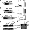
HIV-1 determinants of selective incorporation of cleaved Env into VLPs. (A) A549-rtTA cells were transfected with different ratios (weight/weight) of plasmids expressing the HIV-1 Gag-mCherry fusion protein and HIV-1AD8 Env. Forty-eight hours later, preparations of the cell lysates and virus-like particles (VLPs) were analyzed by Western blotting with a polyclonal goat anti-gp120 antibody. The ratios of gp120 to gp160 in the Western blot were quantitated (right panel). (B) A549-rtTA cells were transfected with plasmids expressing HIV-1 Gag-mCherry and HIV-1AD8 Env and different amounts of a plasmid expressing the HIV-1 Vpu protein (the weight/weight ratios of the Vpu to Env plasmids are shown). The cell lysates and VLPs were analyzed by Western blotting with a goat anti-gp120 antibody (upper left panel) or rabbit anti-Gag (p55/p24/p17) antibody (lower left panel). The ratios of gp120 to gp160 in the Western blot were quantitated (right panel). (C) A549-rtTA cells transiently expressing wild-type (wt) HIV-1AD8 Env or the cytoplasmic tail-deleted Env (Δ712) and, in some cases, the Gag-mCherry protein were used to prepare cell lysates; particles were concentrated from cell supernatants. The cell lysates and particles were analyzed by Western blotting with a goat anti-gp120 antibody. The ratios of cleaved gp120 to uncleaved Env in the Western blot were quantitated. (D and E) A549-rtTA cells transiently expressing Gag-mCherry and wild-type (wt) or mutant HIV-1AD8 Envs were used to prepare cell lysates; VLPs were concentrated from cell supernatants. The cell lysates and VLPs were analyzed by Western blotting with a goat anti-gp120 antibody. Each of the results shown in the figure panels was reproduced in an independent experiment.
FIG 3
FIG 3
Effect of Env proteolytic processing on incorporation into VLPs. A549-rtTA cells were transfected with a plasmid expressing the wild-type (wt) HIV-1JR-FL Env or the cleavage-defective Env(−) mutant and with a plasmid expressing Gag-mCherry or the pcDNA control, as indicated. Seventy-two hours after transfection and doxycycline induction, the cell lysates and VLPs were mock treated (no Rx) or treated with PNGase F or Endo Hf. The cell lysates and VLPs were analyzed by Western blotting with a goat anti-gp120 antibody (upper panels) or rabbit anti-Gag (p55/p24/p17) antibody (lower panels). The Envs deglycosylated by PNGase F are indicated by red arrows, and those deglycosylated by Endo Hf are indicated by green arrows (Endo H-R and Endo H-S indicate Endo Hf-resistant and -sensitive gp160 glycoproteins, respectively). The results of a typical experiment are shown.
FIG 4
FIG 4
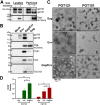
Characterization of stable A549 cells inducibly expressing HIV-1AD8 Env and/or Gag-mCherry. (A) A549-Env and A549-Gag/Env cells were induced with doxycycline to express HIV-1AD8 Env or both Gag-mCherry and Env, respectively. The cell lysates and particles pelleted from filtered tissue culture medium were analyzed by Western blotting with a goat anti-gp120 antibody (upper panel) or a rabbit anti-Gag (p55/p24/p17) antibody (lower panel). Note that extracellular vesicles pelleted from the culture medium of the A549-Env cells (no Gag-mCherry) exhibit a gp120/gp160 ratio similar to that of cell lysates; this contrasts with the higher gp120/gp160 ratio in VLPs prepared from the medium of A549-Gag/Env cells. (B) The medium of doxycycline-treated A549-Env, A549-Gag/Env, and A549-Gag cells was collected, filtered, and subjected to ultracentrifugation. Medium from A549-Env cells without doxycycline induction was studied in parallel (Mock). Pelleted samples were normalized by total protein level (estimated by silver staining) and analyzed by Western blotting with antibodies against the indicated viral and host cell proteins. (C) Extracellular vesicles were prepared by ultracentrifugation of the medium of A549-Env cells, and VLPs were prepared from the media of A549-Gag and A549-Gag/Env cells in parallel. The particles were incubated with the PGT121 or PGT151 bNAbs. The bound antibodies were detected with an immunogold-coupled anti-human IgG antibody, and labeled particles were imaged by electron microscopy. Bar, 100 nm. (D) Gold dots and extracellular particles were counted. The differences in the number of gold dots per particle were evaluated by Student’s t tests. *, P < 0.05; ***, P < 0.001. Data are representative of those obtained in at least two independent experiments.
FIG 5
FIG 5
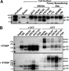
Immunoprecipitation of untreated and cross-linked Env on the cell surface. (A) A549-Gag/Env cells were incubated with the indicated monoclonal antibodies or surface labeled with biotin. The cells were then washed and lysed. Antibody-Env complexes were captured on protein A-Sepharose beads. The biotinylated cell surface proteins were affinity purified on a NeutrAvidin agarose resin. The precipitated Envs were analyzed by Western blotting with a goat anti-gp120 antibody. (B) A549-Gag/Env cells were cross-linked with DTSSP and then incubated with the indicated monoclonal antibody or with human immunoglobulin Gs (hIgGs). The cells were then washed and lysed. Antibody-Env complexes were captured on protein A-Sepharose beads. The captured Envs were analyzed on nonreducing (−DTT) and reducing (+DTT) gels and by Western blotting with a goat anti-gp120 antibody. The results shown in this figure are representative of those obtained in two independent experiments.
FIG 6
FIG 6
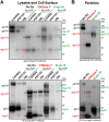
Glycosylation of cleaved and uncleaved Env in cell lysates, on the cell surface, and on VLPs. (A) Envs in the lysates of A549-Gag/Env cells or precipitated from the cell surface (Surf IP) by the PGT151 or 19b antibodies were treated with PNGase F or Endo Hf or were mock treated (No Rx). The Envs were run on SDS-polyacrylamide gels under reducing conditions and analyzed by Western blotting with the indicated antibodies. The deglycosylated gp160 and gp120 (dgp160 and dgp120, respectively) proteins are indicated by arrows (red, PNGase F-treated sample; green, Endo Hf-treated sample). Note that in the Western blot with the anti-gp120 antibody (upper panel), the heavy chains of the precipitated PGT151 and 19b antibodies create background bands that migrate near the deglycosylated gp120 band. (B) A549-Gag/Env supernatants were used to prepare VLPs, which were lysed and mock treated (No Rx) or treated with PNGase F or Endo Hf. The deglycosylated Envs were analyzed by Western blotting and are indicated by arrows, as described above. The results shown in this figure are representative of those obtained in three independent experiments.
FIG 7
FIG 7
Effect of brefeldin A (BFA) on HIV-1 Env synthesis, processing, glycosylation, and incorporation into VLPs. A549-Gag/Env cells were treated with 60 ng/ml brefeldin A 6 h after induction with doxycycline. (A) The cells were lysed, and the cell lysates were mock treated (No Rx) or treated with PNGase F or Endo Hf. The Envs were then separated by SDS-PAGE and analyzed by Western blotting with the indicated antibodies. The deglycosylated proteins are indicated by red arrows for PNGase F-treated samples and green arrows for Endo Hf-treated samples. (B) Untreated or brefeldin A-treated A549-Gag/Env cells were used for precipitation of Envs on the cell surface by the indicated antibodies. The precipitated antibody-Env complexes were captured on protein A-Sepharose beads and analyzed by Western blotting with a goat anti-gp120 antibody. (C) The medium of A549-Gag/Env cells treated with brefeldin A or mock treated was used to prepare VLPs. The VLPs were lysed and analyzed by Western blotting with the indicated antibodies. The results shown in this figure are representative of those obtained in two independent experiments.
FIG 8
FIG 8
Composition of detergent-resistant membranes. A549-Env cells were induced with doxycycline and homogenized. The clarified cell homogenate was made 1% in Triton X-100 and analyzed on a 20-to-40% (wt/vol) iodixanol gradient. After centrifugation, fractions were collected and analyzed by Western blotting for Env and flotillin-1. The flotillin-1 in fractions 1 to 3 serves as a marker for detergent-resistant membranes.
FIG 9
FIG 9
Two pathways of HIV-1 Env trafficking to the cell surface. Env glycoproteins synthesized in the endoplasmic reticulum oligomerize and are modified by high-mannose glycans. Some of these Envs traffic through the conventional secretory pathway to the Golgi, where complex glycans are added at some of the Env glycosylation sites. Proteolytic processing of gp160 occurs in the Golgi, with various degrees of efficiency. A small population of Env passing through the Golgi may escape cleavage. These Golgi Envs are transported to the cell surface, from which they are potentially incorporated into virions, either directly (e.g., for cytoplasmic tail-deleted Env) or following endocytosis. The characteristics of Envs transported to the cell surface through the conventional secretory pathway are listed in the green box. Another subset of Envs in the endoplasmic reticulum is transported to the cell surface via the unconventional Golgi bypass pathway. This Golgi bypass population accounts for the majority of the uncleaved Env on the surface of some Env-expressing cells. The characteristics of these Envs are listed in the red box.

Similar articles

Cited by

References

    1. Wyatt R, Sodroski J. 1998. The HIV-1 envelope glycoproteins: fusogens, antigens, and immunogens. Science 280:1884–1888. doi:10.1126/science.280.5371.1884. - DOI - PubMed
    1. Karlsson Hedestam GB, Fouchier RA, Phogat S, Burton DR, Sodroski J, Wyatt RT. 2008. The challenges of eliciting neutralizing antibodies to HIV-1 and to influenza virus. Nat Rev Microbiol 6:143–155. doi:10.1038/nrmicro1819. - DOI - PubMed
    1. Hoxie JA. 2010. Toward an antibody-based HIV-1 vaccine. Annu Rev Med 61:135–152. doi:10.1146/annurev.med.60.042507.164323. - DOI - PubMed
    1. Haynes BF, Shaw GM, Korber B, Kelsoe G, Sodroski J, Hahn BH, Borrow P, McMichael AJ. 2016. HIV-host interactions: implications for vaccine design. Cell Host Microbe 19:292–303. doi:10.1016/j.chom.2016.02.002. - DOI - PMC - PubMed
    1. Fauci AS. 2016. An HIV vaccine: mapping uncharted territory. JAMA 316:143–144. doi:10.1001/jama.2016.7538. - DOI - PubMed

Publication types

Substances