. 2020 Oct 31;8(1):177.
doi: 10.1186/s40478-020-01060-y.

RNA dependent suppression of C9orf72 ALS/FTD associated neurodegeneration by Matrin-3

Affiliations

RNA dependent suppression of C9orf72 ALS/FTD associated neurodegeneration by Matrin-3

Nandini Ramesh et al. Acta Neuropathol Commun. .

Abstract

The most common genetic cause of amyotrophic lateral sclerosis (ALS) is a GGGGCC (G4C2) hexanucleotide repeat expansions in first intron of the C9orf72 gene. The accumulation of repetitive RNA sequences can mediate toxicity potentially through the formation of intranuclear RNA foci that sequester key RNA-binding proteins (RBPs), and non-ATG mediated translation into toxic dipeptide protein repeats. However, the contribution of RBP sequestration to the mechanisms underlying RNA-mediated toxicity remain unknown. Here we show that the ALS-associated RNA-binding protein, Matrin-3 (MATR3), colocalizes with G4C2 RNA foci in patient tissues as well as iPSC-derived motor neurons harboring the C9orf72 mutation. Hyperexpansion of C9 repeats perturbed subcellular distribution and levels of endogenous MATR3 in C9-ALS patient-derived motor neurons. Interestingly, we observed that ectopic expression of human MATR3 strongly mitigates G4C2-mediated neurodegeneration in vivo. MATR3-mediated suppression of C9 toxicity was dependent on the RNA-binding domain of MATR3. Importantly, we found that expression of MATR3 reduced the levels of RAN-translation products in mammalian cells in an RNA-dependent manner. Finally, we have shown that knocking down endogenous MATR3 in C9-ALS patient-derived iPSC neurons decreased the presence of G4C2 RNA foci in the nucleus. Overall, these studies suggest that MATR3 genetically modifies the neuropathological and the pathobiology of C9orf72 ALS through modulating the RNA foci and RAN translation.


Conflict of interest statement

The authors declare that they have no competing interests

Figures
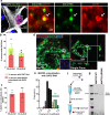
Fig. 1
Fig. 1
MATR3 colocalizes with pathogenic G4C2 RNA foci in C9-ALS iPSC-derived neurons and in post-mortem brain tissue. a Representative confocal image of colocalization (white arrows) between G4C2 RNA foci (green) and MATR3 protein (red) in C9-ALS patient-derived iPSCs differentiated into motor neurons (iPSC-MN), indicated by MAP2 staining (grey). Dotted-white box represents a single G4C2 foci that colocalized with MATR3 and represented in high magnification images to the right. b Quantification of percentage neurons that are positive for G4C2 foci (green bars) in two independent C9-ALS patient iPSC-MNs. Average  % neurons that are positive for G4C2 foci in C9-ALS #1 = 58% and in C9-ALS #2 = 48%. Among them, over half of the neurons also showed co-localization between MATR3 and G4C2 RNA foci (red bars). n = 3 differentiations. c Quantification of percentage G4C2 foci per neuron that colocalized with MATR3. Average  % G4C2 foci that colocalized with MATR3 in C9-ALS #1 = 75% and in C9-ALS #2 = 80%. n = 3 differentiations. d Representative images of immunohistochemical analysis of MATR3 signal (green) in motor cortex neurons from and C9-ALS patient tissue. RNA FISH analysis performed to examine co-localization between G4C2 RNA foci (red) and MATR3 (green) in nuclei of C9-ALS patient tissue cells. Maximum intensity projection (left) and single plane (right) representative images are shown. Inset within image on left represents overlay between Dapi (blue) and G4C2 foci (red). e Moderate (yellow) to strong (green) Pearson’s coefficients indicate co-localization between RNA foci and MATR3 signal. n = 70 G4C2 RNA foci from 3 C9-ALS patient tissues. f Diagrammatic representation of (G4C2) × 10 pull-down assay. Nuclear lysates from HEK293T cells transiently transfected with either FLAG-MATR3 or FLAG-MATR3-ΔRRM2 were incubated with biotinylated G4C210 RNA. The protein-G4C2 RNA complex was pulled down with streptavidin and then separated by SDS-PAGE. g Immunoblot of biotin-G4C210 pull-down fraction probed for FLAG-MATR3 showed physical interaction between MATR3 and G4C2 RNA. The interaction was moderately diminished between MATR3-ΔRRM2 and G4C2 RNA. Error bars indicate S.E.M
Fig. 2
Fig. 2
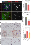
MATR3 levels and sub-cellular localization are perturbed in C9-ALS patient neurons and in post-mortem brain tissue. a Representative confocal images of control and C9-ALS patient-derived iPSCs that were differentiated to neurons (represented by ChAT+, green) and stained for endogenous MATR3 (red/gray). b Quantification of endogenous MATR3 immunofluorescence levels in ChAT+ neurons revealed significantly lower MATR3 levels in C9-ALS iPSC-MNs compared to that in control iPSC-MNs (Unpaired t-test) n = 2-3 independent differentiations from each independent control and C9-ALS iPSC-derived MN. iPSC-MNs from control: Ctrl #1 (yellow circle), Ctrl #2 (green circle), Ctrl #3 (cyan circle). iPSC-MNs from C9-ALS: C9-ALS #1 (gray circle), C9-ALS #2 (blue circle), C9-ALS #3 (purple circle). c Quantification of MATR3 mRNA fold change in control and C9-ALS iPSC MNs showed significantly reduced MATR3 mRNA levels in C9-ALS iPSC-MNs compared to that in control (Mann–Whitney U-test). n = 3 independent differentiations from each independent control and C9-ALS iPSC-derived MN. iPSC-MNs from control: Ctrl #1 (yellow circle), Ctrl #2 (green circle). iPSC-MNs from C9-ALS: C9-ALS #1 (gray circle), C9-ALS #3 (purple circle). d Representative IHC staining for MATR3 in entorhinal cortex sections from control, C9-ALS and sporadic (non-C9) ALS patient post-mortem brains. MATR3 was predominantly nuclear in control and sporadic ALS tissues, whereas in C9-ALS patient tissue, MATR3 also showed intense cytoplasmic signal (blue arrows). Scale = 50 µm. e Quantification of percentage of neurons with cytoplasmic MATR3 revealed increased percentage of neurons with cytoplasmic MATR3 staining in C9-ALS compared to control (Unpaired t-test). Control: 3 cases (n = 330; 135; 93); C9-ALS: 6 cases (n = 93; 485; 217; 310; 500; 251) and non-C9 sporadic-ALS: 3 cases (n = 316; 197; 402). Error bars indicate S.E.M. *p value < 0.05; ***p value < 0.001; ****p value < 0.0001
Fig. 3
Fig. 3
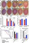
MATR3 is a strong modifier of C9orf72 G4C2-hexanucleotide repeat expansion (HRE)-mediated neurodegeneration in vivo. a Representative images of Drosophila eyes showing G4C2 hexanucleotide repeat expansion mediated eye degeneration in transgenic flies expressing 3 repeats (3R), 30 repeats (30R), 36 repeats (36R) and 58 repeats (58R) in the Drosophila eyes driven by GMR-gal4 driver. Flies expressing G4C2-3R had a comparable eye phenotype to control (GMR-GAL4; EGFP). Flies expressing G4C2-30R, 36R and 58R repeats exhibited signs of external eye degeneration including ommatidial fusion, bristle disorganization, depigmentation, and necrotic patches (arrow). These flies were expressing the G4C2 repeat expansion in the background of UAS-EGFP (control transgene) to account for GAL4 dilution. b Transgenic expression of human MATR3 in the G4C2 flies significantly ameliorated the external eye degenerative phenotypes and restored ommatidial structure and pigmentation. c Quantification of eye phenotypes showed statistically significant rescue in eye degeneration in G4C2-HRE flies upon expression of MATR3 (Kruskal–Wallis test). n ≥ 50 flies per genotype. d Quantification of mRNA levels of G4C2-GFP in heads from flies expressing G4C2-30R and 58R that are GFP-tagged, revealed no change in G4C2-GFP mRNA levels upon MATR3 expression (Kruskal–Wallis test). n = 5 per genotype. e Kaplan–Meier survival curve of flies conditionally expressing G4C2-30R in adult neurons, driven by ElavGS-GAL4 driver, showed significant reduction in longevity (left) and in median survival (right) (Log-Rank Mantel Cox test). n = 100 flies per genotype. f Neuronal expression of G4C2-30R caused profound motor dysfunction in adults. Quantification of the percentage of flies that can climb in 30 s indicated severe locomotion defects in G4C2 30R-expressing flies. Expression of MATR3 partially rescued the motor defects (One-way ANOVA). n ≥ 30 flies per genotype. Error bars indicate S.E.M. *p value < 0.05; **p value < 0.01; ***p value < 0.001; ****p value < 0.0001
Fig. 4
Fig. 4
RRM2 domain of MATR3 required to mediate G4C2-HRE toxicity in vivo. a Schematic of 847 amino acid (aa)-long MATR3 protein and its functional domains: two RNA-recognition motifs (RRM1/2) and two zinc-finger domains (ZF1/2). b Schematic of MATR3 with each functional domain deleted to generate deletion mutants: ΔRRM1, ΔRRM2, ΔZF1, ΔZF2. c Representative images of Drosophila eyes from flies co-expressing G4C2-30R with MATR3 (full-length) or deletion variants (ΔRRM1, ΔRRM2, ΔZF1, ΔZF2). Zoom panels emphasize the degree of ommatidial disorganization and depigmentation. MATR3-ΔRRM2 suppresses G4C2-30R toxicity to a lesser degree compared to MATR3 wildtype, indicated by increased degenerative phenotypes including de-pigmentation and ommatidial fusion compared to G4C2-30R + MATR3 (full-length). d Quantification of eye degeneration revealed no difference in G4C2-30R + MATR3-ΔRRM2 compared to G4C2-30R alone (Kruskal–Wallis test) n ≥ 50 per genotype e Kaplan–Meier survival curve of adult flies showed that neuronal expression of MATR3-ΔRRM2 mutant in G4C2-30R flies did not modify reduced survival defect in G4C2-30R flies (Log Rank Mantel Cox test) n = 100 flies per genotype. Error bars indicate S.E.M. *p value < 0.05, ****p value < 0.001
Fig. 5
Fig. 5
MATR3 overexpression mitigates DPR production from G4C260 transcripts in an RNA-dependent manner. a Representative confocal images of HEK293T cells co-transfected with G4C260-Dendra2 RAN translation reporter plasmid and either FLAG (empty vector), FLAG-MATR3 or FLAG-MATR3-ΔRRM2 plasmids. G4C260 expression resulted in formation of G4C2 RNA foci (red) and, further, the G4C260 transcripts also undergo RAN translation in a subset of cells to produce dipeptide repeats (DPRs), indicated by Dendra2 signal (green) that is in frame with polyGR (white arrows). b Quantification of the percentage of cells that produce GR-Dendra2 (green) RAN product showed that overexpression of FLAG-MATR3 (gray) significantly reduced RAN translation of G4C260 transcript to GR-Dendra2 RAN product. Overexpression of FLAG-MATR3-ΔRRM2 did not impact GR-Dendra2 production in these cells (One-way ANOVA) n = 15-18 per group. c Quantification of G4C260-Dendra2 mRNA levels in cells expressing FLAG (empty vector) or FLAG-MATR3 showed no significant difference (One-way ANOVA) n = 5 per group. d Schematic of previously-published codon-optimized approach to produce dipeptide protein repeats by canonical translation of non-repeat RNA (i.e. not carrying GGGGCC repeats). e Representative images of Drosophila eyes and f quantification of external eye degeneration from flies expressing codon-optimized DPRs: GR36, PR36, GA36 and GP36. Flies expressing PR36 and GR36 developed significant external eye degeneration that is more severe is GR36 compared to PR36. Flies expressing either GA36 or GP36 did not show any external eye degeneration. Expression of MATR3 in these flies did not modify the phenotypes (Kruskal–Wallis test) n ≥ 50 per genotype. Error bars indicate S.E.M. **p value < 0.01
Fig. 6
Fig. 6
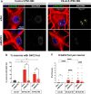
G4C2 RNA foci modulated by MATR3 in C9-ALS patient iPSC-MNs. a Representative confocal images of control (left) and C9-ALS patient (right) iPSC-MNs, indicated by MAP2 (red), transfected with scrambled siRNA or MATR3 siRNAs. FISH-IF was performed to stain nuclear G4C2 RNA foci (green; yellow arrowheads) in these neurons. b, c Quantification of the percentage of neurons that are G4C2 foci-positive (b), and number of G4C2 foci per neuron (c) in iPSC-MNs from one control (Ctrl #1) and two independent C9-ALS (C9-ALS #1, C9-ALS #2). Knocking down MATR3 significantly reduced the percentage of neurons that form G4C2 foci (b) and also reduced number of G4C2 foci per neuron (c) compared to scrambled-control in both C9-ALS iPSC-MNs (Kruskal–Wallis test). n = 24-50 neurons Each data point (circle) represents data from neurons pooled from 3 independent differentiations. Error bars indicate S.E.M. ****p value < 0.0001

Similar articles

Cited by

References

    1. Brown RH, Al-Chalabi A (2017) Amyotrophic lateral sclerosis. 10.1056/NEJMra1603471. Epub ahead of print 12 July 2017
    1. Al-Chalabi A, Hardiman O, Kiernan MC, et al. Amyotrophic lateral sclerosis: moving towards a new classification system. Lancet Neurol. 2016;15:1182–1194. doi: 10.1016/S1474-4422(16)30199-5. - DOI - PubMed
    1. DeJesus-Hernandez M, Mackenzie IR, Boeve BF, et al. Expanded GGGGCC hexanucleotide repeat in noncoding region of C9ORF72 causes chromosome 9p-linked FTD and ALS. Neuron. 2011;72:245–256. doi: 10.1016/j.neuron.2011.09.011. - DOI - PMC - PubMed
    1. Renton AE, Majounie E, Waite A, et al. A hexanucleotide repeat expansion in C9ORF72 is the cause of chromosome 9p21-linked ALS-FTD. Neuron. 2011;72:257–268. doi: 10.1016/j.neuron.2011.09.010. - DOI - PMC - PubMed
    1. Smith BN, Newhouse S, Shatunov A, et al. The C9ORF72 expansion mutation is a common cause of ALS ± FTD in Europe and has a single founder. Eur J Hum Genet. 2013;21:102–108. doi: 10.1038/ejhg.2012.98. - DOI - PMC - PubMed

Publication types

MeSH terms