. 2020 Oct 31;43(10):889-897.
doi: 10.14348/molcells.2020.0182.

K-Ras-Activated Cells Can Develop into Lung Tumors When Runx3-Mediated Tumor Suppressor Pathways Are Abrogated

Affiliations

K-Ras-Activated Cells Can Develop into Lung Tumors When Runx3-Mediated Tumor Suppressor Pathways Are Abrogated

You-Soub Lee et al. Mol Cells. .

Abstract

K-RAS is frequently mutated in human lung adenocarcinomas (ADCs), and the p53 pathway plays a central role in cellular defense against oncogenic K-RAS mutation. However, in mouse lung cancer models, oncogenic K-RAS mutation alone can induce ADCs without p53 mutation, and loss of p53 does not have a significant impact on early K-RAS-induced lung tumorigenesis. These results raise the question of how K-RAS-activated cells evade oncogene surveillance mechanisms and develop into lung ADCs. RUNX3 plays a key role at the restriction (R)-point, which governs multiple tumor suppressor pathways including the p14ARF-p53 pathway. In this study, we found that K-RAS activation in a very limited number of cells, alone or in combination with p53 inactivation, failed to induce any pathologic lesions for up to 1 year. By contrast, when Runx3 was inactivated and K-RAS was activated by the same targeting method, lung ADCs and other tumors were rapidly induced. In a urethane-induced mouse lung tumor model that recapitulates the features of K-RAS-driven human lung tumors, Runx3 was inactivated in both adenomas (ADs) and ADCs, whereas K-RAS was activated only in ADCs. Together, these results demonstrate that the R-point-associated oncogene surveillance mechanism is abrogated by Runx3 inactivation in AD cells and these cells cannot defend against K-RAS activation, resulting in the transition from AD to ADC. Therefore, K-RAS-activated lung epithelial cells do not evade oncogene surveillance mechanisms; instead, they are selected if they occur in AD cells in which Runx3 has been inactivated.

Keywords: K-Ras; Runx3; cancer initiation; lung cancer; p53.


Conflict of interest statement

CONFLICT OF INTEREST

The authors have no potential conflicts of interest to disclose.

Figures
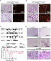
Fig. 1
Fig. 1. Runx3 inactivation, but not p53 inactivation, enables immediate proliferation of K-Ras–activated lung epithelial cells.
(A and B) KPT and KRT mice were targeted with Ad-Cre (2.5 × 10⁷ PFU/mouse, as described by DuPage et al., 2009), and Tomato-positive lung epithelial cells were assessed after 10 days. Clusters of Tomato-positive cells were detected 10 days after infection only in KRT mice lungs. Enlarged images of the boxed regions are shown (lower panels). (C) Schematic representation of the structures of K-RasLoxP-STOP-LoxP-G12D (K-Ras*), Runx3flox, p53flox, and Cretm/ERT1 (CreERT1) alleles. Cre recombinase activates K-Ras by removal of a knocked-in STOP transcriptional cassette from the K-RasLoxP-STOP-LoxP-G12D allele and inactivates the p53flox and Runx3flox alleles by deletion of exons. Survival curves of KR, R-CreERT1, K-CreERT1, KP-CreERT1, and KR-CreERT1 mice in the absence of tamoxifen are shown. The median survival of KR-CreERT1 mice was 48 days. No mice of other genotypes died within 50 weeks after birth (the duration of the experiment). (D) Hematoxylin/eosin (HE) staining of the lungs of K-CreERT1, KP-CreERT1, and R-CreERT1 mice (6 months after birth). A magnified image of the boxed region is shown on the right. Adenomatous lesions in a R-CreERT1 lung are indicated by dotted circles. HE staining of the lung tumors of KR-CreERT1 mice (2 and 8 weeks after birth).
Fig. 2
Fig. 2. The combination of K-Ras activation and Runx3 inactivation is the minimum molecular requirement for induction of lung tumor formation.
(A) Microscopic images of lungs of T-CreERT2 and KT-CreERT2 mouse (6 months after birth) stained with anti-Tomato antibody. Tomato-positive cells (red) are indicated by arrows. (B) HE staining of the lungs of KPT-CreERT2 and KRT-CreERT2 mice (6 months after birth). (C and D) Microscopic images of lungs of KPT-CreERT2 mice and KRT-CreERT2 mice (6 months after birth) stained with HE or anti-Tomato antibody. Magnified images of the boxed regions are shown (lower panels). Small clusters of Tomato-positive cells were detected in KPT-CreERT2 mice, but did not develop into cancers. By contrast, Tomato-positive cells in KRT-CreERT2 mice developed into cancers.
Fig. 3
Fig. 3. KR-CreERT1 mice develop lymphomas and skin cancers, as well as lung ADs/ADCs.
(A) Gross and microscopic images of a skin cancer that developed in a KR-CreERT1 mouse. (B) Gross and microscopic images of the spleen and thymus of WT and KR-CreERT1 mice. Histological analysis revealed that the cortex–medulla boundary in the enlarged thymuses was obscure and filled with undifferentiated T-cells, indicating thymic lymphoma. (C) HE-stained gross images and magnified microscopic images of thymus and lung of KRT-CreERT2 mice (6 months after birth). Lung-invading tumor cells were morphologically identical to thymic lymphoma cells, indicating that the lung-invading tumors originated from a thymic lymphoma. (D) Targeting of the K-Ras* and Runx3flox alleles by leaky activation of CreERT1 in tumors was verified by genomic PCR. L, lung ADC; Th, thymus; Sp, spleen; Sk, skin cancer. Targeting of the K-Ras* and Runx3flox alleles by leaky activation of CreERT2 in tumors was verified by genomic PCR. M, DNA size marker; L, lung ADC; Th, thymus; LT, lung infiltrated thymic lymphoma.
Fig. 4
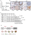
Fig. 4. Sequential molecular events associated with the development of lung AD and progression to ADC.
(A) Mice (FVB strain) were intraperitoneally injected with urethane at a dose of 500 mg/kg body weight. Growth of cancer in mouse lungs vs. time after urethane injection is shown. Five mice were used for each time point. Ave, average diameter (mm) of cancer; SD, standard deviation. (B) Mouse lung ADs that developed 20 weeks (20W) after urethane injection and lung ADCs that developed 58 weeks (58W) after injection were analyzed by immunohistochemical (IHC) staining with anti-phospho-Erk (p-Erk) and anti-Runx3 antibodies. Runx3 expression was markedly downregulated in both ADs and ADCs relative to adjacent normal regions. By contrast, Erk was activated only in ADCs. Nor, normal. (C) WT mouse lung and mouse lung ADs developed at 20 weeks (20W, diameter < 1 mm), relatively small lung ADCs (58W-S, diameter ≒ 2 mm) and relatively large lung ADCs (58W-L, diameter ≒ 3 mm) developed at 58 weeks, and large lung ADCs (68W-L, diameter ≒ 6 mm) developed at 68 weeks after urethane injection were obtained. Four ADs or ADCs from each group were analyzed by whole-exon sequencing. Among the known major oncogenes and tumor suppressors involved in lung cancer, only K-Ras mutations were detected. Amino acid changes in K-Ras and the ratio of mutation signal to total signal are shown. d, diameter; ratio, ratio of oncogenic K-Ras–mutated allele relative to total K-Ras. Bar diagram demonstrating average ratios of K-Ras mutation in each group of ADs/ADCs. P = P value. (D) Schematic representation of the molecular events associated with the development of lung ADs and progression into ADCs. Runx3↓ and K-Ras↑ indicate Runx3 inactivation and K-Ras activation, respectively. Human AAH and BAC correspond to mouse lung ADs.

Similar articles

Cited by

References

    1. Canon J., Rex K., Saiki A.Y., Mohr C., Cooke K., Bagal D., Gaida K., Holt T., Knutson C.G., Koppada N., et al. The clinical KRAS(G12C) inhibitor AMG 510 drives anti-tumour immunity. Nature. 2019;575:217–223. doi: 10.1038/s41586-019-1694-1. - DOI - PubMed
    1. Chi X.Z., Lee J.W., Lee Y.S., Park I.Y., Ito Y., Bae S.C. Runx3 plays a critical role in restriction-point and defense against cellular transformation. Oncogene. 2017;36:6884–6894. doi: 10.1038/onc.2017.290. - DOI - PMC - PubMed
    1. Drosten M., Guerra C., Barbacid M. Genetically engineered mouse models of K-Ras-driven lung and pancreatic tumors: validation of therapeutic targets. Cold Spring Harb. Perspect. Med. 2018;8:a031542. doi: 10.1101/cshperspect.a031542. - DOI - PMC - PubMed
    1. DuPage M., Dooley A.L., Jacks T. Conditional mouse lung cancer models using adenoviral or lentiviral delivery of Cre recombinase. Nat. Protoc. 2009;4:1064–1072. doi: 10.1038/nprot.2009.95. - DOI - PMC - PubMed
    1. Feldser D.M., Kostova K.K., Winslow M.M., Taylor S.E., Cashman C., Whittaker C.A., Sanchez-Rivera F.J., Resnick R., Bronson R., Hemann M.T., et al. Stage-specific sensitivity to p53 restoration during lung cancer progression. Nature. 2010;468:572–575. doi: 10.1038/nature09535. - DOI - PMC - PubMed

MeSH terms