. 2020 Oct 22;25(21):4888.
doi: 10.3390/molecules25214888.

Subcellular Location of Tirapazamine Reduction Dramatically Affects Aerobic but Not Anoxic Cytotoxicity

Affiliations

Subcellular Location of Tirapazamine Reduction Dramatically Affects Aerobic but Not Anoxic Cytotoxicity

Chris P Guise et al. Molecules. .

Abstract

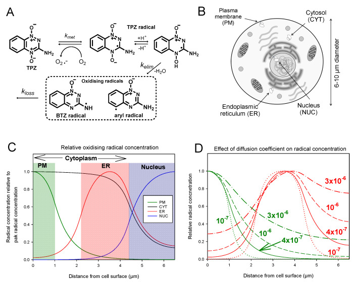
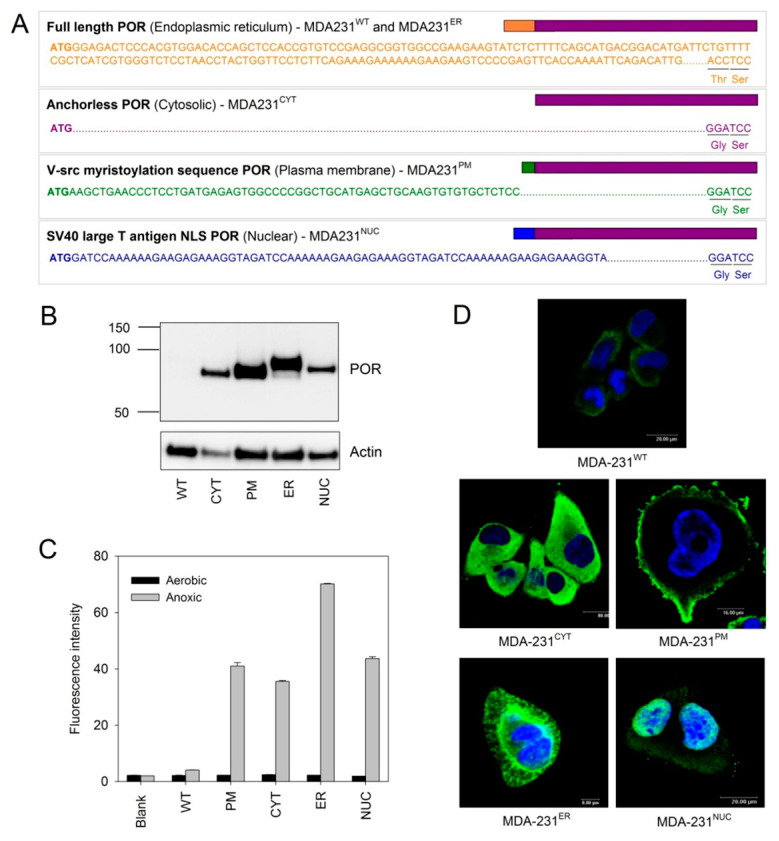
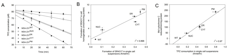
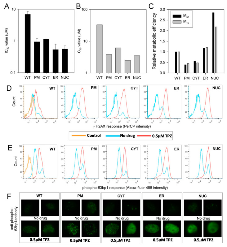
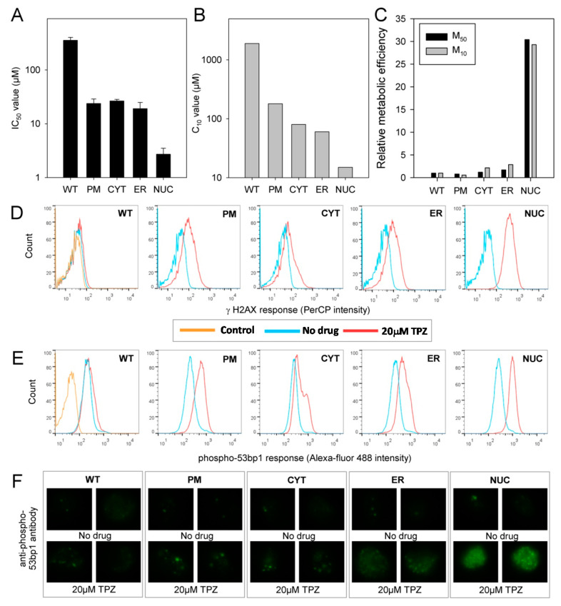
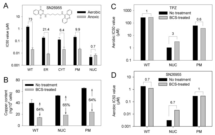
Hypoxia is an adverse prognostic feature of solid cancers that may be overcome with hypoxia-activated prodrugs (HAPs). Tirapazamine (TPZ) is a HAP which has undergone extensive clinical evaluation in this context and stimulated development of optimized analogues. However the subcellular localization of the oxidoreductases responsible for mediating TPZ-dependent DNA damage remains unclear. Some studies conclude only nuclear-localized oxidoreductases can give rise to radical-mediated DNA damage and thus cytotoxicity, whereas others identify a broader role for endoplasmic reticulum and cytosolic oxidoreductases, indicating the subcellular location of TPZ radical formation is not a critical requirement for DNA damage. To explore this question in intact cells we engineered MDA-231 breast cancer cells to express the TPZ reductase human NADPH: cytochrome P450 oxidoreductase (POR) harboring various subcellular localization sequences to guide this flavoenzyme to the nucleus, endoplasmic reticulum, cytosol or inner surface of the plasma membrane. We show that all POR variants are functional, with differences in rates of metabolism reflecting enzyme expression levels rather than intracellular TPZ concentration gradients. Under anoxic conditions, POR expression in all subcellular compartments increased the sensitivity of the cells to TPZ, but with a fall in cytotoxicity per unit of metabolism (termed 'metabolic efficiency') when POR is expressed further from the nucleus. However, under aerobic conditions a much larger increase in cytotoxicity was observed when POR was directed to the nucleus, indicating very high metabolic efficiency. Consequently, nuclear metabolism results in collapse of hypoxic selectivity of TPZ, which was further magnified to the point of reversing O2 dependence (oxic > hypoxic sensitivity) by employing a DNA-affinic TPZ analogue. This aerobic hypersensitivity phenotype was partially rescued by cellular copper depletion, suggesting the possible involvement of Fenton-like chemistry in generating short-range effects mediated by the hydroxyl radical. In addition, the data suggest that under aerobic conditions reoxidation strictly limits the TPZ radical diffusion range resulting in site-specific cytotoxicity. Collectively these novel findings challenge the purported role of intra-nuclear reductases in orchestrating the hypoxia selectivity of TPZ.

Keywords: DNA damage-response; DNA-targeted cytotoxin; cell membrane; cell nucleus; cytochrome P450 oxidoreductase; endoplasmic reticulum; hypoxia-activated prodrug; tirapazamine.


Conflict of interest statement

The authors declare no conflict of interest. The funders had no role in the design of the study; in the collection, analyses, or interpretation of data; in the writing of the manuscript, or in the decision to publish the results.

Figures

Figure 1
Figure 1
Reaction-diffusion modelling predicts non-isotropic distribution of cytotoxic TPZ radicals following TPZ activation under anoxia in sub-cellular compartments: (A) Scheme showing the metabolic activation of TPZ, which undergoes one-electron reduction to generate the oxygen-sensitive radical anion. The protonated radical eliminates water to generate reactive oxidizing benzotriazinyl (BTZ) and aryl radicals, in the absence of oxygen; (B) Schematic representation showing sources of one-electron TPZ reduction explored in the present study; (C) Reaction-diffusion modelling of the intracellular steady state TPZ-derived oxidizing radicals (relative to peak concentration) when generated in different subcellular compartments (PM green, ER red, CYT black, NUC blue), assuming an intracellular diffusion coefficient, D, of 4 × 10⁻⁷ cm2 s−1 for TPZ and its radicals; (D) Model results for D = 10−7 cm2 s−1 (dotted line), 4 × 10⁻⁷ cm2 s−1 (solid line), 10−6 cm2 s−1 (dashed line) or, 3 × 10⁻⁶ cm2 s−1 (dot-dash line) for activation at the PM (green) or ER (red).
Figure 2
Figure 2
Generation of clonal MDA-231 cell lines expressing POR in different subcellular locations: (A) Details of the localization sequences used to direct POR to different subcellular compartments; (B) Western blot analysis showing expression levels and size differences of POR in the transfected cell lines; (C) Flow cytometry detection of POR enzyme activity in the transfected cell lines with an oxygen-sensitive fluorogenic POR substrate (FSL61). Results are the median fluorescence intensity of triplicate samples ± SEM. Blank is cell-free control; (D) Immunofluorescence microscopy showing the location of POR expression (green) in the MDA-231 cell lines. Nuclei are stained blue (Hoechst 33258).
Figure 3
Figure 3
Expression of POR in different subcellular compartments results in increased TPZ activation: (A) Consumption of TPZ under anoxic conditions in parental and transfected MDA-231 cells. Results are the average of duplicate experiments, error bars indicate the range; (B) Correlation between the rates of anoxic formation of the non-toxic mono-N-oxide metabolite SR4317 in single cell suspensions (mean of duplicate samples) versus whole cell homogenates (mean of duplicate experiments). Error bars indicate the range in data; (C) Correlation between POR activity (rate of cytochrome C reduction in whole cell lysates; mean of duplicate experiments) and the rate of anoxic TPZ consumption in single cell suspensions (mean of duplicate experiments).
Figure 4
Figure 4
TPZ can achieve DNA damage under anoxia irrespective of the subcellular compartment of activation: (A) Sensitivity of parental and transfected MDA-231 cell lines to TPZ under anoxic conditions as determined by antiproliferative assay. IC50 values show the mean of 2–3 independent experiments, error bars show the SEM; (B) Sensitivity of parental and transfected MDA-231 cell lines to TPZ under anoxic conditions as determined by clonogenic assay. The concentration of TPZ required to reduce clonogenic survival by 90% (C10 values) were calculated in triplicate per drug concentration; (C) Relative metabolic efficiency in MDA-231 transfectants (extent to which metabolism contributes to cytotoxicity). M50 = 1/(IC50 × kmet) of anoxic TPZ metabolism as derived from Figure 2A. M10 = 1/(C10 × kmet) of anoxic TPZ metabolism as derived from Figure 2A. The M50 and M10 values were normalized to the value for MDA-231WT cells; (D) Flow cytometry analysis of γ-H2AX levels in parental and transfected MDA-231 cells following a 4 h anoxic exposure to 0.5 µM TPZ and a 1 h drug-free recovery period under aerobic conditions. Control: no antibody labelling; (E) Flow cytometry analysis of phospho-53bp1 levels in parental and transfected MDA-231 cells following a 4 h anoxic exposure to 0.5 µM TPZ and a 1 h drug-free recovery period under aerobic conditions. Control: secondary antibody only; (F) Immunofluoresence microscopy showing phospho-53bp1 foci formation following a four-hour anoxic exposure to 0.5 µM TPZ and a 1 h drug-free recovery period under aerobic conditions.
Figure 5
Figure 5
TPZ cytotoxicity under aerobic conditions displays major bias towards cells with POR localized in the nuclear compartment: (A) Sensitivity of parental and transfected MDA-231 cell lines to TPZ under aerobic conditions as determined by antiproliferative assay. IC50 values show the mean of 2-4 independent experiments, error bars show the SEM; (B) Sensitivity of parental and transfected MDA-231 cell lines to TPZ under aerobic conditions as determined by clonogenic assay. C10 values are derived from a single experiment (triplicate plates per drug concentration); (C) Relative metabolic efficiency in MDA-231 transfectants (extent to which productive metabolism contributes to cytotoxicity). M50 = 1/(IC50 × kmet) of anoxic TPZ activity as derived from Figure 3A. M10 = 1/(C10 × kmet) of anoxic TPZ activity as derived from Figure 3A). The M50 and C10 values were normalised to the value for MDA-231WT cells; (D) Flow cytometry analysis of γ-H2AX levels in parental and transfected MDA-231 cells following a 4 h aerobic exposure to 20 µM TPZ and a 1 h drug-free recovery period under aerobic conditions. Control is no antibody labelling; (E) Flow cytometry analysis of phospho-53bp1 levels in parental and transfected MDA-231 cells following a 4 h aerobic exposure to 20 µM TPZ and a 1 h drug-free recovery period under aerobic conditions. Control is secondary antibody only; (F) Immunofluoresence microscopy showing phospho-53bp1 foci formation following a four hour aerobic exposure to 20 µM TPZ and a 1 h drug-free recovery period under aerobic conditions.
Figure 6
Figure 6
Aerobic toxicity of TPZ and an analogue with increased affinity for DNA is mediated by a copper-dependent mechanism in MDA-231NUC cells: (A) Sensitivity of parental and transfected MDA-231 cell lines to SN26955, which contains a TPZ moiety linked to a DNA intercalator, under aerobic conditions as determined by antiproliferative assay. IC50 values show the mean of 3-4 independent experiments, error bars show the SEM; numbers are the HCR values. (B) Quantitation of the copper content of cells incubated for 7 days in normal media or media supplemented with the copper chelating agent bathocuproine (BCS) as determined by ICP-MS analysis. (C) Effects on chronic BCS exposure on the aerobic sensitivity of parental, MDA-231NUC or MDA-231PM cells to TPZ as determined by anti-proliferative assay. (D) Effects on chronic BCS exposure on the aerobic sensitivity of parental, MDA-231NUC or MDA-231PM cells to SN26955 as determined by anti-proliferative assay.

Similar articles

Cited by

References

    1. Vaupel P., Mayer A. Hypoxia in cancer: Significance and impact on clinical outcome. Cancer Metastasis Rev. 2007;26:225–239. doi: 10.1007/s10555-007-9055-1. - DOI - PubMed
    1. Dhani N., Fyles A., Hedley D., Milosevic M. The clinical significance of hypoxia in human cancers. Semin. Nucl. Med. 2015;45:110–121. doi: 10.1053/j.semnuclmed.2014.11.002. - DOI - PubMed
    1. Hammond E.M., Asselin M.C., Forster D., O’Connor J.P., Senra J.M., Williams K.J. The meaning, measurement and modification of hypoxia in the laboratory and the clinic. Clin. Oncol. (R. Coll. Radiol.) 2014;26:277–288. doi: 10.1016/j.clon.2014.02.002. - DOI - PubMed
    1. Harris A.L. Hypoxia—A key regulatory factor in tumour growth. Nat. Rev. Cancer. 2002;2:38–47. doi: 10.1038/nrc704. - DOI - PubMed
    1. Bennett M.H., Feldmeier J., Smee R., Milross C. Hyperbaric oxygenation for tumour sensitisation to radiotherapy. Cochrane Database Syst. Rev. 2012;4:Art. No.: CD005007. doi: 10.1002/14651858.CD005007.pub3. - DOI - PMC - PubMed

MeSH terms

LinkOut - more resources