. 2020 Nov;49(11):320-334.
doi: 10.1038/s41684-020-00659-x. Epub 2020 Oct 20.

Experimental murine arteriovenous fistula model to study restenosis after transluminal angioplasty

Affiliations

Experimental murine arteriovenous fistula model to study restenosis after transluminal angioplasty

Chuanqi Cai et al. Lab Anim (NY). 2020 Nov.

Abstract

Percutaneous transluminal angioplasty (PTA) is a very common interventional treatment for treating stenosis in arteriovenous fistula (AVF) used for hemodialysis vascular access. Restenosis occurs after PTA, resulting in vascular lumen loss and a decrease in blood flow. Experimental animal models have been developed to study the pathogenesis of stenosis, but there is no restenosis model after PTA of stenotic AVF in mice. Here, we describe the creation of a murine model of restenosis after angioplasty of a stenosis in an AVF. The murine restenosis model has several advantages, including the rapid development of restenotic lesions in the vessel after angioplasty and the potential to evaluate endovascular and perivascular therapeutics for treating restenosis. The protocol includes a detailed description of the partial nephrectomy procedure to induce chronic kidney disease, the AVF procedure for development of de novo stenosis and the angioplasty treatment associated with progression of restenosis. We monitored the angioplasty-treated vessel for vascular patency and hemodynamic changes for a period of 28 d using ultrasound. Vessels were collected at different time points and processed for histological analysis and immunostaining. This angioplasty model, which can be performed with basic microvascular surgery skills, could be used to identify potential endovascular and perivascular therapies to reduce restenosis after angioplasty procedures.


Figures
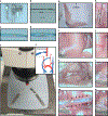
Fig. 1 |
Fig. 1 |. Materials required and technical procedures for CKD, AVF and angioplasty procedures.
a, Different sizes of microvascular clamps for different vessel diameters: the smallest one is only for clamping the vein, and the others are used for arterial and EJV clamping. b, Different sutures for ligating and creating the vascular anastomosis: the 6–0 silk suture is for ligation of the distal end of the EJV, 8–0 is for ligation of the proximal end of the EJV and the 11–0 suture is for AVF anastomosis and closure of the puncture used for inserting the angioplasty catheter. c, Deflated 1.25 mm × 6 mm balloon catheter. d, Inflated balloon catheter at 14 atm pressure. e, Experimental setup before anesthetizing the mouse. f, Schematic of AVF creation and angioplasty procedures. g, Curved incision in the right neck paralleling the trachea. h, The right EJV should be dissected ≥10 mm in length. i, The REJV and LCCA are flushed with heparinized saline before creating the AVF anastomosis. j, After the AVF anastomosis has been created, red pulsatile arterial blood flow is observed in the outflow vein. k, Two weeks after creating the AVF anastomosis, the arterialized EJV has an enlarged diameter. l, After clamping the inflow and outflow vessels, the vessels are flushed with heparinized saline through the puncture site. m, The angioplasty is performed at 14 atm for 30 s. n, The angioplasty puncture site is closed using an 11–0 suture. R, Right side; L, left side. Black arrows indicate the inflated balloon catheter, and the dashed circles indicate the puncture site. Animal procedures in this work were approved by the Mayo Clinic Institutional Animal Care and Use Committee.
Fig. 2 |
Fig. 2 |. Time course of histomorphometric changes after the angioplasty procedure.
a, Time course of CKD surgery, AVF and angioplasty procedures. b, Representative of H&E section of an arterialized EJV after an angioplasty or sham procedure at D0, D14 and D28. At D0, the sham vessel shows obvious arterialization; at D0-post, the vessel shows a reduced neointimal area and cells in the neointimal and media area. At D14, the sham vessel has abundant cellular deposition in the neointimal area, whereas the angioplasty-treated vessel shows less neointima. At D28, no difference can be seen in the neointimal area between the sham- and angioplasty-treated vessels. c, Representative Verhoeff–Van Gieson (VVG) staining in a normal contralateral vein, sham and angioplasty-treated vessel at D14. In the normal jugular vein, a single layer of IEL is poorly formed. At D14, in the sham vessels, an obvious IEL is shown between the neointimal area and media area, whereas the angioplasty-treated vessel has an intermittent IEL. d, Doppler ultrasound shows that before the angioplasty procedure, peak velocity is similar between sham- and angioplasty-treated vessels. At D14, angioplasty-treated vessels have a significant increase in peak velocity compared to sham vessels. No significant difference is observed at D21 and D28. e, At D14, angioplasty-treated vessels have significantly larger vessel diameters than do sham vessels, with no difference at D28. f, At D0, angioplasty-treated vessels show a significant decrease in the vessel lumen area compared to sham vessels, but a significant increase at D14 with no difference at D28. g, There was a significant decrease in the neointimal area/media area (N/M) ratio in angioplasty-treated vessels compared to sham vessels at D0, D14 and D28. Each time point represents the mean ± s.e.m. of 5–12 animals for each group. Two-way ANOVA with Bonferroni’s correction was performed by Graph Pad Prism version 8 (GraphPad Software Inc.). Significant differences are indicated: NS, not significant; *P < 0.05; **P < 0.01; ***P < 0.001; #P < 0.0001. Solid arrows indicate IEL. Scale bars: 50 μm. ADV, adventitia; D, day; L, lumen; D0-post, day 0 post angioplasty; D14, day 14 post angioplasty; D28, day 28 post angioplasty.
Fig. 3 |
Fig. 3 |. Immunostaining for endothelial cells (CD31+ cells) at different time points.
a, Representative CD31 staining of sham- and angioplasty-treated vessels at D0-post, D14 and D28. At D0, D14 and D28, sham vessels show a discontinuous endothelium layer. There is no endothelial layer in angioplasty-treated vessels at D0-post. At D14, angioplasty-treated vessels have intact endothelium. At D28, CD31+ vessels are present in the neointimal area of both sham- and angioplasty-treated vessels. b, Semiquantitative analysis shows a significant decrease in the average CD31 index at D0-post compared to shams, but a significant increase in the index in angioplasty-treated vessels compared to sham controls at D14, with no difference at D28. each time point represents the mean ± s.e.m. of five to seven animals for each group. Two-way ANOVA with Bonferroni’s correction was performed by Graph Pad Prism version 8 (GraphPad Software Inc.). Significant differences are indicated: #, P < 0.0001. Black solid arrows indicate microvessels, and dashed red lines indicate endothelium layers. Scale bars: 25 μm. ADV, adventitia; L, lumen; NS, not significant.
Fig. 4 |
Fig. 4 |. Immunostaining for smooth muscle cells (α-SMA+ cells) at different time points.
a, Representative α-SMA staining of sham- and angioplasty-treated vessels at D0-post, D14 and D28. At D0-post, there are rare α-SMA+ cells in the vessel wall. More α-SMA+ cells can be seen in the sham vessels compared to angioplasty-treated vessels, located in the neointimal and media areas at D14 and D28. b Semiquantitative analysis shows a significant decrease in α-SMA index in angioplasty-treated vessels compared to sham controls at D14, with no difference at D28. Each time point represents the mean ± s.e.m. of five to seven animals for each group. Two-way ANOVA with Bonferroni’s correction was performed by Graph Pad Prism version 8 (GraphPad Software Inc.). Significant differences are indicated: ***P < 0.001. Red solid arrows indicate positive cells. Scale bars: 25 μm. ADV, adventitia; L, lumen; NS, not significant.
Fig. 5 |
Fig. 5 |. Immunostaining for cellular proliferation (Ki-67) at different time points.
a, Representative Ki-67 staining of sham- and angioplasty-treated vessels at D0-post, D14 and D28. At D0-post, there are more Ki-67+ cells in sham vessels than in angioplasty-treated vessels. At D14, there are abundant Ki-67+ cells in sham vessels, but fewer Ki-67+ cells can be seen in angioplasty-treated vessels. There are no differences between the two groups at D28. b, Semiquantitative analysis shows significant decrease in the Ki-67 index in angioplasty-treated vessels compared to sham controls at D0-post and at D14, with no difference at D28. Each time point represents the mean ± s.e.m. of five to seven animals for each group. Two-way ANOVA with Bonferroni’s correction was performed by Graph Pad Prism version 8 (GraphPad Software Inc.). Significant differences are indicated: P < 0.05; **P < 0.01. Red solid arrows indicate positive cells. Scale bars: 25 μm. ADV, adventitia; L, lumen; NS, not significant.
Fig. 6 |
Fig. 6 |. Drug delivery using perivascular and endovascular methods in the murine angioplasty model.
a, Representative image of a successful angioplasty procedure. b, After the angioplasty, a drug aimed at preventing restenosis can be allowed to dwell in the lumen with occlusion of the vessel. After 5–30 min, the suture can be loosened after removing the residual drug and flushed with heparinized saline. Finally, the puncture site is closed before the subsequent unclamping process. c, After the angioplasty procedure but before incision closure, perivascular drug delivery to the vessel wall can be performed using, for example, biocompatible poly lactic-co-glycolic acid nanoparticles loaded with drugs in hydrogel delivered to the outflow vein. Animal procedures in this work were approved by the Mayo Clinic Institutional Animal Care and Use Committee.

Similar articles

Cited by

References

    1. Al-Jaishi AA et al. Patency rates of the arteriovenous fistula for hemodialysis: a systematic review and meta-analysis. Am. J. Kidney Dis 63, 464–478 (2014). - PubMed
    1. Brahmbhatt A, Remuzzi A, Franzoni M. & Misra S. The molecular mechanisms of hemodialysis vascular access failure. Kidney Int. 89, 303–316 (2016). - PMC - PubMed
    1. Bountouris I, Kritikou G, Degermetzoglou N. & Avgerinos KI A review of percutaneous transluminal angioplasty in hemodialysis fistula. Int. J. Vasc. Med 2018, 1420136 (2018). - PMC - PubMed
    1. Cai C. et al. Evaluation of venous stenosis angioplasty in a murine arteriovenous fistula model. J. Vasc. Interv. Radiol 30, 1512–1521.e3 (2019). - PMC - PubMed
    1. Cai C. et al. Effect of sex differences in treatment response to angioplasty in a murine arteriovenous fistula model. Am. J. Physiol. Renal Physiol 318, F565–F575 (2020). - PMC - PubMed