. 2020 Oct 20;39(1):221.
doi: 10.1186/s13046-020-01703-x.

Hypoxia-autophagy axis induces VEGFA by peritoneal mesothelial cells to promote gastric cancer peritoneal metastasis through an integrin α5-fibronectin pathway

Affiliations

Hypoxia-autophagy axis induces VEGFA by peritoneal mesothelial cells to promote gastric cancer peritoneal metastasis through an integrin α5-fibronectin pathway

Xiaoxun Wang et al. J Exp Clin Cancer Res. .

Abstract

Background: Peritoneal metastasis (PM) is an important pathological process in the progression of gastric cancer (GC). The metastatic potential of tumor and stromal cells is governed by hypoxia, which is a key molecular feature of the tumor microenvironment. Mesothelial cells also participate in this complex and dynamic process. However, the molecular mechanisms underlying the hypoxia-driven mesothelial-tumor interactions that promote peritoneal metastasis of GC remain unclear.

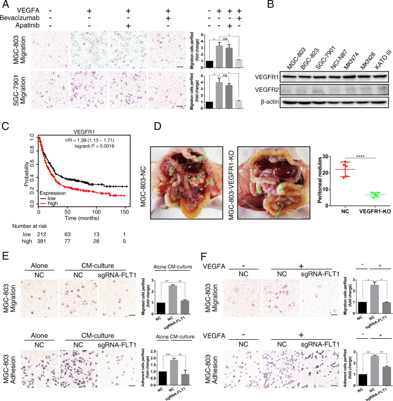
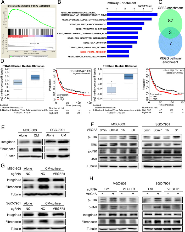
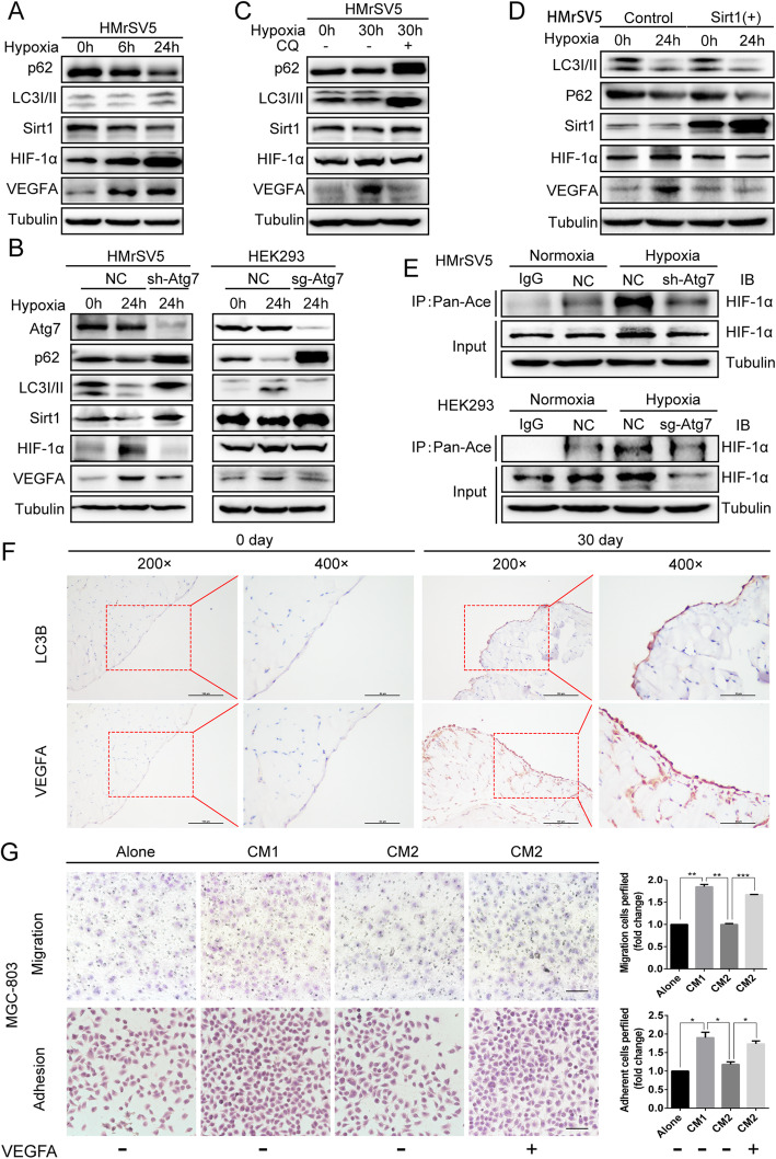
Methods: We determined the hypoxic microenvironment in PM of nude mice by immunohistochemical analysis and screened VEGFA by human growth factor array kit. The crosstalk mediated by VEGFA between peritoneal mesothelial cells (PMCs) and GC cells was determined in GC cells incubated with conditioned medium prepared from hypoxia-treated PMCs. The association between VEGFR1 and integrin α5 and fibronectin in GC cells was enriched using Gene Set Enrichment Analysis and KEGG pathway enrichment analysis. In vitro and xenograft mouse models were used to evaluate the impact of VEGFA/VEGFR1 on gastric cancer peritoneal metastasis. Confocal microscopy and immunoprecipitation were performed to determine the effect of hypoxia-induced autophagy.

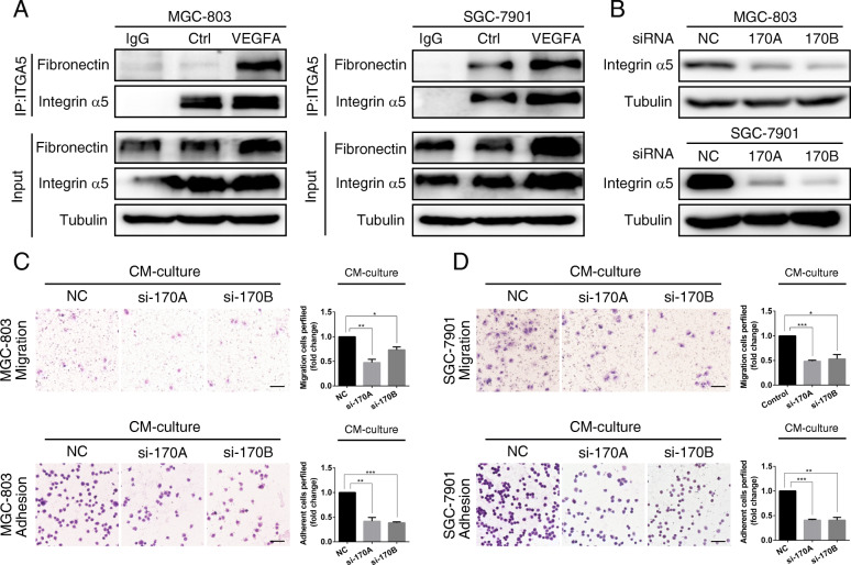
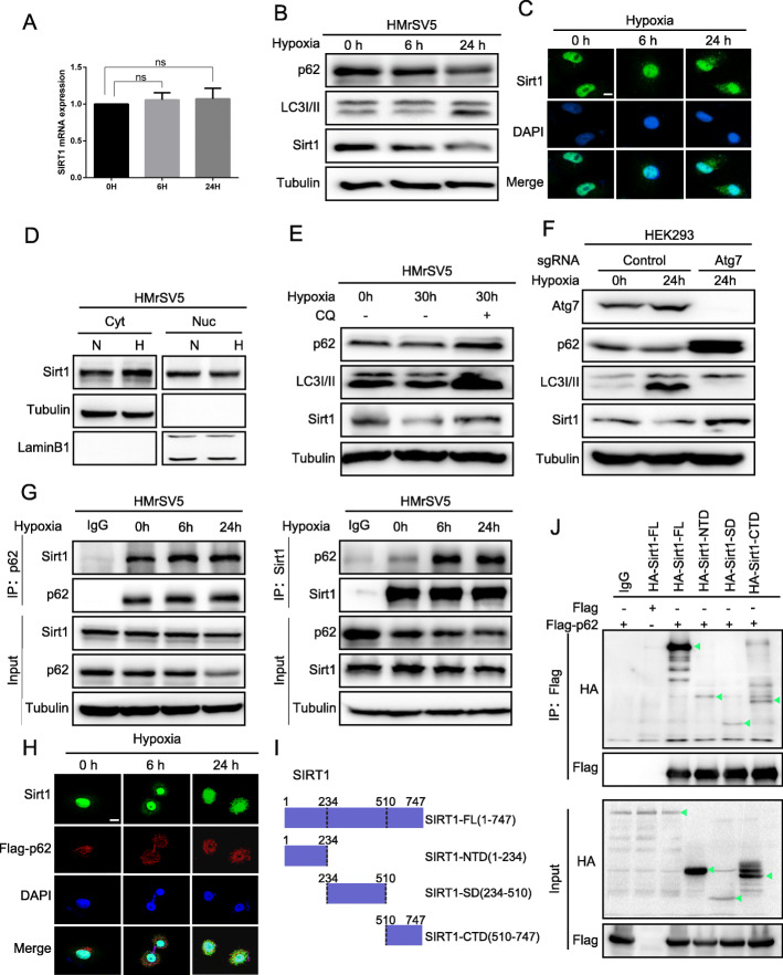
Results: Here we report that in the PMCs of the hypoxic microenvironment, SIRT1 is degraded via the autophagic lysosomal pathway, leading to increased acetylation of HIF-1α and secretion of VEGFA. Under hypoxic conditions, VEGFA derived from PMCs acts on VEGFR1 of GC cells, resulting in p-ERK/p-JNK pathway activation, increased integrin α5 and fibronectin expression, and promotion of PM.

Conclusions: Our findings have elucidated the mechanisms by which PMCs promote PM in GC in hypoxic environments. This study also provides a theoretical basis for considering autophagic pathways or VEGFA as potential therapeutic targets to treat PM in GC.

Keywords: Adhesion; Autophagy; Hypoxia; Migration; VEGFA.


Conflict of interest statement

The authors declare no potential conflicts of interest.

Figures

Fig. 1
Fig. 1
PMCs-derived VEGFA promotes the adhesion and migration of GC cells in a hypoxic microenvironment. a Immunohistochemical analysis of the hypoxia inducible transcription factor HIF-1α in benign mouse peritonea and GC metastatic peritonea. b Growth factor screening analysis identified the secretion of several growth factors. Supernatants were acquired from normal media and conditioned media from hypoxic PMCs. c The level of the VEGFA protein level in the supernatant was determined for mesothelial cells exposed to 0, 6, 24 and 48 h of hypoxia. VEGFA protein level was determined using ELISA. d The effect of hypoxic-conditioned media (CM) and exogenous VEGFA on cell adhesion and migration was determined after 24 h, in the presence or absence of Bevacizumab (Beva) antibody. Representative photographs of adherent and migratory cells (magnification, 200×) are shown. Scale bar represents 100 μm. Error bars represent standard deviation (SD) of the mean, *P < 0.05, **P < 0.01, ***P < 0.001. e MGC-803 cells were intraperitoneally inoculated into nude mice. The experimental group was given intraperitoneal administration of 200 μg of Bevacizumab every other day. The peritoneal nodules (green arrows) were evaluated after 20 days (N = 5 per group). Representative data are shown. ****P < 0.0001
Fig. 2
Fig. 2
VEGFA derived from hypoxic PMCs facilitates GC cell adhesion and migration via VEGFR1. a The effect of exogenous VEGFA on cell migration was observed in the presence of Apatinib or Bevacizumab. Representative photographs of migratory cells are shown. Scale bar represents 100 μm. Error bars represent SD of the mean. *P < 0.05. b Western blot analysis of VEGFR1 protein levels in various GC cells. c Survival analysis for different VEGFR1 expression levels using the KM-Plotter database of GC. d MGC-803 sgRNA-VEGFR1 and sgRNA-NC cells were intraperitoneally inoculated. The metastatic nodules (green arrows) were analyzed after 20 days (N = 5 per group). Representative data are shown. ****P < 0.0001. e and f MGC-803 cells were exposed to CM from hypoxic PMCs or exogenous VEGFA, synchronously with knockout of VEGFR1 using sgRNA-FLT1. Representative photographs of adherent and migratory cells are shown. Scale bar represents 100 μm. Error bars represent SD of the mean. *P < 0.05. **P < 0.01. ***P < 0.001
Fig. 3
Fig. 3
VEGFA derived from hypoxic PMCs promotes the expression of integrin α5/fibronectin via VEGFR1. a and b Gene Set Enrichment Analysis (GSEA) indicating that “FOCAL_ADHESION” is significantly associated with VEGFR1. KEGG pathway enrichment analysis identified the KEGG_FOCAL_ADHESION pathway in CM-treated MGC-803 cells by gene array analysis. c Three genes were identified that overlapped between the gene signature of GSEA enrichment analysis and the KEGG pathway enrichment analysis. d Survival analysis for different integrin α5 and fibronectin expressions using the KM-Plotter database of GC. The Oncomine database showed the differential expression of integrin α5/fibronectin in gastric carcinoma tissues and gastric mucosal tissues. e Immunoblot analysis of integrin α5 and fibronectin after cultivation of GC cells with CM from hypoxic cells. f Immunoblot analysis of p-JNK and p-ERK in GC cells at the indicated time points following the addition of 100 ng/mL VEGFA. g VEGFR1 was knocked out with sg-RNA and the expression of integrin α5 and fibronectin was detected after cultivation of GC cells with CM from hypoxic cells. h VEGFR1 was knocked out with sg-RNA and the expression of integrin α5, fibronectin, p-JNK and p-ERK was determined after exogenous VEGFA application
Fig. 4
Fig. 4
VEGFA derived from hypoxic PMCs promotes adhesion and migration through the expression of integrin α5/fibronectin. a The indicated cells were treated with exogenous VEGFA and immunoprecipitation was performed with an integrin α5 antibody, followed by detection of integrin α5 or fibronectin by immunoblot analysis. b Immunoblotting of integrin α5 was performed in the indicated cells following transfection with si-integrin α5-170A or si-integrin α5-170B for 24 h. c and d MGC-803 and SGC-7901 cells were exposed to CM from hypoxic PMCs, synchronously with knockdown of integrin α5. Representative photographs of adherent and migratory cells are shown. Scale bar represents 100 μm. Bars represent SD of the mean. *P < 0.05. **P < 0.01. ***P < 0.001
Fig. 5
Fig. 5
SIRT1 is degraded by hypoxia-induced autophagy through the p62-SIRT1 autolysosome pathway. a RT-qPCR analysis of SIRT1 mRNA in HMrSV5 cells exposed to hypoxic conditions for 0 h, 6 h, 24 h. b Immunoblot analysis of p62, LC3I/II and SIRT1 at the indicated time points during hypoxia. c Immunofluorescence analysis of SIRT1 localization in hypoxic PMCs after 0 h, 6 h, or 24 h. Scale bar represents 10 μm. d The nuclear-cytosol distribution experiment detected the nuclear and cytoplasmic location of SIRT1 following hypoxia treatment (H), compared to normoxic cells (N). e and f Analysis of p62, LC3I/II and SIRT1 protein levels during hypoxia and in response to treatment with chloroquine (CQ) in HMrSV5 cells or ATG7 knockout in HEK293 cells. g HMrSV5 cells were cultured under hypoxic conditions for 0 h, 6 h, or 24 h. Immunoprecipitation was performed using SIRT1 or p62 antibodies, followed by immunoblotting with antibodies against SIRT1 and p62. h Immunofluorescence confocal microscopy was performed to assess the colocalization of SIRT1 and p62 at different time points during hypoxia. Scale bar represents 10 μm. i and j HEK293 cells were co-transfected with plasmids for Flag-p62 and HA-SIRT1 N terminal domain (NTD, aa 1–234), sirtuin (catalytic) domain (SD, aa 234–510), C terminal domain (CTD, aa 510–747) or full length (FL, aa 1–747). Lysates were immunoprecipitated with anti-Flag antibody
Fig. 6
Fig. 6
Hypoxia-induced autophagy mediated degradation of SIRT1 in PMCs promotes VEGFA secretion through acetylation of HIF-1α. a The levels of p62, LC3I/II, SIRT1, HIF-1α, and VEGFA were observed in HMrSV5 cells cultured in hypoxic conditions for the indicated time. b and c Western blot analysis of p62, LC3I/II, SIRT1, HIF-1α, and VEGFA under hypoxia and in response to treatment with chloroquine (CQ) or knockdown of ATG7 in HMrSV5 cells, or knockout of ATG7 in HEK293. d The levels of LC3I/II, p62, SIRT1, HIF-1α, and VEGFA were analyzed in HMrSV5 cells exposed to normoxia or hypoxia for 24 h with or without overexpression of SIRT1. e ATG7 was knocked out in HEK293 cells and knocked down in HMrSV5 cells, followed by exposure to normoxic or hypoxic conditions for 24 h. Immunoprecipitation was performed with a pan-acetyl antibody followed by immunoblot analysis using antibodies against HIF-1α. f Immunohistochemical analysis of LC3BI/II and VEGFA in benign mouse peritonea and GC metastatic peritonea. G. MGC-803 cells were subjected to normal media or conditioned media (CM1: conditioned media from hypoxic PMCs, CM2: conditioned media of hypoxic shRNA-Atg7 PMCs) and to CM2 with synchronous addition of exogenous VEGFA. Representative photographs of adherent and migratory cells are shown. Scale bars represent 100 μm. Bars represent SD of the mean. *P < 0.05. **P < 0.01. ***P < 0.001

Similar articles

Cited by

References

    1. Bray F, Ferlay J, Soerjomataram I, Siegel RL, Torre LA, Jemal A. Global cancer statistics 2018: GLOBOCAN estimates of incidence and mortality worldwide for 36 cancers in 185 countries. CA Cancer J Clin. 2018;68:394–424. doi: 10.3322/caac.21492. - DOI - PubMed
    1. Van Cutsem E, Sagaert X, Topal B, Haustermans K, Prenen H. Gastric cancer. Lancet. 2016;388:2654–2664. doi: 10.1016/S0140-6736(16)30354-3. - DOI - PubMed
    1. Thomassen I, van Gestel YR, van Ramshorst B, Luyer MD, Bosscha K, Nienhuijs SW, et al. Peritoneal carcinomatosis of gastric origin: a population-based study on incidence, survival and risk factors. Int J Cancer. 2014;134:622–628. doi: 10.1002/ijc.28373. - DOI - PubMed
    1. Natarajan S, Foreman KM, Soriano MI, Rossen NS, Shehade H, Fregoso DR, et al. Collagen remodeling in the hypoxic tumor-Mesothelial niche promotes ovarian Cancer metastasis. Cancer Res. 2019;79:2271–2284. doi: 10.1158/0008-5472.CAN-18-2616. - DOI - PMC - PubMed
    1. Mikula-Pietrasik J, Sosinska P, Kucinska M, Murias M, Maksin K, Malinska A, et al. Peritoneal mesothelium promotes the progression of ovarian cancer cells in vitro and in a mice xenograft model in vivo. Cancer Lett. 2014;355:310–315. doi: 10.1016/j.canlet.2014.09.041. - DOI - PubMed

MeSH terms