

Immunotoxin Screening System: A Rapid and Direct Approach to Obtain Functional Antibodies with Internalization Capacities
- PMID: 33076544
- PMCID: PMC7602748
- DOI: 10.3390/toxins12100658
Immunotoxin Screening System: A Rapid and Direct Approach to Obtain Functional Antibodies with Internalization Capacities
Abstract
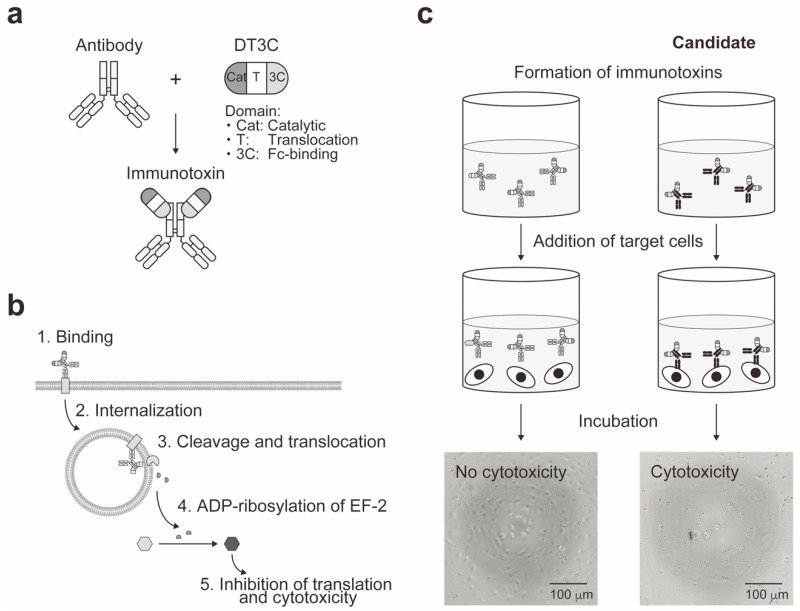
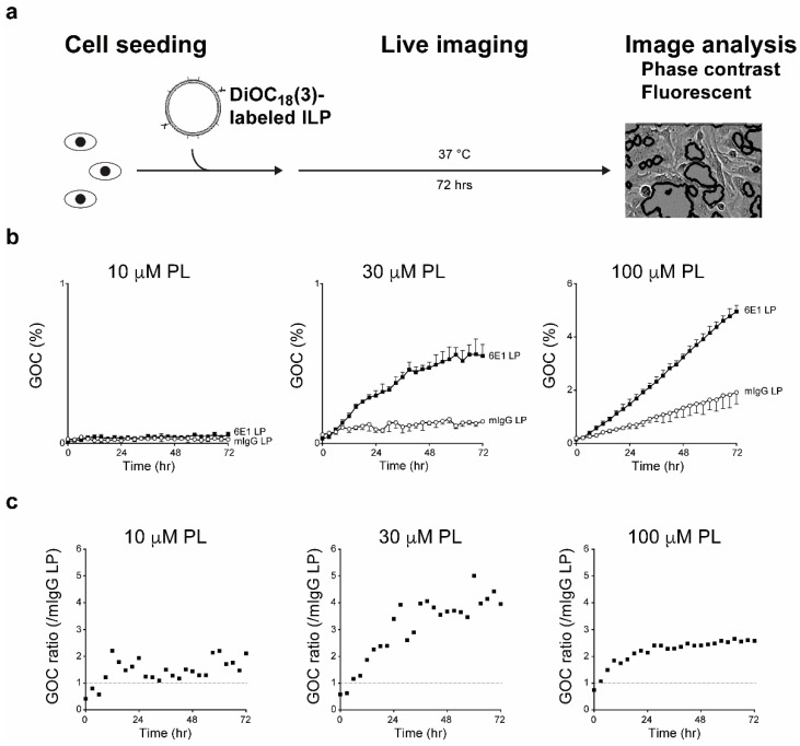
Toxins, while harmful and potentially lethal, have been engineered to develop potent therapeutics including cytotoxins and immunotoxins (ITs), which are modalities with highly selective targeting capabilities. Currently, three cytotoxins and IT are FDA-approved for treatment of multiple forms of hematological cancer, and additional ITs are tested in the clinical trials or at the preclinical level. For next generation of ITs, as well as antibody-mediated drug delivery systems, specific targeting by monoclonal antibodies is critical to enhance efficacies and reduce side effects, and this methodological field remains open to discover potent therapeutic monoclonal antibodies. Here, we describe our application of engineered toxin termed a cell-based IT screening system. This unique screening strategy offers the following advantages: (1) identification of monoclonal antibodies that recognize cell-surface molecules, (2) selection of the antibodies that are internalized into the cells, (3) selection of the antibodies that induce cytotoxicity since they are linked with toxins, and (4) determination of state-specific activities of the antibodies by differential screening under multiple experimental conditions. Since the functional monoclonal antibodies with internalization capacities have been identified successfully, we have pursued their subsequent modifications beyond antibody drug conjugates, resulting in development of immunoliposomes. Collectively, this screening system by using engineered toxin is a versatile platform, which enables straight-forward and rapid selection for discovery of novel functional antibodies.
Keywords: DT3C; antibody drug conjugate; diphtheria toxin; drug delivery; immunoliposome; immunotoxin; monoclonal antibody.
Conflict of interest statement
The authors declare no conflict of interest.
Figures

Similar articles
-
Antibody internalization after cell surface antigen binding is critical for immunotoxin development.Bioconjug Chem. 2009 Oct 21;20(10):1975-82. doi: 10.1021/bc900333j. Epub 2009 Sep 28. Bioconjug Chem. 2009. PMID: 19785403
-
Immunotoxin FN18-CRM9 induces stronger T cell signaling than unconjugated monoclonal antibody FN18.Transplantation. 2001 Aug 15;72(3):496-503. doi: 10.1097/00007890-200108150-00023. Transplantation. 2001. PMID: 11502982
-
Antibody internalization studied using a novel IgG binding toxin fusion.J Immunol Methods. 2007 Apr 10;321(1-2):41-59. doi: 10.1016/j.jim.2007.01.008. Epub 2007 Feb 6. J Immunol Methods. 2007. PMID: 17336321
-
[Recombinant immunotoxins and chimeric toxins for targeted therapy in oncology].Bull Cancer. 1997 Dec;84(12):1135-40. Bull Cancer. 1997. PMID: 9587366 Review. French.
-
Recent development and optimization of pseudomonas aeruginosa exotoxin immunotoxins in cancer therapeutic applications.Int Immunopharmacol. 2021 Jul;96:107759. doi: 10.1016/j.intimp.2021.107759. Epub 2021 May 24. Int Immunopharmacol. 2021. PMID: 34162138 Review.
Cited by
-
Prospects for the use of viral proteins for the construction of chimeric toxins.Arch Virol. 2024 Sep 26;169(10):208. doi: 10.1007/s00705-024-06139-8. Arch Virol. 2024. PMID: 39327316 Review.
-
Intracellular Protein Delivery: Approaches, Challenges, and Clinical Applications.BME Front. 2024 Jan 25;5:0035. doi: 10.34133/bmef.0035. eCollection 2024. BME Front. 2024. PMID: 38282957 Free PMC article. Review.
-
Strategies to mitigate the on- and off-target toxicities of recombinant immunotoxins: an antibody engineering perspective.Antib Ther. 2022 Jun 16;5(3):164-176. doi: 10.1093/abt/tbac014. eCollection 2022 Jul. Antib Ther. 2022. PMID: 35928456 Free PMC article. Review.
-
Drug conjugates for targeting regulatory T cells in the tumor microenvironment: guided missiles for cancer treatment.Exp Mol Med. 2023 Sep;55(9):1996-2004. doi: 10.1038/s12276-023-01080-3. Epub 2023 Sep 1. Exp Mol Med. 2023. PMID: 37653036 Free PMC article. Review.
-
Design of two immunotoxins based rovalpituzumab antibody against DLL3 receptor; a promising potential opportunity.Res Pharm Sci. 2022 Jul 14;17(4):428-444. doi: 10.4103/1735-5362.350243. eCollection 2022 Aug. Res Pharm Sci. 2022. PMID: 36034078 Free PMC article.
References
Publication types
MeSH terms
Substances
Grants and funding
- JP19cm0106231s0102/Japan Agency for Medical Research and Development/International
- JP19dm0107156h0001/Japan Agency for Medical Research and Development/International
- 18K07657/Japan Society for the Promotion of Science/International
- 16K07184/Japan Society for the Promotion of Science/International
- 19K07831/Japan Society for the Promotion of Science/International
LinkOut - more resources
Full Text Sources
