

Interstitial pneumonitis related to trastuzumab deruxtecan, a human epidermal growth factor receptor 2-targeting Ab-drug conjugate, in monkeys
- PMID: 33051938
- PMCID: PMC7734153
- DOI: 10.1111/cas.14686
Interstitial pneumonitis related to trastuzumab deruxtecan, a human epidermal growth factor receptor 2-targeting Ab-drug conjugate, in monkeys
Abstract
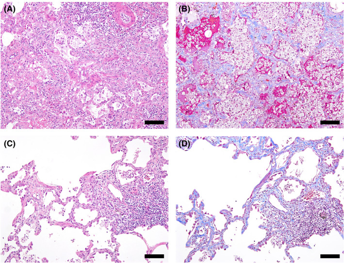
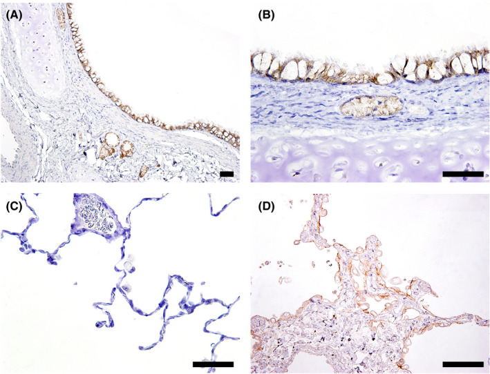
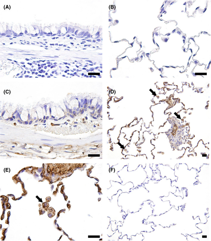
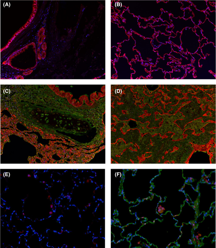
Trastuzumab deruxtecan (T-DXd: DS-8201a) is an anti-human epidermal growth factor receptor 2 (HER2) Ab-drug conjugated with deruxtecan (DXd), a derivative of exatecan. The objective of this study was to characterize T-DXd-induced lung toxicity in cynomolgus monkeys. Trastuzumab deruxtecan was injected i.v. into monkeys once every 3 weeks for 6 weeks (10, 30, and 78.8 mg/kg) or for 3 months (3, 10, and 30 mg/kg). To evaluate the involvement of DXd alone in T-DXd-induced toxicity, DXd monohydrate was given i.v. to monkeys once a week for 4 weeks (1, 3, and 12 mg/kg). Interstitial pneumonitis was observed in monkeys given T-DXd at 30 mg/kg or more. The histopathological features of diffuse lymphocytic infiltrates and slight fibrosis were similar to interstitial lung diseases (ILD)/pneumonitis related to anticancer drugs in patients, with an incidence that was dose-dependent and dose-frequency-dependent. Monkeys receiving DXd monohydrate did not suffer lung toxicity, although the DXd exposure level was higher than that of DXd in the monkeys given T-DXd. The HER2 expression in monkey lungs was limited to the bronchial level, although the lesions were found at the alveolar level. Immunohistochemical analysis confirmed that T-DXd localization was mainly in alveolar macrophages, but not pulmonary epithelial cells. These findings indicate that monkeys are an appropriate model for investigating T-DXd-related ILD/pneumonitis. The results are also valuable for hypothesis generation regarding the possible mechanism of T-DXd-induced ILD/pneumonitis in which target-independent uptake of T-DXd into alveolar macrophages could be involved. Further evaluation is necessary to clarify the mechanism of ILD/pneumonitis in patients with T-DXd therapy.
Keywords: DS-8201a; HER2; cynomolgus monkey; interstitial lung disease; trastuzumab deruxtecan.
© 2020 The Authors. Cancer Science published by John Wiley & Sons Australia, Ltd on behalf of Japanese Cancer Association.
Conflict of interest statement
The authors are employees of Daiichi Sankyo Co., Ltd.
Figures
Similar articles
-
Antitumor Activity and Safety of Trastuzumab Deruxtecan in Patients With HER2-Low-Expressing Advanced Breast Cancer: Results From a Phase Ib Study.J Clin Oncol. 2020 Jun 10;38(17):1887-1896. doi: 10.1200/JCO.19.02318. Epub 2020 Feb 14. J Clin Oncol. 2020. PMID: 32058843 Free PMC article. Clinical Trial.
-
Comprehensive preclinical pharmacokinetic evaluations of trastuzumab deruxtecan (DS-8201a), a HER2-targeting antibody-drug conjugate, in cynomolgus monkeys.Xenobiotica. 2019 Sep;49(9):1086-1096. doi: 10.1080/00498254.2018.1531158. Epub 2019 Jan 4. Xenobiotica. 2019. PMID: 30351177
-
Incidence of pneumonitis/interstitial lung disease induced by HER2-targeting therapy for HER2-positive metastatic breast cancer.Breast Cancer Res Treat. 2020 Aug;183(1):23-39. doi: 10.1007/s10549-020-05754-8. Epub 2020 Jun 26. Breast Cancer Res Treat. 2020. PMID: 32591987 Free PMC article. Review.
-
Trastuzumab Deruxtecan-Induced Interstitial Lung Disease/Pneumonitis in ERBB2-Positive Advanced Solid Malignancies: A Systematic Review.Drugs. 2022 Jun;82(9):979-987. doi: 10.1007/s40265-022-01736-w. Epub 2022 Jun 27. Drugs. 2022. PMID: 35759121 Free PMC article.
-
Trastuzumab-deruxtecan: an investigational agent for the treatment of HER2-positive breast cancer.Expert Opin Investig Drugs. 2020 Sep;29(9):901-910. doi: 10.1080/13543784.2020.1792443. Epub 2020 Jul 23. Expert Opin Investig Drugs. 2020. PMID: 32701032 Review.
Cited by
-
A Review of the Current FDA-Approved Antibody-Drug Conjugates: Landmark Clinical Trials and Indications.Pharmaceut Med. 2024 Jan;38(1):39-54. doi: 10.1007/s40290-023-00505-8. Epub 2023 Nov 29. Pharmaceut Med. 2024. PMID: 38019416 Review.
-
FZ-AD005, a Novel DLL3-Targeted Antibody-Drug Conjugate with Topoisomerase I Inhibitor, Shows Potent Antitumor Activity in Preclinical Models.Mol Cancer Ther. 2024 Oct 1;23(10):1367-1377. doi: 10.1158/1535-7163.MCT-23-0701. Mol Cancer Ther. 2024. PMID: 38940283 Free PMC article.
-
Pooled analysis of drug-related interstitial lung disease and/or pneumonitis in nine trastuzumab deruxtecan monotherapy studies.ESMO Open. 2022 Aug;7(4):100554. doi: 10.1016/j.esmoop.2022.100554. Epub 2022 Aug 11. ESMO Open. 2022. PMID: 35963179 Free PMC article.
-
Clinical Guidance on the Monitoring and Management of Trastuzumab Deruxtecan (T-DXd)-Related Adverse Events: Insights from an Asia-Pacific Multidisciplinary Panel.Drug Saf. 2023 Oct;46(10):927-949. doi: 10.1007/s40264-023-01328-x. Epub 2023 Aug 8. Drug Saf. 2023. PMID: 37552439 Free PMC article. Review.
-
Prognostic and predictive factors for the efficacy and safety of trastuzumab deruxtecan in HER2-positive gastric or gastroesophageal junction cancer.Gastric Cancer. 2025 Jan;28(1):63-73. doi: 10.1007/s10120-024-01560-z. Epub 2024 Nov 2. Gastric Cancer. 2025. PMID: 39487862 Free PMC article.
References
-
- Oh DY, Bang YJ. HER2‐targeted therapies ‐ a role beyond breast cancer. Nat Rev Clin Oncol. 2020;17(1):33‐48. - PubMed
-
- Nakada T, Masuda T, Naito H, et al. Novel antibody drug conjugates containing exatecan derivative‐based cytotoxic payloads. Bioorg Med Chem Lett. 2016;26(6):1542‐1545. - PubMed
-
- Nakada T, Sugihara K, Jikoh T, et al. The latest research and development into the antibody‐drug conjugate, [fam‐] trastuzumab deruxtecan (DS‐8201a), for HER2 cancer therapy. Chem Pharm Bull (Tokyo). 2019;67(3):173‐185. - PubMed
-
- Ogitani Y, Aida T, Hagihara K, et al. DS‐8201a, a novel HER2‐targeting ADC with a novel DNA topoisomerase I inhibitor, demonstrates a promising antitumor efficacy with differentiation from T‐DM1. Clin Cancer Res. 2016;22(20):5097‐5108. - PubMed
MeSH terms
Substances
LinkOut - more resources
Full Text Sources
Other Literature Sources
Medical
Research Materials
Miscellaneous
