. 2020 Oct 7;12(10):3062.
doi: 10.3390/nu12103062.

Fructose Consumption by Adult Rats Exposed to Dexamethasone In Utero Changes the Phenotype of Intestinal Epithelial Cells and Exacerbates Intestinal Gluconeogenesis

Affiliations

Fructose Consumption by Adult Rats Exposed to Dexamethasone In Utero Changes the Phenotype of Intestinal Epithelial Cells and Exacerbates Intestinal Gluconeogenesis

Gizela A Pereira et al. Nutrients. .

Abstract

Fructose consumption by rodents modulates both hepatic and intestinal lipid metabolism and gluconeogenesis. We have previously demonstrated that in utero exposure to dexamethasone (DEX) interacts with fructose consumption during adult life to exacerbate hepatic steatosis in rats. The aim of this study was to clarify if adult rats born to DEX-treated mothers would display differences in intestinal gluconeogenesis after excessive fructose intake. To address this issue, female Wistar rats were treated with DEX during pregnancy and control (CTL) mothers were kept untreated. Adult offspring born to CTL and DEX-treated mothers were assigned to receive either tap water (Control-Standard Chow (CTL-SC) and Dexamethasone-Standard Chow (DEX-SC)) or 10% fructose in the drinking water (CTL-fructose and DEX-fructose). Fructose consumption lasted for 80 days. All rats were subjected to a 40 h fasting before sample collection. We found that DEX-fructose rats have increased glucose and reduced lactate in the portal blood. Jejunum samples of DEX-fructose rats have enhanced phosphoenolpyruvate carboxykinase (PEPCK) expression and activity, higher facilitated glucose transporter member 2 (GLUT2) and facilitated glucose transporter member 5 (GLUT5) content, and increased villous height, crypt depth, and proliferating cell nuclear antigen (PCNA) staining. The current data reveal that rats born to DEX-treated mothers that consume fructose during adult life have increased intestinal gluconeogenesis while recapitulating metabolic and morphological features of the neonatal jejunum phenotype.

Keywords: dexamethasone; fructose; intestinal gluconeogenesis; intrauterine growth restriction (IUGR).


Conflict of interest statement

The authors declare no conflicts of interest, financial or otherwise, associated with this article. The authors are responsible for the writing and content of the article.

Figures
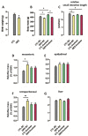
Figure 1
Figure 1
Morphometrical parameters of rats exposed to dexamethasone (DEX) in utero. Body weight was measured at birth (A) and at the end of treatment (B) in nonfasted rats. Fasted rats were euthanized at the end of the 80th day of fructose consumption, and the small intestine length relative to tibia length (C), and fat pads (mesenteric, (D); epidydimal, (E); retroperitoneal, (F)) and liver (G) masses relative to body weight were also measured. Results are presented as mean ± standard error of the mean (S.E.M.). * p < 0.05, ** p < 0.01 (n = 10–20). Offspring born to control (CTL) mothers that received only standard chow (SC) and tap water during adult life (CTL-SC); offspring born to DEX-treated mothers that received only SC and tap water during adult life (DEX-SC); offspring born to CTL mothers that received SC plus 10% fructose during adult life (CTL-fructose); and offspring born to DEX-treated mothers that received SC plus 10% fructose during adult life (DEX-fructose).
Figure 2
Figure 2
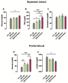
Effects of fructose on biochemical parameters of systemic and portal blood in rats exposed to dexamethasone (DEX) in utero. Systemic blood samples were collected to measure glucose (A), triacylglycerol (B), and total cholesterol (C). Portal hepatic vein samples were collected to measure glucose (D) and lactate (E). Results are presented as mean ± standard error of the mean (S.E.M.). * p < 0.05, ** p < 0.01, *** p < 0.001, **** p < 0.0001 (n = 8–12). Offspring born to control (CTL) mothers that received only standard chow (SC) and tap water during adult life (CTL-SC); offspring born to DEX-treated mothers that received only SC and tap water during adult life (DEX-SC); offspring born to CTL mothers that received SC plus 10% fructose during adult life (CTL-fructose); and offspring born to DEX-treated mothers that received SC plus 10% fructose during adult life (DEX-fructose).
Figure 3
Figure 3
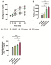
Whole-body and intestinal use of pyruvate as a gluconeogenesis substrate by rats exposed to dexamethasone (DEX) in utero and treated with fructose during adult life. The 40 h fasted rats received an i.p. injection containing sodium pyruvate. The blood from the tail was collected before and 15, 30, 60, and 90 min after intraperitoneal (i.p.) injection for glucose measurements (A) and the area under the curve (AUC) was calculated above each individual baseline (B). Glucose levels were also measured in portal blood at the end of the pyruvate tolerance test (PTT) (C). Results are presented as mean ± standard error of the mean (S.E.M.) * p < 0.05, ** p < 0.01, **** p < 0.0001 (n = 8). Offspring born to control (CTL) mothers that received only standard chow (SC) and tap water during adult life (CTL-SC); offspring born to DEX-treated mothers that received only SC and tap water during adult life (DEX-SC); offspring born to CTL mothers that received SC plus 10% fructose during adult life (CTL-fructose); and offspring born to DEX-treated mothers that received SC plus 10% fructose during adult life (DEX-fructose).
Figure 4
Figure 4
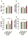
Expression and activity of enzymes involved in intestinal gluconeogenesis. Scraped epithelium of jejunum fragments was isolated and processed for qPCR detection of G6pc (A) and Pck1 (C) gene expression, as well as for maximum activities of the corresponding enzymes G6Pase (B) and PEPCK (D). Results are presented as mean ± standard error of the mean (S.E.M.). * p < 0.05 (n = 6–12). Offspring born to control (CTL) mothers that received only standard chow (SC) and tap water during adult life (CTL-SC); offspring born to dexamethasone (DEX)-treated mothers that received only SC and tap water during adult life (DEX-SC); offspring born to CTL mothers that received SC plus 10% fructose during adult life (CTL-fructose); and offspring born to DEX-treated mothers that received SC plus 10% fructose during adult life (DEX-fructose).
Figure 5
Figure 5
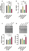
In utero dexamethasone (DEX) exposure alters glucose metabolism and phenotypic features of the jejunum epithelia. Scraped epithelium of jejunum fragments was isolated and processed for measurement of maximum hexokinase activity (A), expression of glutathione S-transferase 1 (Ggt1) by quantitative polymerase chain reaction (qPCR) (B), and Western blot of facilitated glucose transporter member 2 (GLUT2) (C) and facilitated glucose transporter member 5 (GLUT5) (D). Results are presented as mean ± standard error of the mean (S.E.M.). * p < 0.05, *** p < 0.001, **** p < 0.0001 (n = 6–12). Offspring born to control (CTL) mothers that received only standard chow (SC) and tap water during adult life (CTL-SC); offspring born to DEX-treated mothers that received only SC and tap water during adult life (DEX-SC); offspring born to CTL mothers that received SC plus 10% fructose during adult life (CTL-fructose); and offspring born to DEX-treated mothers that received SC plus 10% fructose during adult life (DEX-fructose).
Figure 6
Figure 6
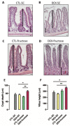
Morphometric analysis of the jejunum wall. The figure shows representative sections of villus and crypts of the four experimental groups (AD). The height of each villus was measured from the top of the villus to the crypt transition (E), and the crypt depth was defined as the invagination between two villi (F). Results are presented as mean ± standard error of the mean (S.E.M.). * p < 0.05, ** p < 0.01 (n = 5). Offspring born to control (CTL) mothers that received only standard chow (SC) and tap water during adult life (CTL-SC); offspring born to dexamethasone (DEX)-treated mothers that received only SC and tap water during adult life (DEX-SC); offspring born to CTL mothers that received SC plus 10% fructose during adult life (CTL-fructose); and offspring born to DEX-treated mothers that received SC plus 10% fructose during adult life (DEX-fructose).
Figure 7
Figure 7
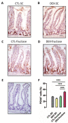
Jejunum was removed for immunohistochemical detection of proliferating cell nuclear antigen (PCNA). The figure shows representative sections of PCNA staining (AD) and negative control sample (E). Sections were used to calculate the percentage of PCNA-positive cells in crypt cells (F). Results are presented as mean ± standard error of the mean (S.E.M.). **** p < 0.0001 (n = 5). Offspring born to control (CTL) mothers that received only standard chow (SC) and tap water during adult life (CTL-SC); offspring born to dexamethasone (DEX)-treated mothers that received only SC and tap water during adult life (DEX-SC); offspring born to CTL mothers that received SC plus 10% fructose during adult life (CTL-fructose); and offspring born to DEX-treated mothers that received SC plus 10% fructose during adult life (DEX-fructose).

Similar articles

Cited by

References

    1. Tappy L., Lê K.A. Metabolic effects of fructose and the worldwide increase in obesity. Physiol. Rev. 2010;90:23–46. doi: 10.1152/physrev.00019.2009. - DOI - PubMed
    1. Faria J.A., de Araújo T.M., Razolli D.S., Ignácio-Souza L.M., Souza D.N., Bordin S., Anhê G.F. Metabolic impact of light phase-restricted fructose consumption is linked to changes in hypothalamic AMPK phosphorylation and melatonin production in rats. Nutrients. 2017;9:332. doi: 10.3390/nu9040332. - DOI - PMC - PubMed
    1. Oron-Herman M., Kamari Y., Grossman E., Yeger G., Peleg E., Shabtay Z., Shamiss A., Sharabi Y. Metabolic syndrome: Comparison of the two commonly used animal models. Am. J. Hypertens. 2008;21:1018–1022. doi: 10.1038/ajh.2008.218. - DOI - PubMed
    1. Bizeau M.E., Thresher J.S., Pagliassotti M.J. A high-sucrose diet increases gluconeogenic capacity in isolated periportal and perivenous rat hepatocytes. Am. J. Physiol. Endocrinol. Metab. 2001;280:E695–E702. doi: 10.1152/ajpendo.2001.280.5.E695. - DOI - PubMed
    1. Merino B., Fernández-Díaz C.M., Cózar-Castellano I., Perdomo G. Intestinal fructose and glucose metabolism in health and disease. Nutrients. 2019;12:94. doi: 10.3390/nu12010094. - DOI - PMC - PubMed

MeSH terms