. 2020 Oct 4;9(10):814.
doi: 10.3390/pathogens9100814.

First Insight into the Modulation of Noncanonical NF-κB Signaling Components by Poxviruses in Established Immune-Derived Cell Lines: An In Vitro Model of Ectromelia Virus Infection

Affiliations

First Insight into the Modulation of Noncanonical NF-κB Signaling Components by Poxviruses in Established Immune-Derived Cell Lines: An In Vitro Model of Ectromelia Virus Infection

Justyna Struzik et al. Pathogens. .

Abstract

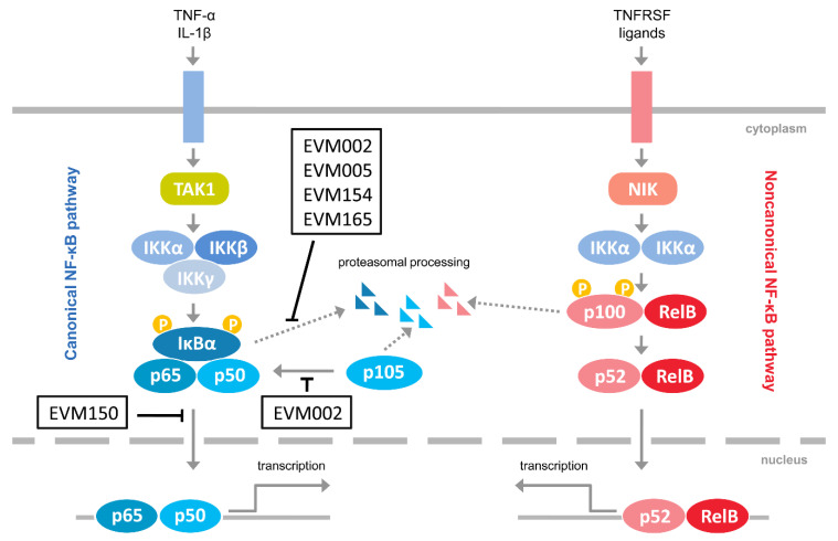
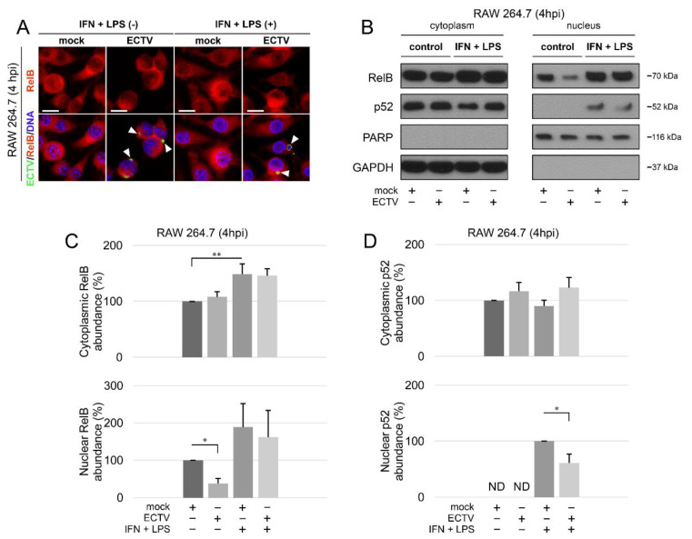
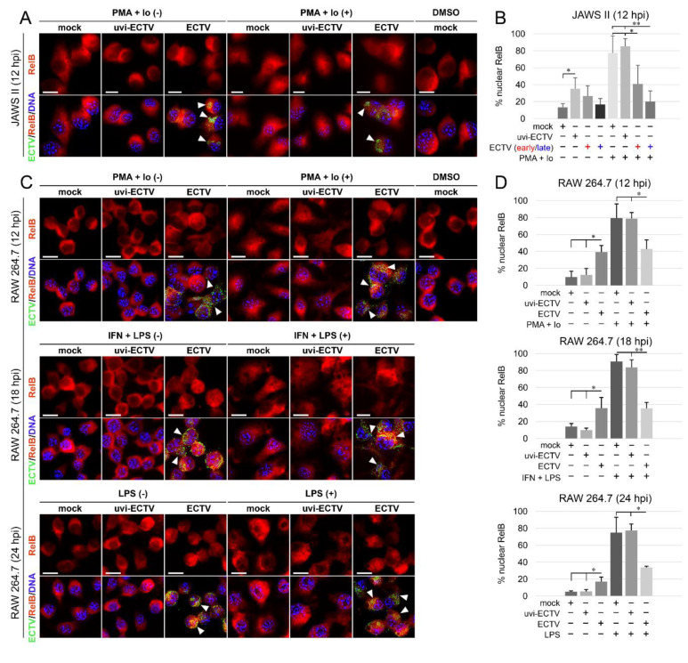
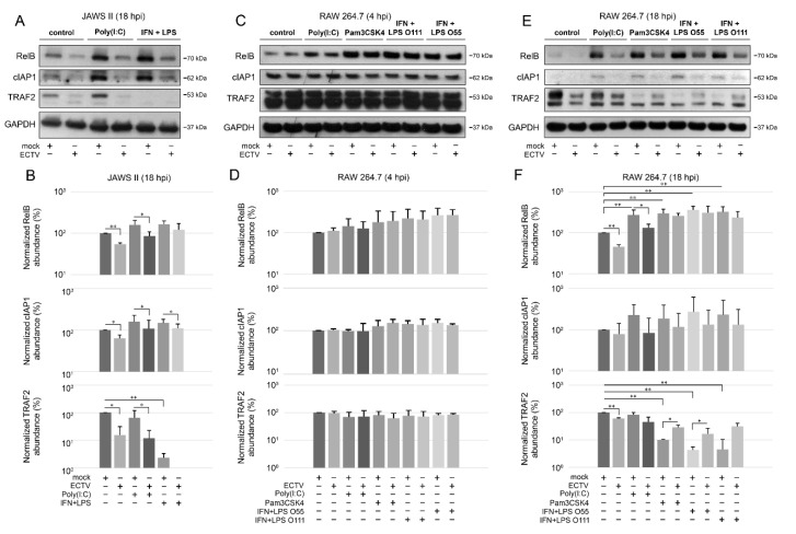
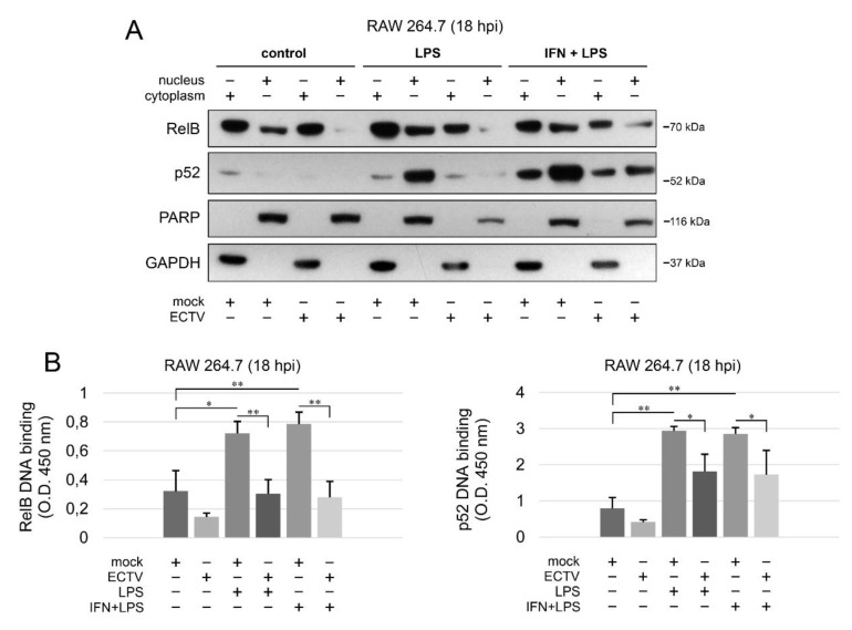
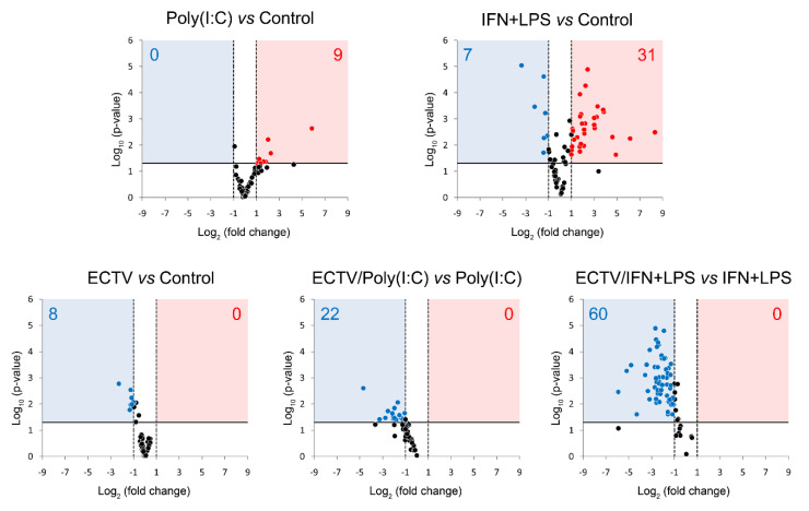
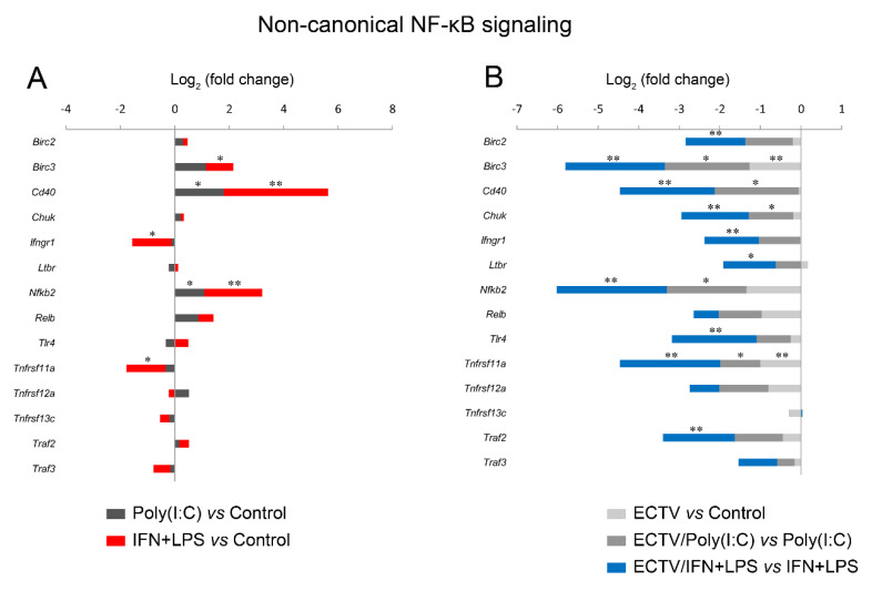
Dendritic cells (DCs) and macrophages are the first line of antiviral immunity. Viral pathogens exploit these cell populations for their efficient replication and dissemination via the modulation of intracellular signaling pathways. Disruption of the noncanonical nuclear factor κ-light-chain-enhancer of activated B cells (NF-κB) signaling has frequently been observed in lymphoid cells upon infection with oncogenic viruses. However, several nononcogenic viruses have been shown to manipulate the noncanonical NF-κB signaling in different cell types. This study demonstrates the modulating effect of ectromelia virus (ECTV) on the components of the noncanonical NF-κB signaling pathway in established murine cell lines: JAWS II DCs and RAW 264.7 macrophages. ECTV affected the activation of TRAF2, cIAP1, RelB, and p100 upon cell treatment with both canonical and noncanonical NF-κB stimuli and thus impeded DNA binding by RelB and p52. ECTV also inhibited the expression of numerous genes related to the noncanonical NF-κB pathway and RelB-dependent gene expression in the cells treated with canonical and noncanonical NF-κB activators. Thus, our data strongly suggest that ECTV influenced the noncanonical NF-κB signaling components in the in vitro models. These findings provide new insights into the noncanonical NF-κB signaling components and their manipulation by poxviruses in vitro.

Keywords: antiviral immunity; dendritic cells; ectromelia virus; macrophages; noncanonical NF-κB signaling.


Conflict of interest statement

The authors declare no conflict of interest. The funders had no role in the design of the study; in the collection, analyses, or interpretation of data; in the writing of the manuscript, or in the decision to publish the results.

Figures

Figure 1
Figure 1
Inhibitors of NF-κB encoded by ectromelia virus (ECTV). The figure represents ECTV-encoded proteins that have been shown to interfere with NF-κB signaling [23,24,25,26]. Pointed arrows indicate activation; blunted arrows indicate inhibition. EVM002, EVM005, EVM154, EVM165, Ank/F-box proteins; EVM150, Kelch repeat, and BTB domain-containing protein 1; IL-1β, interleukin-1β; IKKα, inhibitor κB kinase α subunit; IKKβ, inhibitor κB kinase β subunit; IKKγ, inhibitor κB kinase γ subunit; NIK, NF-κB-inducing kinase; TAK1, transforming growth factor β-activated kinase 1; TNFRSF, tumor necrosis factor receptor superfamily; TNF-α, tumor necrosis factor α.
Figure 2
Figure 2
ECTV inhibits nuclear translocation of RelB and p52 in RAW 264.7 cells at 4 hpi. (A) Immunofluorescence microscopy images of cellular distribution of RelB in mock- and ECTV-infected RAW 264.7 cells. Cells were either unstimulated or stimulated with rmIFN-γ + Escherichia coli LPS O111:B4. RelB—red fluorescence; ECTV antigens—green fluorescence; DNA—blue fluorescence. White arrowheads indicate viral factories. Scale bar—20 μm. (B) Immunoblot analysis of RelB and p52 content in cytoplasmic and nuclear fractions of mock- and ECTV-infected RAW 264.7 cells. The cells were either unstimulated or were stimulated with rmIFN-γ + LPS O111:B4. GAPDH—cytoplasmic loading control, PARP—nuclear loading control. (C) Densitometric quantification of RelB abundance in cytoplasmic and nuclear fractions of mock- and ECTV-infected RAW 264.7 cells. (D) Densitometric quantification of p52 content in cytoplasmic and nuclear fractions of mock- and ECTV-infected RAW 264.7 cells. The data were obtained from three independent biological experiments. * p ≤ 0.05, ** p ≤ 0.01.
Figure 3
Figure 3
ECTV influences nuclear translocation of RelB in JAWS II and RAW 264.7 cells at 12–24 hpi. (A) Immunofluorescence microscopy images of subcellular localization of RelB in control and PMA + Io-treated mock-, uvi-ECTV, and ECTV-infected JAWS II cells at 12 hpi. RelB—red fluorescence, ECTV antigens—green fluorescence, DNA—blue fluorescence. DMSO—solvent control. White arrowheads indicate viral factories. Scale bar—20 μm. (B) Quantification of nuclear RelB-positive cells. Mock-, uvi-ECTV, and ECTV-infected JAWS II cells displaying round (early) and bloated (late) viral factories were analyzed. Graphs represent the percentage of cells displaying a greater intensity of fluorescence in the nucleus compared to control cells. Data were obtained from three independent experiments. A total of 50 cells/condition/experiment was counted (* p ≤ 0.05, ** p ≤ 0.01). (C) Immunofluorescence analysis of RelB subcellular localization in mock-, uvi-ECTV-, and ECTV-infected RAW 264.7 cells. Cells were either unstimulated or stimulated with PMA + Io, rmIFN-γ + Escherichia coli LPS O111:B4, or LPS O111:B4 and analyzed at 12, 18, and 24 hpi, respectively. (D) Quantification of nuclear translocation of RelB in mock-, uvi-ECTV, and ECTV-infected RAW 264.7 cells with bloated viral factories. Graphs represent the mean percentage of RelB-positive cells from three independent experiments. A total of 100 cells/condition/experiment was analyzed (* p ≤ 0.05, ** p ≤ 0.01).
Figure 4
Figure 4
ECTV inhibits phosphorylation of p100 and RelB and affects p100 processing in JAWS II and RAW 264.7 cells. (A) Immunoblot analysis of phosphorylation of RelB in unstimulated or PMA + Io-stimulated mock-, uvi-ECTV-, and ECTV-infected JAWS II cells. (B) Immunoblot analysis of the content of p-RelB and p-p100 in unstimulated or rmIFN-γ + Escherichia coli O111:B4 LPS-stimulated mock- and ECTV-infected JAWS II cells. (C) Immunoblot analysis of p-RelB and p-p100 in unstimulated and Pam3CSK4- or rmIFN-γ + LPS O111:B4-stimulated mock- and ECTV-infected RAW 264.7 cells. (D) Immunoblot analysis of p100/p52 content in mock- and ECTV-infected JAWS II cells treated with poly(I:C) or rmIFN-γ + LPS O111:B4. (E) Densitometric analysis of the p100/p52 ratio and p52 content in JAWS II cells. Data obtained from two independent experiments are shown on histograms with a logarithmic scale. * p ≤ 0.05, ** p ≤ 0.01. GAPDH—loading control. (F) Immunoblot analysis of p100/p52 proteins in mock- and ECTV-infected RAW 264.7 cells at 4 hpi. The cells were left untreated or treated with poly(I:C), Pam3CSK4, rmIFN-γ + E. coli LPS O55:B5, or rmIFN-γ + LPS O111:B4. (G) Densitometric analysis of the p100/p52 ratio and p52 content in RAW 264.7 cells based on the data obtained from two independent experiments. Data are presented on histograms with a logarithmic scale. GAPDH—loading control. (H) Immunoblot analysis of p100/p52 proteins in mock- and ECTV-infected RAW 264.7 cells at 18 hpi. The cells were left untreated or treated with poly(I:C), Pam3CSK4, rmIFN-γ + E. coli LPS O55:B5, or rmIFN-γ + LPS O111:B4. (I) Densitometric analysis of the p100/p52 ratio and p52 content in RAW 264.7 cells based on the data obtained from two independent experiments. Data are presented on histograms with a logarithmic scale. * p ≤ 0.05, ** p ≤ 0.01. GAPDH—loading control.
Figure 5
Figure 5
ECTV influences the expression of components of the noncanonical NF-κB signaling pathway in JAWS II and RAW 264.7 cells. (A) Immunoblots of mock- and ECTV-infected JAWS II cells untreated or treated with poly(I:C) or rmIFN-γ + Escherichia coli LPS O111:B4. (B) Densitometric evaluation of RelB, cIAP1, and TRAF2 expression in JAWS II cells. The analysis was based on the data of two independent biological experiments. The data are shown on histograms with a logarithmic scale. * p ≤ 0.05, ** p ≤ 0.01. GAPDH—loading control. (C) Immunoblots of mock- and ECTV-infected RAW 264.7 cells at 4 hpi. Cells were left untreated or were treated with poly(I:C), Pam3CSK4, rmIFN-γ + E. coli LPS O55:B5, or rmIFN-γ + LPS O111:B4. (D) Densitometric evaluation of RelB, cIAP1, and TRAF2 expression in RAW 264.7 cells. The analysis was based on the data of two independent biological experiments and is plotted on histograms with a logarithmic scale. GAPDH—loading control. (E) Immunoblots of mock- and ECTV-infected RAW 264.7 cells at 18 hpi. Cells were left untreated or were treated with poly(I:C), Pam3CSK4, rmIFN-γ + E. coli LPS O55:B5, or rmIFN-γ + LPS O111:B4. (F) Densitometric evaluation of RelB, cIAP1, and TRAF2 expression in RAW 264.7 cells. The analysis was based on the data of two independent biological experiments and is plotted on histograms with a logarithmic scale. * p ≤ 0.05, ** p ≤ 0.01. GAPDH—loading control.
Figure 6
Figure 6
ECTV counteracts nuclear translocation and DNA binding by RelB and p52. (A) Immunoblot analysis of the content of RelB and p52 in cytoplasmic and nuclear fractions of mock- and ECTV-infected RAW 264.7 cells unstimulated or stimulated with Escherichia coli LPS O111:B4 alone or rmIFN-γ + LPS O111:B4 for 18 h. GAPDH—cytoplasmic loading control, PARP—nuclear loading control. (B) DNA-binding ELISA of RelB and p52. The 5′-GGGACTTTCC-3′ oligonucleotide binding by RelB and p52 was evaluated in nuclear extracts of mock- and ECTV-infected RAW 264.7 cells. The results of the analysis of DNA binding displayed as OD 450 nm values of the analyzed extracts were based on three independent experiments (* p ≤ 0.05, ** p ≤ 0.01).
Figure 7
Figure 7
ECTV downregulates the expression of genes involved in NF-κB signaling. The expression of the analyzed genes was evaluated using RT-qPCR. Volcano plot graphs show the differences in the activation of NF-κB signaling at transcriptional level in mock- and ECTV-infected RAW 264.7 macrophages, untreated or treated with poly(I:C) or rmIFN-γ + Escherichia coli LPS O111:B4 for 18 h. The relative expression of the analyzed transcripts is represented by the X-axis. Changes in gene expression are shown in different colors (red–upregulation, blue—downregulation, black—less than two or nonsignificant change). Fold change cutoff is shown by vertical dotted lines, whereas p-value cutoff is represented by a horizontal solid line. Data were obtained from three independent experiments (p ≤ 0.05).
Figure 8
Figure 8
ECTV inhibits the expression of RelB target genes in RAW 264.7 macrophages upon the induction of RelB nuclear translocation. The expression of the analyzed genes was evaluated using RT-qPCR. (A) The effect of the stimulation of RAW 264.7 cells with rmIFN-γ + Escherichia coli LPS O111:B4 for 18 h on RelB-controlled genes. (B) The influence of ECTV (18 hpi) on RelB target genes. (C) The impact of ECTV on the expression of RelB-dependent genes following RAW 264.7 stimulation with rmIFN-γ + LPS O111:B4 for 18 h. Data represent three independent experiments (* p ≤ 0.05, ** p ≤ 0.01).
Figure 9
Figure 9
ECTV downregulates the genes of the crucial components of the noncanonical NF-κB signaling pathway. The expression of the analyzed genes was evaluated using RT-qPCR. (A) The influence of poly(I:C) and rmIFN-γ + Escherichia coli LPS O111:B4 on the expression of the genes encoding noncanonical NF-κB signaling components. (B) The changes in the expression of the genes related to the noncanonical NF-κB signaling pathway in unstimulated and stimulated RAW 264.7 cells upon ECTV infection. Data were derived from three independent experiments (* p ≤ 0.05, ** p ≤ 0.01).
Figure 10
Figure 10
ECTV inhibits the expression of the genes associated with the noncanonical NF-κB signaling. The expression of the analyzed genes was evaluated using RT-qPCR. (A) Influence of poly(I:C) and rmIFN-γ + Escherichia coli LPS O111:B4 on genes linked to the noncanonical NF-κB activation pathway in RAW 264.7 macrophages following 18 h of cell stimulation. (B) Comparison of mock- and ECTV-infected RAW 264.7 cells after stimulation with poly(I:C) or rmIFN-γ + LPS O111:B4 at 18 hpi. Data were acquired from three independent experiments (* p ≤ 0.05, ** p ≤ 0.01).

Similar articles

Cited by

References

    1. Zhao J., He S., Minassian A., Li J., Feng P. Recent advances on viral manipulation of NF-κB signaling pathway. Curr. Opin. Virol. 2015;15:103–111. doi: 10.1016/j.coviro.2015.08.013. - DOI - PMC - PubMed
    1. Alexopoulou L., Holt A.C., Medzhitov R., Flavell R.A. Recognition of double-stranded RNA and activation of NF-κΒ by Toll-like receptor 3. Nature. 2001;413:732–738. doi: 10.1038/35099560. - DOI - PubMed
    1. Liu T., Zhang L., Joo D., Sun S.C. NF-κB signaling in inflammation. Signal. Transduct. Target. Ther. 2017;2:1–9. doi: 10.1038/sigtrans.2017.23. - DOI - PMC - PubMed
    1. Cheshire J.L., Baldwin A.S., Jr. Synergistic activation of NF-κB by tumor necrosis factor α and γ interferon via enhanced IκBα degradation and de novo IκBβ degradation. Mol. Cell. Biol. 1997;17:6746–6754. doi: 10.1128/MCB.17.11.6746. - DOI - PMC - PubMed
    1. Sun S.C. The non-canonical NF-κB pathway in immunity and inflammation. Nat. Rev. Immunol. 2017;17:545–558. doi: 10.1038/nri.2017.52. - DOI - PMC - PubMed

LinkOut - more resources