Age-Related Hearing Loss in C57BL/6J Mice Is Associated with Mitophagy Impairment in the Central Auditory System
- PMID: 33003463
- PMCID: PMC7584026
- DOI: 10.3390/ijms21197202
Age-Related Hearing Loss in C57BL/6J Mice Is Associated with Mitophagy Impairment in the Central Auditory System
Abstract
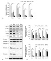
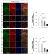
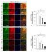
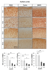
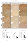
Aging is associated with functional and morphological changes in the sensory organs, including the auditory system. Mitophagy, a process that regulates the turnover of dysfunctional mitochondria, is impaired with aging. This study aimed to investigate the effect of aging on mitophagy in the central auditory system using an age-related hearing loss mouse model. C57BL/6J mice were divided into the following four groups based on age: 1-, 6-, 12-, and 18-month groups. The hearing ability was evaluated by measuring the auditory brainstem response (ABR) thresholds. The mitochondrial DNA damage level and the expression of mitophagy-related genes, and proteins were investigated by real-time polymerase chain reaction and Western blot analyses. The colocalization of mitophagosomes and lysosomes in the mouse auditory cortex and inferior colliculus was analyzed by immunofluorescence analysis. The expression of genes involved in mitophagy, such as PINK1, Parkin, and BNIP3 in the mouse auditory cortex and inferior colliculus, was investigated by immunohistochemical staining. The ABR threshold increased with aging. In addition to the mitochondrial DNA integrity, the mRNA levels of PINK1, Parkin, NIX, and BNIP3, as well as the protein levels of PINK1, Parkin, BNIP3, COX4, LC3B, mitochondrial oxidative phosphorylation (OXPHOS) subunits I-IV in the mouse auditory cortex significantly decreased with aging. The immunofluorescence analysis revealed that the colocalization of mitophagosomes and lysosomes in the mouse auditory cortex and inferior colliculus decreased with aging. The immunohistochemical analysis revealed that the expression of PINK1, Parkin, and BNIP3 decreased in the mouse auditory cortex and inferior colliculus with aging. These findings indicate that aging-associated impaired mitophagy may contribute to the cellular changes observed in an aged central auditory system, which result in age-related hearing loss. Thus, the induction of mitophagy can be a potential therapeutic strategy for age-related hearing loss.
Keywords: age-related hearing loss; auditory cortex; mitochondria; mitophagy; presbycusis.
Conflict of interest statement
The authors declare no conflict of interest.
Figures

Similar articles
-
Reduced mitophagy in the cochlea of aged C57BL/6J mice.Exp Gerontol. 2020 Aug;137:110946. doi: 10.1016/j.exger.2020.110946. Epub 2020 May 5. Exp Gerontol. 2020. PMID: 32387126
-
Urolithin A prevents age-related hearing loss in C57BL/6J mice likely by inducing mitophagy.Exp Gerontol. 2024 Nov;197:112589. doi: 10.1016/j.exger.2024.112589. Epub 2024 Sep 24. Exp Gerontol. 2024. PMID: 39307249
-
Mitochondrial DNA 3,860-bp Deletion Increases with Aging in the Auditory Nervous System of C57BL/6J Mice.ORL J Otorhinolaryngol Relat Spec. 2019;81(2-3):92-100. doi: 10.1159/000499475. Epub 2019 May 24. ORL J Otorhinolaryngol Relat Spec. 2019. PMID: 31129670
-
Genetic influences on susceptibility of the auditory system to aging and environmental factors.Scand Audiol Suppl. 1992;36:1-39. Scand Audiol Suppl. 1992. PMID: 1488615 Review.
-
Hearing loss-related altered neuronal activity in the inferior colliculus.Hear Res. 2024 Aug;449:109033. doi: 10.1016/j.heares.2024.109033. Epub 2024 May 20. Hear Res. 2024. PMID: 38797036 Review.
Cited by
-
The Role and Research Progress of Mitochondria in Sensorineural Hearing Loss.Mol Neurobiol. 2024 Sep 18. doi: 10.1007/s12035-024-04470-4. Online ahead of print. Mol Neurobiol. 2024. PMID: 39292339 Review.
-
The Role of Molecular and Cellular Aging Pathways on Age-Related Hearing Loss.Int J Mol Sci. 2024 Sep 7;25(17):9705. doi: 10.3390/ijms25179705. Int J Mol Sci. 2024. PMID: 39273652 Free PMC article. Review.
-
Age-related differences in affective behaviors in mice: possible role of prefrontal cortical-hippocampal functional connectivity and metabolomic profiles.Front Aging Neurosci. 2024 Mar 8;16:1356086. doi: 10.3389/fnagi.2024.1356086. eCollection 2024. Front Aging Neurosci. 2024. PMID: 38524115 Free PMC article.
-
Traditional Chinese medicine for the prevention and treatment of presbycusis.Heliyon. 2023 Nov 28;9(12):e22422. doi: 10.1016/j.heliyon.2023.e22422. eCollection 2023 Dec. Heliyon. 2023. PMID: 38076135 Free PMC article. Review.
-
Macrophage Depletion Protects Against Cisplatin-Induced Ototoxicity and Nephrotoxicity.bioRxiv [Preprint]. 2023 Nov 17:2023.11.16.567274. doi: 10.1101/2023.11.16.567274. bioRxiv. 2023. Update in: Sci Adv. 2024 Jul 26;10(30):eadk9878. doi: 10.1126/sciadv.adk9878. PMID: 38014097 Free PMC article. Updated. Preprint.
References
MeSH terms
Substances
Grants and funding
LinkOut - more resources
Full Text Sources
Medical

