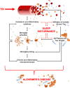
The Bidirectional Relationship Between Sleep and Inflammation Links Traumatic Brain Injury and Alzheimer's Disease
- PMID: 32982677
- PMCID: PMC7479838
- DOI: 10.3389/fnins.2020.00894
The Bidirectional Relationship Between Sleep and Inflammation Links Traumatic Brain Injury and Alzheimer's Disease
Abstract
Traumatic brain injury (TBI) and Alzheimer's disease (AD) are diseases during which the fine-tuned autoregulation of the brain is lost. Despite the stark contrast in their causal mechanisms, both TBI and AD are conditions which elicit a neuroinflammatory response that is coupled with physical, cognitive, and affective symptoms. One commonly reported symptom in both TBI and AD patients is disturbed sleep. Sleep is regulated by circadian and homeostatic processes such that pathological inflammation may disrupt the chemical signaling required to maintain a healthy sleep profile. In this way, immune system activation can influence sleep physiology. Conversely, sleep disturbances can exacerbate symptoms or increase the risk of inflammatory/neurodegenerative diseases. Both TBI and AD are worsened by a chronic pro-inflammatory microenvironment which exacerbates symptoms and worsens clinical outcome. Herein, a positive feedback loop of chronic inflammation and sleep disturbances is initiated. In this review, the bidirectional relationship between sleep disturbances and inflammation is discussed, where chronic inflammation associated with TBI and AD can lead to sleep disturbances and exacerbated neuropathology. The role of microglia and cytokines in sleep disturbances associated with these diseases is highlighted. The proposed sleep and inflammation-mediated link between TBI and AD presents an opportunity for a multifaceted approach to clinical intervention.
Keywords: Alzheimer’s disease; concussion; cytokines; inflammation; microglia; neurodegeneration; sleep; traumatic brain injury.
Copyright © 2020 Green, Ortiz, Wonnacott, Williams and Rowe.
Figures


Similar articles
-
Experimental diffuse brain injury and a model of Alzheimer's disease exhibit disease-specific changes in sleep and incongruous peripheral inflammation.J Neurosci Res. 2021 Apr;99(4):1136-1160. doi: 10.1002/jnr.24771. Epub 2020 Dec 14. J Neurosci Res. 2021. PMID: 33319441 Free PMC article.
-
Traumatic Brain Injury Causes Chronic Cortical Inflammation and Neuronal Dysfunction Mediated by Microglia.J Neurosci. 2021 Feb 17;41(7):1597-1616. doi: 10.1523/JNEUROSCI.2469-20.2020. Epub 2021 Jan 15. J Neurosci. 2021. PMID: 33452227 Free PMC article.
-
Immune-endocrine interactions in the pathophysiology of sleep-wake disturbances following traumatic brain injury: A narrative review.Brain Res Bull. 2022 Jul;185:117-128. doi: 10.1016/j.brainresbull.2022.04.017. Epub 2022 May 7. Brain Res Bull. 2022. PMID: 35537569 Review.
-
Age exacerbates the CCR2/5-mediated neuroinflammatory response to traumatic brain injury.J Neuroinflammation. 2016 Apr 18;13(1):80. doi: 10.1186/s12974-016-0547-1. J Neuroinflammation. 2016. PMID: 27090212 Free PMC article.
-
The Putative Role of Neuroinflammation in the Interaction between Traumatic Brain Injuries, Sleep, Pain and Other Neuropsychiatric Outcomes: A State-of-the-Art Review.J Clin Med. 2023 Feb 23;12(5):1793. doi: 10.3390/jcm12051793. J Clin Med. 2023. PMID: 36902580 Free PMC article. Review.
Cited by
-
Sleep deprivation and NLRP3 inflammasome: Is there a causal relationship?Front Neurosci. 2022 Dec 22;16:1018628. doi: 10.3389/fnins.2022.1018628. eCollection 2022. Front Neurosci. 2022. PMID: 36620464 Free PMC article. Review.
-
Possible Neuropathology of Sleep Disturbance Linking to Alzheimer's Disease: Astrocytic and Microglial Roles.Front Cell Neurosci. 2022 Jun 9;16:875138. doi: 10.3389/fncel.2022.875138. eCollection 2022. Front Cell Neurosci. 2022. PMID: 35755779 Free PMC article. Review.
-
Impaired sleep is associated with tau deposition on 18F-flortaucipir PET and accelerated cognitive decline, accounting for medications that affect sleep.J Neurol Sci. 2024 Mar 15;458:122927. doi: 10.1016/j.jns.2024.122927. Epub 2024 Feb 8. J Neurol Sci. 2024. PMID: 38341949
-
Self-reported quantity and quality of sleep in children and adolescents with a chronic condition compared to healthy controls.Eur J Pediatr. 2023 Jul;182(7):3139-3146. doi: 10.1007/s00431-023-04980-8. Epub 2023 Apr 26. Eur J Pediatr. 2023. PMID: 37099091 Free PMC article.
-
The importance of including both sexes in preclinical sleep studies and analyses.Sci Rep. 2024 Oct 15;14(1):23622. doi: 10.1038/s41598-024-70996-1. Sci Rep. 2024. PMID: 39406742 Free PMC article.
References
-
- Aisen P. S. (2002). Evaluation of selective COX-2 inhibitors for the treatment of Alzheimer’s disease. J. Pain Symptom Manage. 23 4(Suppl.), S35–S40. - PubMed
-
- Asahi M., Wang X., Mori T., Sumii T., Jung J. C., Moskowitz M. A., et al. (2001). Effects of matrix metalloproteinase-9 gene knock-out on the proteolysis of blood-brain barrier and white matter components after cerebral ischemia. J. Neurosci. 21 7724–7732. 10.1523/jneurosci.21-19-07724.2001 - DOI - PMC - PubMed
Publication types
LinkOut - more resources
Full Text Sources

