Intracellular pH Regulation of Skeletal Muscle in the Milieu of Insulin Signaling
- PMID: 32977552
- PMCID: PMC7598285
- DOI: 10.3390/nu12102910
Intracellular pH Regulation of Skeletal Muscle in the Milieu of Insulin Signaling
Abstract
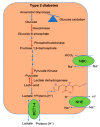
Type 2 diabetes (T2D), along with obesity, is one of the leading health problems in the world which causes other systemic diseases, such as cardiovascular diseases and kidney failure. Impairments in glycemic control and insulin resistance plays a pivotal role in the development of diabetes and its complications. Since skeletal muscle constitutes a significant tissue mass of the body, insulin resistance within the muscle is considered to initiate the onset of diet-induced metabolic syndrome. Insulin resistance is associated with impaired glucose uptake, resulting from defective post-receptor insulin responses, decreased glucose transport, impaired glucose phosphorylation, oxidation and glycogen synthesis in the muscle. Although defects in the insulin signaling pathway have been widely studied, the effects of cellular mechanisms activated during metabolic syndrome that cross-talk with insulin responses are not fully elucidated. Numerous reports suggest that pathways such as inflammation, lipid peroxidation products, acidosis and autophagy could cross-talk with insulin-signaling pathway and contribute to diminished insulin responses. Here, we review and discuss the literature about the defects in glycolytic pathway, shift in glucose utilization toward anaerobic glycolysis and change in intracellular pH [pH]i within the skeletal muscle and their contribution towards insulin resistance. We will discuss whether the derangements in pathways, which maintain [pH]i within the skeletal muscle, such as transporters (monocarboxylate transporters 1 and 4) and depletion of intracellular buffers, such as histidyl dipeptides, could lead to decrease in [pH]i and the onset of insulin resistance. Further we will discuss, whether the changes in [pH]i within the skeletal muscle of patients with T2D, could enhance the formation of protein aggregates and activate autophagy. Understanding the mechanisms by which changes in the glycolytic pathway and [pH]i within the muscle, contribute to insulin resistance might help explain the onset of obesity-linked metabolic syndrome. Finally, we will conclude whether correcting the pathways which maintain [pH]i within the skeletal muscle could, in turn, be effective to maintain or restore insulin responses during metabolic syndrome.
Keywords: carnosine; chronic kidney disease; diabetes; glycolysis; histidyl dipeptides; insulin signaling; intracellular pH; obesity.
Conflict of interest statement
The authors declare no conflict of interest.
Figures


Similar articles
-
Metabolism and insulin signaling in common metabolic disorders and inherited insulin resistance.Dan Med J. 2014 Jul;61(7):B4890. Dan Med J. 2014. PMID: 25123125 Review.
-
Role of reduced insulin-stimulated bone blood flow in the pathogenesis of metabolic insulin resistance and diabetic bone fragility.Med Hypotheses. 2016 Aug;93:81-6. doi: 10.1016/j.mehy.2016.05.008. Epub 2016 May 12. Med Hypotheses. 2016. PMID: 27372862
-
p38 MAPK in Glucose Metabolism of Skeletal Muscle: Beneficial or Harmful?Int J Mol Sci. 2020 Sep 4;21(18):6480. doi: 10.3390/ijms21186480. Int J Mol Sci. 2020. PMID: 32899870 Free PMC article. Review.
-
Insulin signaling and glucose transport in insulin resistant human skeletal muscle.Cell Biochem Biophys. 2007;48(2-3):103-13. doi: 10.1007/s12013-007-0030-9. Cell Biochem Biophys. 2007. PMID: 17709880 Review.
-
The Baf60c/Deptor pathway links skeletal muscle inflammation to glucose homeostasis in obesity.Diabetes. 2014 May;63(5):1533-45. doi: 10.2337/db13-1061. Epub 2014 Jan 23. Diabetes. 2014. PMID: 24458360 Free PMC article.
Cited by
-
Trehalose-Carnosine Prevents the Effects of Spinal Cord Injury Through Regulating Acute Inflammation and Zinc(II) Ion Homeostasis.Cell Mol Neurobiol. 2023 May;43(4):1637-1659. doi: 10.1007/s10571-022-01273-w. Epub 2022 Sep 19. Cell Mol Neurobiol. 2023. PMID: 36121569 Free PMC article.
-
Exploring protein relative relations in skeletal muscle proteomic analysis for insights into insulin resistance and type 2 diabetes.Sci Rep. 2024 Jul 31;14(1):17631. doi: 10.1038/s41598-024-68568-4. Sci Rep. 2024. PMID: 39085321 Free PMC article.
-
Ionophore Ability of Carnosine and Its Trehalose Conjugate Assists Copper Signal in Triggering Brain-Derived Neurotrophic Factor and Vascular Endothelial Growth Factor Activation In Vitro.Int J Mol Sci. 2021 Dec 16;22(24):13504. doi: 10.3390/ijms222413504. Int J Mol Sci. 2021. PMID: 34948299 Free PMC article.
-
Hidden chronic metabolic acidosis of diabetes type 2 (CMAD): Clues, causes and consequences.Rev Endocr Metab Disord. 2023 Aug;24(4):735-750. doi: 10.1007/s11154-023-09816-2. Epub 2023 Jun 28. Rev Endocr Metab Disord. 2023. PMID: 37380824 Review.
-
Investigating Celastrol's Anti-DCM Targets and Mechanisms via Network Pharmacology and Experimental Validation.Biomed Res Int. 2022 Jul 5;2022:7382130. doi: 10.1155/2022/7382130. eCollection 2022. Biomed Res Int. 2022. Retraction in: Biomed Res Int. 2023 Dec 29;2023:9874346. doi: 10.1155/2023/9874346 PMID: 35845929 Free PMC article. Retracted.
References
-
- Lillioja S., Mott D.M., Howard B.V., Bennett P.H., Yki-Jarvinen H., Freymond D., Nyomba B.L., Zurlo F., Swinburn B., Bogardus C. Impaired Glucose Tolerance as a Disorder of Insulin Action. Longitudinal and cross-sectional studies in Pima Indians. N. Eng. J. Med. 1988;318:1217–1225. doi: 10.1056/NEJM198805123181901. - DOI - PubMed
Publication types
MeSH terms
Substances
Grants and funding
LinkOut - more resources
Full Text Sources
Medical

