. 2020 Dec;26(12):1075-1085.
doi: 10.1016/j.cardfail.2020.09.009. Epub 2020 Sep 18.

Novel BAG3 Variants in African American Patients With Cardiomyopathy: Reduced β-Adrenergic Responsiveness in Excitation-Contraction

Affiliations

Novel BAG3 Variants in African American Patients With Cardiomyopathy: Reduced β-Adrenergic Responsiveness in Excitation-Contraction

Arthur M Feldman et al. J Card Fail. 2020 Dec.

Abstract

Background: We reported 3 novel nonsynonymous single nucleotide variants of Bcl2-associated athanogene 3 (BAG3) in African Americans with heart failure (HF) that are associated with a 2-fold increase in cardiac events (HF hospitalization, heart transplantation, or death).

Methods and results: We expressed BAG3 variants (P63A, P380S, and A479V) via adenovirus-mediated gene transfer in adult left ventricular myocytes isolated from either wild-type (WT) or cardiac-specific BAG3 haploinsufficient (cBAG3+/-) mice: the latter to simulate the clinical situation in which BAG3 variants are only found on 1 allele. Compared with WT myocytes, cBAG3+/- myocytes expressed approximately 50% of endogenous BAG3 levels and exhibited decreased [Ca2+]i and contraction amplitudes after isoproterenol owing to decreased L-type Ca2+ current. BAG3 repletion with WT BAG3 but not P380S, A479V, or P63A/P380S variants restored contraction amplitudes in cBAG3+/- myocytes to those measured in WT myocytes, suggesting excitation-contraction abnormalities partly account for HF in patients harboring these mutants. Because P63A is near the WW domain (residues 21-55) and A479V is in the BAG domain (residues 420-499), we expressed BAG3 deletion mutants (Δ1-61 and Δ421-575) in WT myocytes and demonstrated that the BAG but not the WW domain was involved in enhancement of excitation-contraction by isoproterenol.

Conclusions: The BAG3 variants contribute to HF in African American patients partly by decreasing myocyte excitation-contraction under stress, and that both the BAG and PXXP domains are involved in mediating β-adrenergic responsiveness in myocytes.

Keywords: BAG3; adenovirus-mediated gene transfer; dilated cardiomyopathy; excitation–contraction coupling; isolated adult cardiac myocytes.


Conflict of interest statement

Declaration of competing interest K.K. is a board member, scientific advisor, and holds equity in Excision Biotherapeutics, a biotech start-up that has licensed the viral gene editing technology from Temple University for commercial development and clinical trials. A.M.F. and K.K. have a pending US patent #611934,483 for BAG3 as a target for heart failure therapy. A.M.F. and J.Y.C. have a pending US patent #621205,990 for BAG3 composition and methods. Exclusive rights to the patents have been optioned by Temple University to Renovacor, Inc. A.M.F. and J.Y.C. hold equity in Renovacor, Inc.

Figures

Figure 1.
Figure 1.. Consensus protein-protein binding domains in BAG3.
BAG3 serves as a co-chaperone for both the constituitively expressed and the inducible heat shock protein 70 (Hsc70/Hsp70): its BAG domain binding with the ATPase binding site of Hsc70/Hsp70. Hsp70-BAG3 interactions mediate chaperone assisted autophagy by shuttling client proteins along the cellular micro-tubular apparatus that are dedicated to eliminating mis-folded proteins. The BAG domain also couples with the anti-apoptotic protein Bcl-2 resulting in inhibition of apoptosis. Two highly conserved IPV (Ile-Pro-Val) motifs allow BAG3 to interact with the small heat shock proteins HspB8 and HspB6 to support macroautophagy, a process in which mis-folded or damaged proteins and organelles (e.g. mitochondria) are sequestered in autophagosomes and degraded whereas the second IPV domain binds to αβ-crystallin with the subsequent inhibition of protein aggregation. The WW domain plays a pivotal role in chaperone-assisted selective autophagy or CASA, a tension-induced autophagy pathway that is essential for mechano-transduction in muscle and immune cells, . The WW region also couples with the PXXP region to modify the three-dimensional structure of the protein. The proline-rich PXXP region also facilitates binding to the SH3 (Src homology 3)-containing protein phospholipase C-γ (PLC-γ) with subsequent stimulation of invasion, adhesion and migration of cancer cells, and the retrograde transport of mis-folded proteins to peri-nuclear aggresomes.
Figure 2.
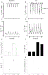
Figure 2.. Abnormal Ca2+ homeostasis and reduced contraction in cardiac-specific haplo-insufficient BAG3 murine myocytes.
[Ca2+]i and contraction were measured in field-stimulated (2 Hz, 37°C) LV myocytes isolated from 8-wk old WT and cBAG3+/− mice (Methods). Representative traces of contraction at baseline (A) and after 1 μM isoproterenol (Iso)(B), and [Ca2+]i transients at baseline (C) and after Iso (D) of WT (solid lines) and cBAG3+/− (dashed lines) myocytes are shown. E, G and H: Means ± SE of [Ca2+]i transient amplitudes (E), systolic and diastolic [Ca2+]i (G) and t1/2 of [Ca2+]i transient decline (H) from 11 WT and 13 cBAG3+/− myocytes (2 mice each) at baseline and 15 WT and 19 cBAG3+/− myocytes (2 mice each) after 1 μM Iso. For systolic and diastolic [Ca2+]i, error bars are not shown if they fall within the boundaries of the symbols. F: Means ± SE of contraction amplitudes from 21 WT and 16 cBAG3+/− myocytes (3 WT and 2 cBAG3+/− mice) at baseline and 19 WT and 12 cBAG3+/− myocytes after Iso. For E, F, G: *p<0.008, group (WT vs. cBAG3+/−) × Iso interaction effect. For H: *p=0.0425, WT vs. cBAG3+/−.
Figure 3.
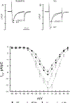
Figure 3.. Reduced L-type Ca2+ current (ICa) in cardiac-specific haplo-insufficient BAG3 myocytes.
ICa was measured in myocytes isolated from 8-wk old WT and cBAG3+/− myocytes (Methods). A and B: Representative traces of ICa (0 mV) from WT and cBAG3+/− myocytes before (A) and after (B) 1 μM Iso. C: IV relationship of ICa in WT (▄, baseline; □, after Iso; n=7 cells from 3 mice) and cBAG3+/− (●, baseline; ○, after Iso; n=5 cells from 3 mice) myocytes. 3-way ANOVA indicates significant group (WT vs. cBAG3+/−, p<0.0001), voltage (p<0.0001), Iso (p<0.0001), group × voltage (p=0.0425) and group × Iso (p=0.0021) interaction effects.
Figure 4.
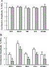
Figure 4.. Non-synonymous, single nucleotide BAG3 variants in African-American patients with heart failure do not enhance β-adrenergic responsiveness compared to WT BAG3.
LV myocytes isolated from 8-wk old WT (open bars) and cBAG3+/− (hatched bars) mice were infected with adenovirus (Adv) expressing GFP, WT BAG3, P380S, A479V or P63A/P380S BAG3 variants and cultured for 24h before contraction (2 Hz, 37°C) measurements, both before and after 1 μM Iso (Methods). A. Means ± SE of baseline contraction amplitudes [% resting cell length (RCL)] measured in WT and cBAG3+/− myocytes. B. Means ± SE of contraction amplitudes after Iso. Note the difference in Y-axis scales. For WT-GFP, there are 20 myocytes at baseline and 15 myocytes after Iso (n=6 mice); for cBAG3+/−-GFP, there are 19 myocytes at baseline and 17 myocytes after Iso (n=6 mice); for WT-BAG3, there are 14 myocytes at baseline and 14 myocytes after Iso (n=5 mice); for cBAG3+/−-BAG3, there are 16 myocytes at baseline and 21 myocytes after Iso (n=5 mice); for WT-P380S, there are 7 myocytes at baseline and 8 myocytes after Iso (n=3 mice); for cBAG3+/−-P380S, there are 8 myocytes at baseline and 8 myocytes after Iso (n=2 mice); for WT-A479V, there are 17 myocytes at baseline and 22 myocytes after Iso (n=3 mice); for cBAG3+/−-A479V, there are 18 myocytes at baseline and 19 myocytes after Iso (n=3 mice); for WT-P63A/P380S, there are 7 myocytes at baseline and 7 myocytes after Iso (n= 1 mouse); and for cBAG3+/−-P63A/P380S, there are 5 myocytes at baseline and 6 myocytes after Iso (n=1 mouse). 3-way ANOVA indicate significant group (WT vs. cBAG3+/−; p=0.0002), Adv (p<0.0001), Iso (p<0.0001) and group × Iso interaction (p=0.0003) effects. *p<0.002, BAG3 vs. GFP, P380S, A479V or P63A/P380S by subgroup analysis. #p=0.0003, group (WT vs. cBAG3+/−) × Iso interaction effects.
Figure 5.
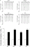
Figure 5.. BAG3 but not WW domain is required for enhanced contraction amplitudes after isoproterenol.
LV myocytes isolated from 8 wk-old WT mice were infected with adenovirus (Adv) expressing GFP, WT BAG3, WW domain (residues 62 to 575 and abbreviated as 575) or BAG domain (residues 1 to 420 and abbreviated as 420) deletion mutants and cultured for 24h before contraction (2 Hz, 37°C) measurements (Methods). A. Representative traces of contraction for GFP, BAG3, 420 and 575 myocytes, both before (dotted lines) and after (solid lines) 1 μM isoproterenol (Iso). B. Means ± SE of contraction amplitudes [% resting cell length (RCL)] before (open bars) and after Iso (solid bars). For GFP, there are 11 myocytes at baseline and 13 myocytes after Iso (n=4 mice); for BAG3, there are 14 myocytes at baseline and 12 myocytes after Iso (n=5 mice); for 420, there are 10 myocytes at baseline and 14 myocytes after Iso (n=2 mice); and for 575, there are 9 myocytes at baseline and 12 myocytes after Iso (n=3 mice). 2-way ANOVA of all 4 groups indicate significant Adv (p=0.0021), Iso (p<0.0001) and Adv × Iso (p=0.0011) interaction effects. *p<0.02, GFP vs. BAG3 or GFP vs. 575 by subgroup analysis (p≤0.01 is statistically significant after Bonferroni adjustment); #p<0.001, BAG3 vs. 420 by subgroup analysis.
Figure 6.
Figure 6.. BAG3 but not WW domain is required for higher systolic [Ca2+]i and larger [Ca2+]i transient amplitudes after isoproterenol.
LV myocytes isolated from 8 wk-old WT mice were infected with adenovirus (Adv) expressing GFP, WT BAG3, WW domain (575) or BAG domain (420) deletion mutants and cultured for 24h before [Ca2+]i (2 Hz, 37°C) measurements (Methods). A. Representative traces of [Ca2+]i transients for GFP, BAG3, 420 and 575 myocytes, both at baseline (dotted lines) and after 1 μM Iso (solid lines). B. Means ± SE of systolic (●) and diastolic (○) [Ca2+]i at baseline and after Iso (I). Error bars are not shown if they fall within the boundaries of the symbols. For GFP, there are 13 myocytes at baseline and 10 myocytes after Iso (n=2 mice); for BAG3, there are 9 myocytes at baseline and 14 myocytes after Iso (n=3 mice); for 420, there are 9 myocytes at baseline and 9 myocytes after Iso (n=2 mice); and for 575, there are 12 myocytes at baseline and 13 myocytes after Iso (n=3 mice). 2-way ANOVA of systolic [Ca2+]i from all 4 groups indicate significant Adv (p=0.0001), Iso (p<0.0001) and Adv × Iso (p=0.0006) interaction effects. *p≤0.005, GFP vs. BAG3 or GFP vs. 575 by subgroup analysis (p≤0.01 is statistically significant after Bonferroni adjustment). C. Means ± SE of [Ca2+]i transient amplitudes (% increase in Fura2 signal) at baseline (open bars) and after Iso (solid bars). 2-way ANOVA of all 4 groups indicate significant Adv (p=0.0002), Iso (p<0.0001) and Adv × Iso (p=0.0003) interaction effects. *p≤0.0009, GFP vs. BAG3 or GFP vs. 575 by subgroup analysis (p≤0.01 is statistically significant after Bonferroni adjustment).
Figure 7.
Figure 7.. BAG3 but not WW domain is required for enhanced L-type Ca2+ current (ICa) after isoproterenol.
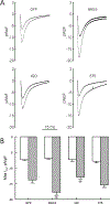
LV myocytes isolated from 8 wk-old WT mice were infected with adenovirus (Adv) expressing GFP, WT BAG3, WW domain (575) or BAG domain (420) deletion mutants and cultured for 24h before ICa measurements (Methods). A. Representative ICa traces at 0 mV for GFP, BAG3, 420 and 575 myocytes, both at baseline (black) and after 1 μM Iso (red). B. Means ± SE of maximal ICa amplitudes (0 mV) before (open bars) and after Iso (hatched bars). For GFP, there are 7 myocytes each at baseline and after Iso (n=5 mice); for BAG3, there are 4 myocytes each at baseline and after Iso (n=3 mice); for 420, there are 6 myocytes each at baseline and after Iso (n=3 mice); and for 575, there are 6 myocytes each at baseline and after Iso (n=3 mice). 2-way ANOVA of all 4 groups indicate significant Adv (p=0.0158), Iso (p<0.0001) and Adv × Iso (p=0.0078) interaction effects. *p<0.01, BAG3 vs. GFP or BAG3 vs. 420 by subgroup analysis (p≤0.01 is statistically significant after Bonferroni adjustment).

Similar articles

Cited by

References

    1. Knezevic T, Myers VD, Gordon J, Tilley DG, Sharp TE 3rd, Wang J, Khalili K, Cheung JY and Feldman AM. BAG3: a new player in the heart failure paradigm. Heart Fail Rev. 2015;20:423–434. - PMC - PubMed
    1. Myers VD, Tomar D, Madesh M, Wang J, Song J, Zhang XQ, Gupta MK, Tahrir FG, Gordon J, McClung JM, Kontos CD, Khalili K, Cheung JY and Feldman AM. Haplo-insufficiency of Bcl2-associated athanogene 3 in mice results in progressive left ventricular dysfunction, β-adrenergic insensitivity, and increased apoptosis. Journal of cellular physiology. 2018;233:6319–6326. - PMC - PubMed
    1. Su F, Myers VD, Knezevic T, Wang J, Gao E, Madesh M, Tahrir FG, Gupta MK, Gordon J, Rabinowitz J, Ramsey FV, Tilley DG, Khalili K, Cheung JY and Feldman AM. Bcl-2-associated athanogene 3 protects the heart from ischemia/reperfusion injury. JCI Insight. 2016;1:e90931. - PMC - PubMed
    1. Tahrir FG, Knezevic T, Gupta MK, Gordon J, Cheung JY, Feldman AM and Khalili K. Evidence for the role of BAG3 in mitochondrial quality control in cardiomyocytes. Journal of cellular physiology. 2017;232:797–805. - PMC - PubMed
    1. Gupta MK, Gordon J, Glauser GM, Myers VD, Feldman AM, Cheung JY and Khalili K. Lamin B is a target for selective nuclear PQC by BAG3: implication for nuclear envelopathies. Cell death & disease. 2019;10:23. - PMC - PubMed