Review
. 2020 Sep 15;10(9):1838.
doi: 10.3390/nano10091838.

Engineered Extracellular Vesicles: Tailored-Made Nanomaterials for Medical Applications

Affiliations
Review

Engineered Extracellular Vesicles: Tailored-Made Nanomaterials for Medical Applications

Kenny Man et al. Nanomaterials (Basel). .

Abstract

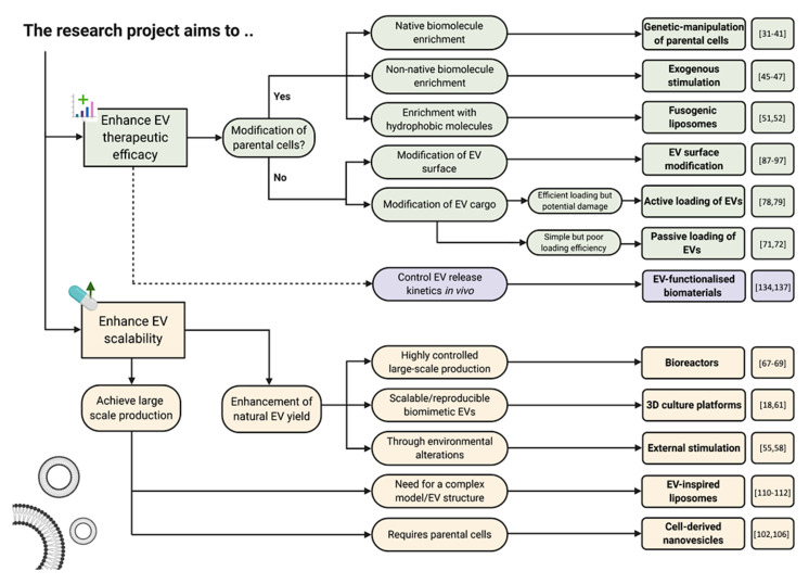
Extracellular vesicles (EVs) are emerging as promising nanoscale therapeutics due to their intrinsic role as mediators of intercellular communication, regulating tissue development and homeostasis. The low immunogenicity and natural cell-targeting capabilities of EVs has led to extensive research investigating their potential as novel acellular tools for tissue regeneration or for the diagnosis of pathological conditions. However, the clinical use of EVs has been hindered by issues with yield and heterogeneity. From the modification of parental cells and naturally-derived vesicles to the development of artificial biomimetic nanoparticles or the functionalisation of biomaterials, a multitude of techniques have been employed to augment EVs therapeutic efficacy. This review will explore various engineering strategies that could promote EVs scalability and therapeutic effectiveness beyond their native utility. Herein, we highlight the current state-of-the-art EV-engineering techniques with discussion of opportunities and obstacles for each. This is synthesised into a guide for selecting a suitable strategy to maximise the potential efficacy of EVs as nanoscale therapeutics.

Keywords: EV engineering; EV-functionalised biomaterials; artificial EVs; exosomes; extracellular vesicles; microvesicles; nanomedicine; regenerative medicine.


Conflict of interest statement

The authors declare no conflict of interest.

Figures

Figure 1
Figure 1
Biogenesis of extracellular vesicles (EVs) and potential applications of these nanoparticles in tissue engineering and regenerative medicine.
Figure 2
Figure 2
Engineering EVs by parental cell modification. (A) Schematic representation of methods to engineer EVs through parental cell genetic modification, stimulation with exogenous molecules, the use of fusogenic liposomes or environmental stimulation. (B) EVs derived from miR-126 overexpressing mesenchymal stem cells (MSCs) significantly increased vessel formation in a mice model. Reproduced from [31], with permission from John Wiley and Sons, 2016. *, p < 0.05 compared with control; #, p < 0.05 compared with CS. (C) CD9-human antigen R (HuR) enriched miR-155 into EVs with the miRNA efficiently delivered to the recipient cells, demonstrated by significantly increased miR-155 expression in the human monocytic cell line THP1. Reproduced from [34], with permission from American Chemical Society, 2019. **, p < 0.01; ***, p < 0.001. (D) Platelet-derived growth factor (PDGF)-stimulated EVs (+PDGF-EVs) exhibited substantially increased angiogenesis when compared to untreated EVs (+b-EVs). Black arrows indicate vessel formation. Adapted from [46], under the creative commons licence, 2014. *, p < 0.05 compared with control; #, p < 0.05 compared to b-Evs. (E) Reduced tumour volume observed in liposome-fused EVs (MFL + laser) when compared to liposomes treatment and laser irradiation alone. Reproduced from [51], with permission from American Chemical Society, 2015. ***, p < 0.001.
Figure 3
Figure 3
Methods of promoting EV scalability through environmental stimulation. (A) Schematic representation of strategies utilised to achieve scalability in EV manufacturing, such as the use of external stimulation, 3D culture platforms and bioreactor systems. (B) Hypoxic conditions enhanced the yield of EVs derived from endothelial cells compared to normoxic conditions. Reproduced with permission from [55], Elsevier, 2017. ***, p < 0.001. (C) 100-fold increase in EV quantity from MSCs cultured as spheroids with shaking during culture. Reproduced from [18], with permission from Springer Nature, 2018. *, p < 0.05. (D) Microcarrier-based 3D culture system combined with tangential flow filtration (3D-TIFF) enhanced MSCs yield 140-fold compared to cells culture in 2D conditions. Reproduced from [68], under the creative commons licence, 2018. (E) A hollow fibre bioreactor promoted EV yield 4-fold compared to 2D culture. Reproduced from [69], under the creative commons licence, 2016.
Figure 4
Figure 4
The use of cell-derived nanovesicles (CDNs) as an EV-mimetic system. (A) Schematic illustration of the process to fabricate CDNs from whole cells. (B) Macrophage-derived CDNs loaded with doxorubicin traffic to tumour tissue (upper panel) and reduced tumour growth compared to free-doxorubicin (lower panel). Reproduced from [102], with permission from American Chemical Society, 2013. *, p < 0.05; **, p < 0.01 compared to PBS. (C) CDN-derived from hepatocytes stimulated proliferation in vitro (upper panel) and liver regeneration in vivo (lower panel). Reproduced from [106], under the creative commons licence, 2018. NS, not significant; *, p < 0.05; **, p < 0.01.
Figure 5
Figure 5
The use of EV-inspired liposomes as therapeutic nanoparticles. (A) Schematic illustration representing the general strategy in mimicking the composition and therapeutic potency of naturally-derived EVs with EV-inspired liposomes (EVLs). (B) Liposomes incorporated with MHC Class I peptide complexes and ligands to facilitate adhesion, early/late activation and survival T cell receptors (upper panel). Incorporation of superparamagnetic particles allows for the tracking of these EVLs in vivo (lower panel). Reproduced from [110], with permission from Elsevier, 2009. (C) Liposome conjugated with APO2L/TRAIL (LUV-APO2L) substantially reduced inflammation within a rabbit model of antigen-induced arthritis model. Reproduced from [112], with permission from John Wiley and Sons, 2010. *, p < 0.05.
Figure 6
Figure 6
Therapeutic administration of EVs to the site of injury. (A) Schematic representation of the local delivery of EVs within an injectable biomaterial resulting in increased retention and therapeutic efficacy compared to delivery in solution. (B) Release kinetics of EVs from different delivery strategies. Burst release of EVs delivered in solutions, while sustained delivery observed from EV-encapsulated biomaterial systems. Adapted from [118], under the creative commons licence, 2018.
Figure 7
Figure 7
Strategies to immobilise EVs within biomaterials. Methods of EVs incorporation within biomaterials include physical entrapment, extracellular matrix (ECM)-immobilisation, electrostatic interactions and covalent conjugation.
Figure 8
Figure 8
Encapsulation of EVs within different hydrogel systems. (A) MSC-derived exosomes with an injectable hydrogel for hindlimb ischemia treatment. EVs encapsulated within chitosan hydrogel demonstrated enhanced retention at the site of injury when compared to local delivery with EVs alone. Adapted from [134], with permission from American Chemical Society, 2018. *, p < 0.05 compared to PBS; #, p < 0.05 compared to Exo. (B) Superior reparation of articular full-thickness rabbit defects treated with in situ forming EV-functionalised hydrogel (EHG) when compared to hydrogel alone (HG), pre-formed EV-hydrogel (Pre-EHG), EV delivery alone (Inj-Exos). Adapted from [136], with permission from Royal Society of Chemistry, 2017. (C) Development of two technologies capable of controlling the release kinetics of encapsulated EVs. Reproduced from [137], with permission from John Wiley and Sons, 2019. *, p < 0.05.
Figure 9
Figure 9
Schematic guide providing an overview of the engineering approaches and technologies currently used to enhance the therapeutic efficacy and scalability of EVs to improve their clinical utility.

Similar articles

Cited by

References

    1. Hourd P., Chandra A., Medcalf N., Williams D.J. StemBook. Harvard Stem Cell Institute; Cambridge, UK: 2008. Regulatory challenges for the manufacture and scale-out of autologous cell therapies. - DOI - PubMed
    1. Yanez-Mo M., Siljander P.R.M., Andreu Z., Zavec A.B., Borras F.E., Buzas E.I., Buzas K., Casal E., Cappello F., Carvalho J., et al. Biological properties of extracellular vesicles and their physiological functions. J. Extracell Vesicles. 2015;4 doi: 10.3402/jev.v4.27066. - DOI - PMC - PubMed
    1. Raposo G., Stoorvogel W. Extracellular vesicles: Exosomes, microvesicles, and friends. J. Cell Biol. 2013;200:373–383. doi: 10.1083/jcb.201211138. - DOI - PMC - PubMed
    1. Williams C., Rodriguez-Barrueco R., Silva J.M., Zhang W.J., Hearn S., Elemento O., Paknejad N., Manova-Todorova K., Welte K., Bromberg J., et al. Double-stranded DNA in exosomes: A novel biomarker in cancer detection. Cell Res. 2014;24:766–769. doi: 10.1038/cr.2014.44. - DOI - PMC - PubMed
    1. Zaborowski M.P., Balaj L., Breakefield X.O., Lai C.P. Extracellular Vesicles: Composition, Biological Relevance, and Methods of Study. Bioscience. 2015;65:783–797. doi: 10.1093/biosci/biv084. - DOI - PMC - PubMed

LinkOut - more resources