

doi: 10.1039/d0cc05092j.
Epub 2020 Sep 17.
Responsive fluorescent nucleotides serve as efficient substrates to probe terminal uridylyl transferase
Affiliations
- PMID: 32939524
- PMCID: PMC7611084
- DOI: 10.1039/d0cc05092j
Item in Clipboard
Responsive fluorescent nucleotides serve as efficient substrates to probe terminal uridylyl transferase
Chem Commun (Camb).
.
Abstract
We repurposed a terminal uridylyl transferase enzyme to site-specifically label RNA with microenvironment sensing fluorescent nucleotide mimics, which in turn provided direct read-outs to estimate the binding affinities of the enzyme to RNA and nucleotide substrates. This enzyme-probe system provides insights into the catalytic cycle, and can facilitate the development of discovery platforms to identify robust enzyme inhibitors.
Conflict of interest statement
Conflicts of interest
The authors declare no conflict of interest.
Figures
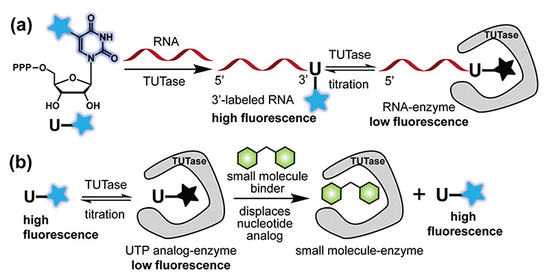
(a) TUTase efficiently incorporates UTP analogs (1 and
2, see Fig. 2) to generate
3′-end labeled RNA ONs exhibiting high fluorescence. Labeled RNA upon
titration with the enzyme results in progressive quenching in fluorescence,
which enables the determination of the binding constant. (b) The analog
(e.g., 2) bound to the enzyme shows low
fluorescence and the addition of a small molecule (e.g., UTP) displaces the
analog resulting in an enhancement in fluorescence, which constitutes a simple
turn-on fluorescent platform for the identification of TUTase
binders/inhibitors.
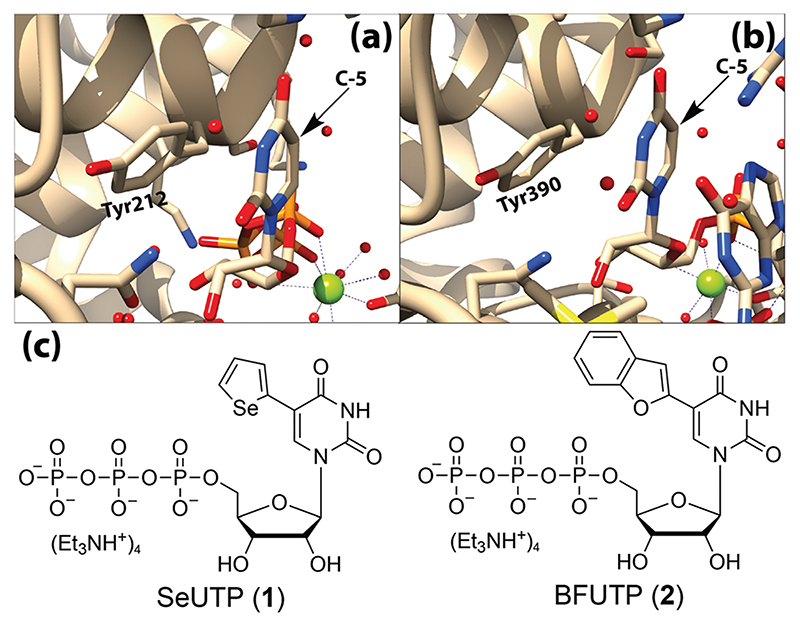
(a) Crystal structure of SpCID1 bound to UTP showing uracil stacking with
Tyr212 (PDB: 4FH5) (b) Crystal structure of DmTailor bound to RNA showing the
last nucleobase, uracil stacking with Tyr390 (PDB: 6I0V). Vacant space around the
C5-position of uracil can been seen in the structures. (c) Microenvironment
sensing UTP analogs used in terminal uridylation reactions.
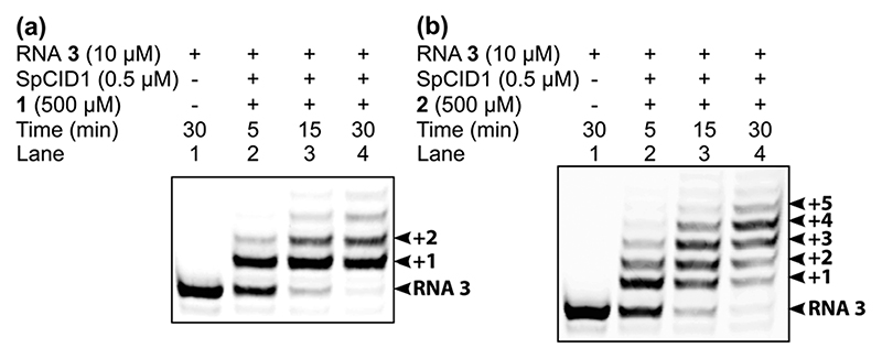
3′ -Terminal uridylation of 5′-FAM-labeled RNA ON 3
with (a) SeUTP 1 and (b) BFUTP 2 using SpCID1. Arrows
indicate the number of insertions at the 3’-end.
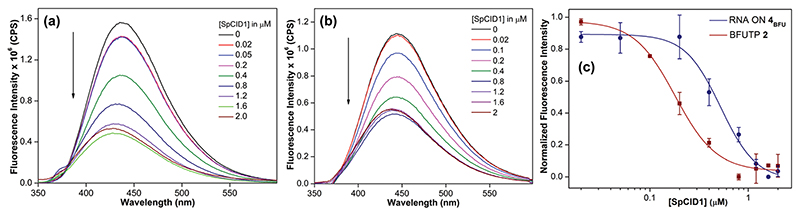
Emission spectra of (a) BFU-labeled RNA ON 4BFU and (b)
BFUTP 2 (200 nM) as a function of increasing concentration of
SpCID1 (0–2 μM). (c) A plot of normalized fluorescence intensity
at λem = 435 nm versus [SpCID1] showing the
curve fit for the binding of 4BFU and 2 to
SpCID1. See ESI† for details.
Similar articles
-
Enzymatic Functionalization of RNA Oligonucleotides by Terminal Uridylyl Transferase Using Fluorescent and Clickable Nucleotide Analogs.Chem Asian J. 2024 Sep 16;19(18):e202400475. doi: 10.1002/asia.202400475. Epub 2024 Aug 19. Chem Asian J. 2024. PMID: 38949615
-
Structural plasticity of Cid1 provides a basis for its distributive RNA terminal uridylyl transferase activity.Nucleic Acids Res. 2015 Mar 11;43(5):2968-79. doi: 10.1093/nar/gkv122. Epub 2015 Feb 20. Nucleic Acids Res. 2015. PMID: 25712096 Free PMC article.
-
Elements of nucleotide specificity in the Trypanosoma brucei mitochondrial RNA editing enzyme RET2.J Chem Inf Model. 2012 May 25;52(5):1308-18. doi: 10.1021/ci3001327. Epub 2012 May 7. J Chem Inf Model. 2012. PMID: 22512810 Free PMC article.
-
Determinants of substrate specificity in RNA-dependent nucleotidyl transferases.Biochim Biophys Acta. 2008 Apr;1779(4):206-16. doi: 10.1016/j.bbagrm.2007.12.003. Epub 2007 Dec 14. Biochim Biophys Acta. 2008. PMID: 18177750 Free PMC article. Review.
-
RNA-specific ribonucleotidyl transferases.RNA. 2007 Nov;13(11):1834-49. doi: 10.1261/rna.652807. Epub 2007 Sep 13. RNA. 2007. PMID: 17872511 Free PMC article. Review.
Cited by
-
RNA Probes for Visualization of Sarcin/ricin Loop Depurination without Background Fluorescence.Chem Asian J. 2022 Dec 14;17(24):e202201077. doi: 10.1002/asia.202201077. Epub 2022 Nov 18. Chem Asian J. 2022. PMID: 36321802 Free PMC article.
-
Incorporation and Utility of a Responsive Ribonucleoside Analogue in Probing the Conformation of a Viral RNA Motif by Fluorescence and 19 F NMR Spectroscopy.Chembiochem. 2022 Feb 4;23(3):e202100601. doi: 10.1002/cbic.202100601. Epub 2021 Dec 7. Chembiochem. 2022. PMID: 34821449 Free PMC article.
References
MeSH terms
Substances
Grants and funding
LinkOut - more resources
Full Text Sources