miR-23a-3p-abundant small extracellular vesicles released from Gelma/nanoclay hydrogel for cartilage regeneration
- PMID: 32939233
- PMCID: PMC7480606
- DOI: 10.1080/20013078.2020.1778883
miR-23a-3p-abundant small extracellular vesicles released from Gelma/nanoclay hydrogel for cartilage regeneration
Abstract
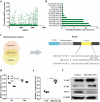
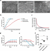
Articular cartilage has limited self-regenerative capacity and the therapeutic methods for cartilage defects are still dissatisfactory in clinic. Recent studies showed that exosomes derived from mesenchymal stem cells promoted chondrogenesis by delivering bioactive substances to the recipient cells, indicating exosomes might be a novel method for repairing cartilage defect. Herein, we investigated the role and mechanism of human umbilical cord mesenchymal stem cells derived small extracellular vesicles (hUC-MSCs-sEVs) on cartilage regeneration. In vitro results showed that hUC-MSCs-sEVs promoted the migration, proliferation and differentiation of chondrocytes and human bone marrow mesenchymal stem cells (hBMSCs). MiRNA microarray showed that miR-23a-3p was the most highly expressed among the various miRNAs contained in hUC-MSCs-sEVs. Our data revealed that hUC-MSCs-sEVs promoted cartilage regeneration by transferring miR-23a-3p to suppress the level of PTEN and elevate expression of AKT. Moreover, we fabricated Gelatin methacrylate (Gelma)/nanoclay hydrogel (Gel-nano) for sustained release of sEVs, which was biocompatible and exhibited excellent mechanical property. In vivo results showed that hUC-MSCs-sEVs containing Gelma/nanoclay hydrogel (Gel-nano-sEVs) effectively promoted cartilage regeneration. These results indicated that Gel-nano-sEVs have a promising capacity to stimulate chondrogenesis and heal cartilage defects, and also provided valuable data for understanding the role and mechanism of hUC-MSCs-sEVs in cartilage regeneration.
Keywords: cartilage defect; gelma/nanoclay hydrogel; hUC-MSCs; miR-23a-3p; small extracellular vesicles.
© 2020 The Author(s). Published by Informa UK Limited, trading as Taylor & Francis Group on behalf of The International Society for Extracellular Vesicles.
Figures


Similar articles
-
Small Extracellular Vesicles Released from Bioglass/Hydrogel Scaffold Promote Vascularized Bone Regeneration by Transferring miR-23a-3p.Int J Nanomedicine. 2022 Dec 9;17:6201-6220. doi: 10.2147/IJN.S389471. eCollection 2022. Int J Nanomedicine. 2022. PMID: 36531118 Free PMC article.
-
Congenital microtia patients: the genetically engineered exosomes released from porous gelatin methacryloyl hydrogel for downstream small RNA profiling, functional modulation of microtia chondrocytes and tissue-engineered ear cartilage regeneration.J Nanobiotechnology. 2022 Mar 28;20(1):164. doi: 10.1186/s12951-022-01352-6. J Nanobiotechnology. 2022. PMID: 35346221 Free PMC article.
-
Comparison of Curative Effect of Human Umbilical Cord-Derived Mesenchymal Stem Cells and Their Small Extracellular Vesicles in Treating Osteoarthritis.Int J Nanomedicine. 2021 Dec 16;16:8185-8202. doi: 10.2147/IJN.S336062. eCollection 2021. Int J Nanomedicine. 2021. PMID: 34938076 Free PMC article.
-
Benchtop Isolation and Characterisation of Small Extracellular Vesicles from Human Mesenchymal Stem Cells.Mol Biotechnol. 2021 Sep;63(9):780-791. doi: 10.1007/s12033-021-00339-2. Epub 2021 Jun 1. Mol Biotechnol. 2021. PMID: 34061307 Review.
-
Mesenchymal stem cell-derived small extracellular vesicles and bone regeneration.Basic Clin Pharmacol Toxicol. 2021 Jan;128(1):18-36. doi: 10.1111/bcpt.13478. Epub 2020 Sep 22. Basic Clin Pharmacol Toxicol. 2021. PMID: 32780530 Free PMC article. Review.
Cited by
-
Injectable Nano-Micro Composites with Anti-bacterial and Osteogenic Capabilities for Minimally Invasive Treatment of Osteomyelitis.Adv Sci (Weinh). 2024 Mar;11(12):e2306964. doi: 10.1002/advs.202306964. Epub 2024 Jan 17. Adv Sci (Weinh). 2024. PMID: 38234236 Free PMC article.
-
New strategies for the treatment of intervertebral disc degeneration: cell, exosome, gene, and tissue engineering.Am J Transl Res. 2022 Nov 15;14(11):8031-8048. eCollection 2022. Am J Transl Res. 2022. PMID: 36505274 Free PMC article. Review.
-
Mesenchymal Stem Cell-Derived Extracellular Vesicles for Osteoarthritis Treatment: Extracellular Matrix Protection, Chondrocyte and Osteocyte Physiology, Pain and Inflammation Management.Cells. 2021 Oct 26;10(11):2887. doi: 10.3390/cells10112887. Cells. 2021. PMID: 34831109 Free PMC article. Review.
-
Exosome-based strategy for degenerative disease in orthopedics: Recent progress and perspectives.J Orthop Translat. 2022 Jul 11;36:8-17. doi: 10.1016/j.jot.2022.05.009. eCollection 2022 Sep. J Orthop Translat. 2022. PMID: 35891923 Free PMC article. Review.
-
Hydrogels for Treatment of Different Degrees of Osteoarthritis.Front Bioeng Biotechnol. 2022 Jun 6;10:858656. doi: 10.3389/fbioe.2022.858656. eCollection 2022. Front Bioeng Biotechnol. 2022. PMID: 35733529 Free PMC article. Review.
References
-
- Iannone F, Lapadula G.. Phenotype of chondrocytes in osteoarthritis. Biorheology. 2008;45(3–4):411–18. - PubMed
-
- Yin L, Wu Y, Yang Z, et al. Characterization and application of size-sorted zonal chondrocytes for articular cartilage regeneration. Biomaterials. 2018;165:66–78. - PubMed
-
- Kon E, Filardo G, Di Martino A, et al. ACI and MACI. Journal of Knee Surgery. 2012;25(1):017–022. - PubMed
-
- Basad E, Ishaque B, Bachmann G, et al. J.J.K.s. Steinmeyer, sports traumatology, arthroscopy, Matrix-induced autologous chondrocyte implantation versus microfracture in the treatment of cartilage defects of the knee: a 2-year randomised study. Knee Surg Sports Traumatol Arthrosc18 2010;18(4):519–527. - PubMed
LinkOut - more resources
Full Text Sources
Research Materials

