. 2020 Nov 23;94(24):e01542-20.
doi: 10.1128/JVI.01542-20. Print 2020 Nov 23.

Cryo-electron Microscopy Structure, Assembly, and Mechanics Show Morphogenesis and Evolution of Human Picobirnavirus

Affiliations

Cryo-electron Microscopy Structure, Assembly, and Mechanics Show Morphogenesis and Evolution of Human Picobirnavirus

Álvaro Ortega-Esteban et al. J Virol. .

Abstract

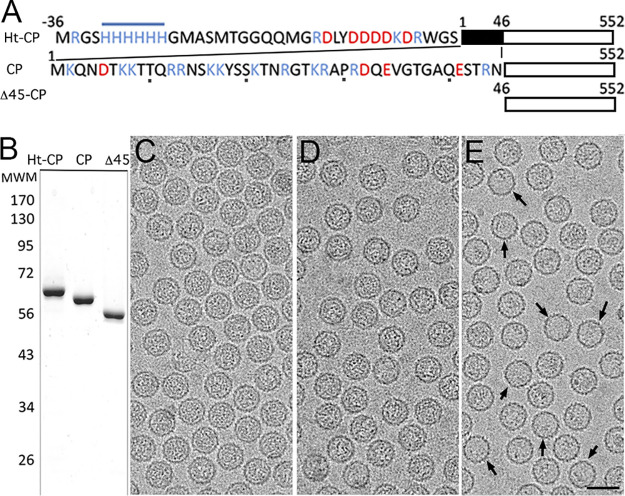
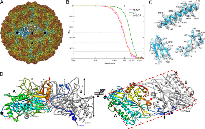
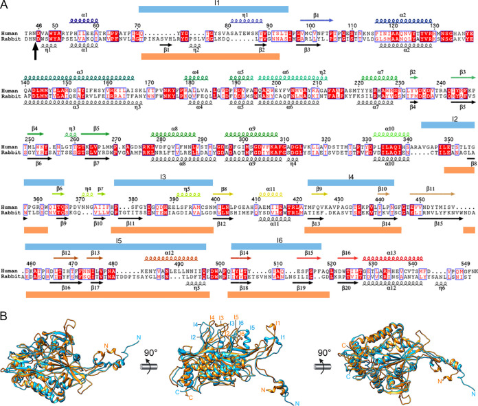
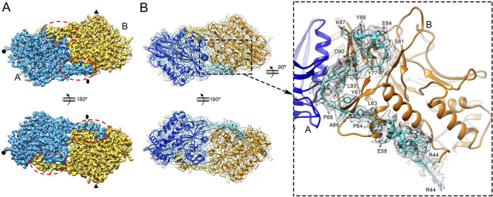
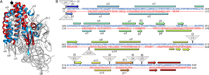
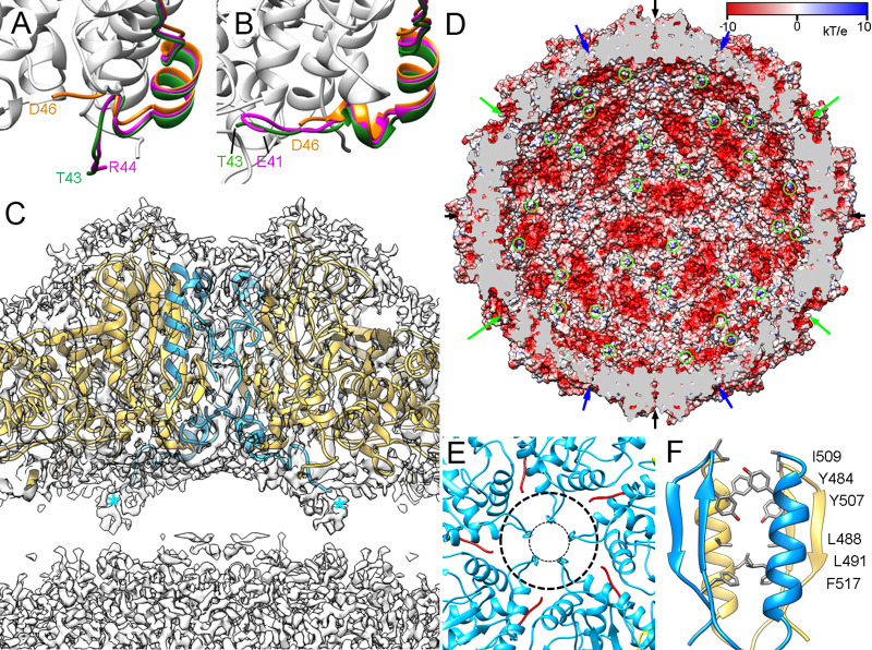
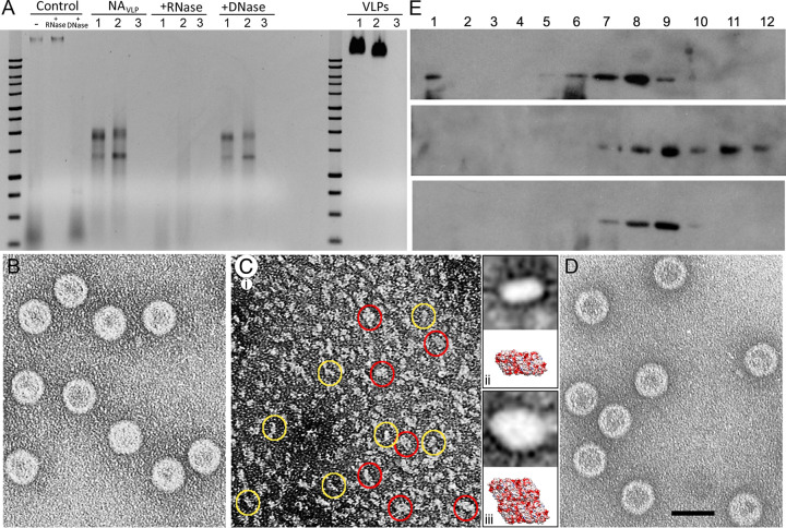
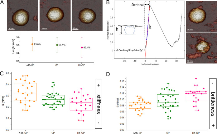
Despite their diversity, most double-stranded-RNA (dsRNA) viruses share a specialized T=1 capsid built from dimers of a single protein that provides a platform for genome transcription and replication. This ubiquitous capsid remains structurally undisturbed throughout the viral cycle, isolating the genome to avoid triggering host defense mechanisms. Human picobirnavirus (hPBV) is a dsRNA virus frequently associated with gastroenteritis, although its pathogenicity is yet undefined. Here, we report the cryo-electron microscopy (cryo-EM) structure of hPBV at 2.6-Å resolution. The capsid protein (CP) is arranged in a single-shelled, ∼380-Å-diameter T=1 capsid with a rough outer surface similar to that of dsRNA mycoviruses. The hPBV capsid is built of 60 quasisymmetric CP dimers (A and B) stabilized by domain swapping, and only the CP-A N-terminal basic region interacts with the packaged nucleic acids. hPBV CP has an α-helical domain with a fold similar to that of fungal partitivirus CP, with many domain insertions in its C-terminal half. In contrast to dsRNA mycoviruses, hPBV has an extracellular life cycle phase like complex reoviruses, which indicates that its own CP probably participates in cell entry. Using an in vitro reversible assembly/disassembly system of hPBV, we isolated tetramers as possible assembly intermediates. We used atomic force microscopy to characterize the biophysical properties of hPBV capsids with different cargos (host nucleic acids or proteins) and found that the CP N-terminal segment not only is involved in nucleic acid interaction/packaging but also modulates the mechanical behavior of the capsid in conjunction with the cargo.IMPORTANCE Despite intensive study, human virus sampling is still sparse, especially for viruses that cause mild or asymptomatic disease. Human picobirnavirus (hPBV) is a double-stranded-RNA virus, broadly dispersed in the human population, but its pathogenicity is uncertain. Here, we report the hPBV structure derived from cryo-electron microscopy (cryo-EM) and reconstruction methods using three capsid protein variants (of different lengths and N-terminal amino acid compositions) that assemble as virus-like particles with distinct properties. The hPBV near-atomic structure reveals a quasisymmetric dimer as the structural subunit and tetramers as possible assembly intermediates that coassemble with nucleic acids. Our structural studies and atomic force microscopy analyses indicate that hPBV capsids are potentially excellent nanocages for gene therapy and targeted drug delivery in humans.

Keywords: 3D cryo-EM; capsid protein; dsRNA virus; hPBV; virus assembly.


Figures

FIG 1
FIG 1
Biochemical and cryo-EM analysis of hPBV CP variants. (A) Scheme of hPBV Ht-CP, CP, and Δ45-CP used, indicating N-terminal sequences and sizes. Acidic residues, red; basic residues, blue. (B) Coomassie blue-stained SDS-PAGE gel of Ht-CP, CP, and Δ45-CP used for cryo-EM data acquisition. Molecular size markers (MWM; Da [103]) are on the left. (C to E) Cryo-electron micrographs of Ht-CP (C), CP (D), and Δ45-CP (E) capsids. Arrows (E) indicate empty capsids. Bar, 50 nm.
FIG 2
FIG 2
Three-dimensional cryo-EM reconstruction of hPBV capsids at 2.6-Å resolution. (A) Radially color-coded atomic model of hPBV capsid viewed along a 2-fold axis. Protruding dimers (orange) are visible in the T=1 lattice. The atomic structure of a CP dimer of subunits A (blue) and B (yellow) is shown. Symbols indicate icosahedral symmetry axes. (B) Fourier shell correlation (FSC) resolution curve for hPBV. Resolutions based on the 0.5, 0.3, and 0.143 criteria are indicated. For the 0.143 threshold, values for the CP capsid were at 2.63 Å and those for Δ45-CP and Ht-CP capsids were at 2.8 Å. (C) Regions of the cryo-EM density map (gray mesh), with atomic models of helix α3 (residues 140 to 166), a β sheet (residues 473 to 478 and 503 to 516), and a loop (residues 73 to 96). Atomic models are shown as ribbons and sticks, with amino acid residues labeled. (D) Ribbon diagram of the CP dimer (side view, left; top view, right). The A subunit is rainbow-colored from blue (N terminus [N-term]) to red (C terminus [C-term]); the B subunit is in gray. CP domains are indicated (S, shell; P, protruding). The thick dashed line highlights the parallelogram shape. Black symbols indicate icosahedral symmetry axes; the red oval and thin dashed line indicate the local 2-fold symmetry axis.
FIG 3
FIG 3
Structural homology of hPBV and rPBV CP. (A) Sequence and secondary structure elements of CP-A, rainbow-colored from blue (N terminus [N-term]) to red (C terminus [C-term]). The N-terminal 42-residue segment is not visible; the aligned sequence and secondary structure elements are shown for rPBV (black). A vertical black arrow indicates the scissile bond in rPBV CP; blue and orange boxes indicate insertions in human and rabbit CP, respectively (I1 to I6). (B) Superimposed rabbit (orange) and human (blue) CP monomers.
FIG 4
FIG 4
Domain-swapped organization of CP dimers in the hPBV capsid. (A) Cryo-EM density map of the CP dimer (outer surface, top; inner surface, bottom). The A and B subunits are blue and yellow, respectively. Dashed ovals indicate the N-terminal regions swapped between the dimers. (B) Atomic models of the A and B subunits and corresponding density (mesh) (view and color code as in panel A). Swapped regions of A and B subunits are highlighted in light blue and yellow, respectively. The closeup (side view) on the right shows the A-swapped Arg44-Ile95 region (light blue) with its cryo-EM density (gray mesh).
FIG 5
FIG 5
Structural homology of the hPBV and partitivirus capsid proteins. (A) Superimposed hPBV (blue) and PsV-F (red) CP (white regions indicate nonsuperimposed regions for both CP). (B) Sequence alignment of hPBV CP (blue) and PsV-F CP (red) resulting from the Dali structural alignment. The α helices (rectangles) and β strands (arrows) are rainbow-colored from blue (N terminus [N-term]) to red (C terminus [C-term]) for each CP. Triangles represent nonaligned segments or insertions (I1 to I6; sizes [in kilobases] are given in the triangles).
FIG 6
FIG 6
CP N-terminal ends and pore structure in the hPBV capsid. (A and B) Close-up of the visible N-terminal (A) and C-terminal (B) segments of CP (green), Ht-CP (pink), and Δ45-CP (orange). The last visible residue is indicated. (C) Magnified view of a 50-Å-thick slab around an icosahedral 2-fold axis. Atomic model regions of subunits A (blue) and B (yellow) are superimposed on the density map (contoured at 0.5 σ above the mean density). Asterisks indicate N-terminal ends of two A subunits. (D) The hPBV capsid inner surface is represented with electrostatic potentials, showing the distribution of negative (red) and positive (blue) charges. The internal cargo was removed computationally. Arrows indicate capsid pores at the 5-fold (green), 3-fold (blue), and 2-fold (black) axes. The N-terminal ends of A subunits are indicated (green circles). (E) Pores at the 5-fold axes (top view). His135 defines a 9-Å pore opening (inner dashed circle); a rearrangement in the Met131-Tyr138 loop enlarges this pore to 23 Å (outer dashed circle). (F) Numerous polar (top) and hydrophobic (bottom) interactions between local 2-fold-related A and B subunits.
FIG 7
FIG 7
Encapsidated nucleic acids in hPBV are RNase sensitive. Disassembly and reassembly of the T=1 hPBV capsid is reversible. (A) Structurally unaltered hPBV Ht-CP (lanes 1), CP (lanes 2), and Δ45-CP (lanes 3) VLP were analyzed by agarose gel electrophoresis and detected by ethidium bromide staining (VLPs). The same amount of total protein was analyzed for each sample. A 1- kb DNA ladder (10,000 to 250 bp; Promega) was used as a molecular weight marker. Purified nucleic acids from disrupted VLP (NAVLP) were treated with RNase (+RNase) or DNase (+DNase). Total nucleic acids of HEK293T cells (Control), which contain rRNA (bottom smear) and plasmid DNA (top band), were used as controls. (B to D) Disassembly/reassembly of the hPBV CP capsid. Electron microscopy and negative staining of purified hPBV CP (B), disassembled capsids (C), and reassembled capsids (D). Two major subassemblies (yellow and red circles) were detected in the disassembled material (C, panel i). Classification and alignment of these subassemblies resulted in average images of ∼63 by 112 (C, panel ii, top) and ∼105 by 140 Å (C, panel iii, top), with dimensions and morphology similar to those of a CP dimer (C, panel ii, bottom) or tetramer (C, panel iii, bottom). (E) hPBV CP capsids (top), disassembled material (center), and reassembled particles (bottom) were purified by ultracentrifugation on sucrose gradients, collected in 12 fractions, and analyzed by SDS-PAGE and Western blotting using anti-CP antibodies. The sedimentation direction was from right to left, with fraction 12 at the gradient top.
FIG 8
FIG 8
Biophysical properties of hPBV capsids determined by AFM indentations. (A) (Top) AFM topographies of hPBV Δ45-CP (left), CP (center), and Ht-CP (right) particles. (Bottom) Height of hPBV Δ45-CP (orange), CP (green), and Ht-CP (pink) particles after adsorption. Percentages indicate the ratio between average height as measured by AFM and nominal height based on cryo-EM maps. (B) (Left) A force-versus-indentation curve (FIC) of a CP hPBV capsid. Particle stiffness (k) and critical deformation (δcritical) can be calculated from the nanoindentation. (Inset) Critical strain (εcritical), which provides information about particle deformation before breakage, is defined as the ratio between the critical indentation and particle height. (Right) AFM topographies of the hPBV capsid before (top) and after (bottom) breakage. (C and D) Comparison of the average stiffness (C) and fragility (D) of hPBV Δ45-CP (orange), CP (green), and Ht-CP (pink) particles.

Similar articles

Cited by

References

    1. Patton JT. 2008. Segmented double-stranded RNA viruses. Structure and molecular biology. Caister Academic Press, Norfolk, United Kingdom.
    1. He Y, Shivakoti S, Ding K, Cui Y, Roy P, Zhou ZH. 2019. In situ structures of RNA-dependent RNA polymerase inside bluetongue virus before and after uncoating. Proc Natl Acad Sci U S A 116:16535–16540. doi:10.1073/pnas.1905849116. - DOI - PMC - PubMed
    1. Ding K, Celma CC, Zhang X, Chang T, Shen W, Atanasov I, Roy P, Zhou ZH. 2019. In situ structures of rotavirus polymerase in action and mechanism of mRNA transcription and release. Nat Commun 10:2216. doi:10.1038/s41467-019-10236-7. - DOI - PMC - PubMed
    1. Cui Y, Zhang Y, Zhou K, Sun J, Zhou ZH. 2019. Conservative transcription in three steps visualized in a double-stranded RNA virus. Nat Struct Mol Biol 26:1023–1034. doi:10.1038/s41594-019-0320-0. - DOI - PMC - PubMed
    1. Lahiri I, Xu J, Han BG, Oh J, Wang D, DiMaio F, Leschziner AE. 2019. 3.1A structure of yeast RNA polymerase II elongation complex stalled at a cyclobutane pyrimidine dimer lesion solved using streptavidin affinity grids. J Struct Biol 207:270–278. doi:10.1016/j.jsb.2019.06.004. - DOI - PMC - PubMed

Publication types

LinkOut - more resources