. 2020 Nov 13;295(46):15767-15781.
doi: 10.1074/jbc.RA120.015278. Epub 2020 Sep 11.

Glycerophosphodiesterase 3 (GDE3) is a lysophosphatidylinositol-specific ectophospholipase C acting as an endocannabinoid signaling switch

Affiliations

Glycerophosphodiesterase 3 (GDE3) is a lysophosphatidylinositol-specific ectophospholipase C acting as an endocannabinoid signaling switch

Fabienne Briand-Mésange et al. J Biol Chem. .

Abstract

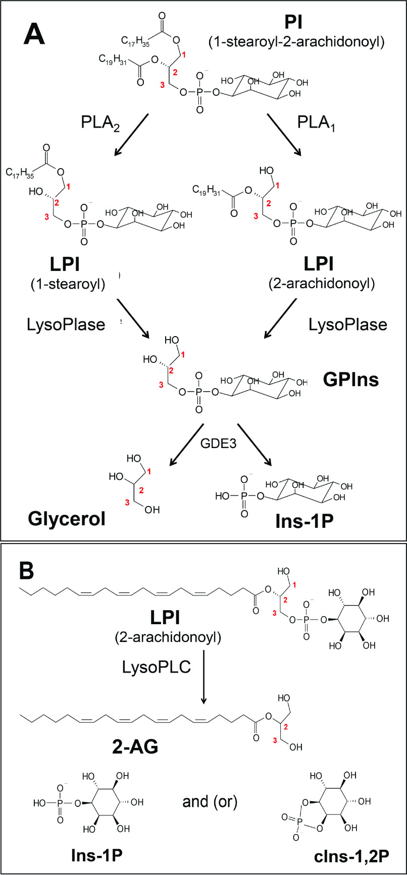
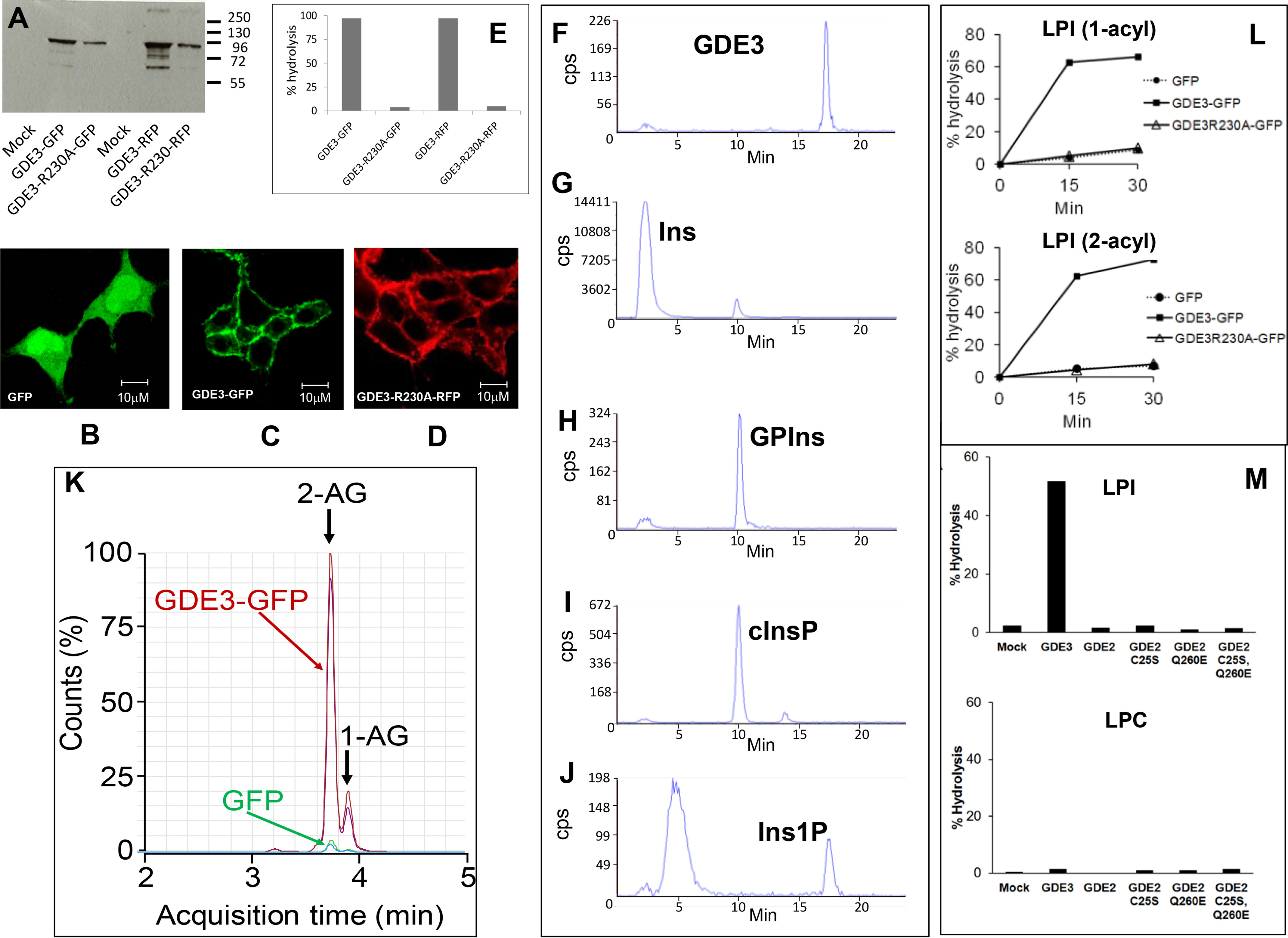
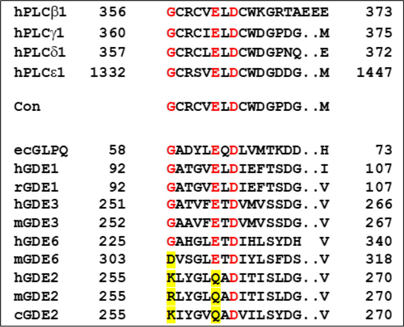
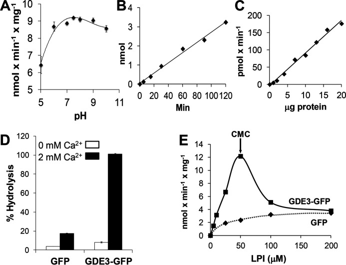
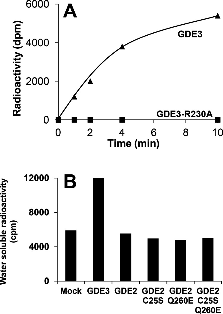
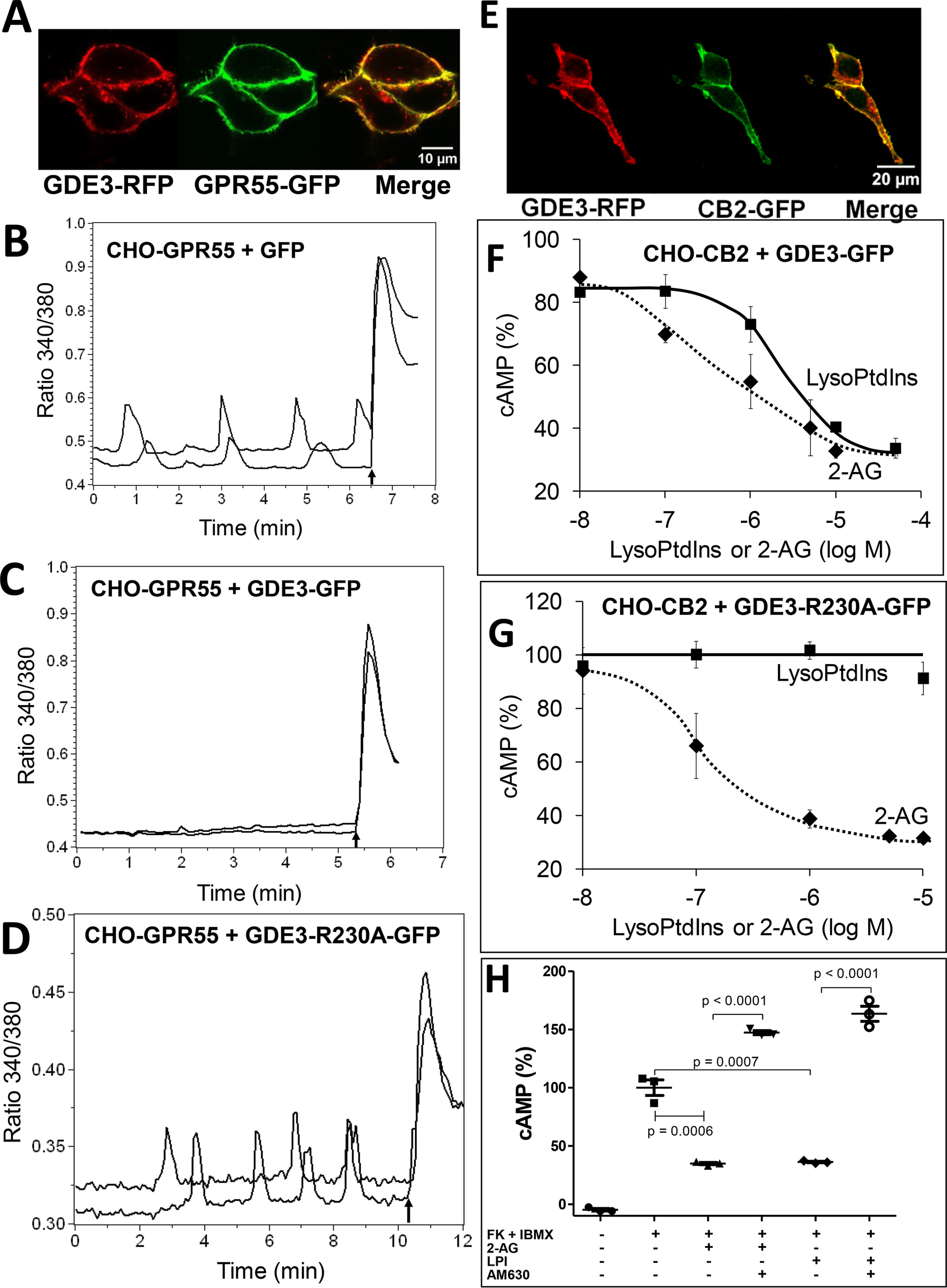
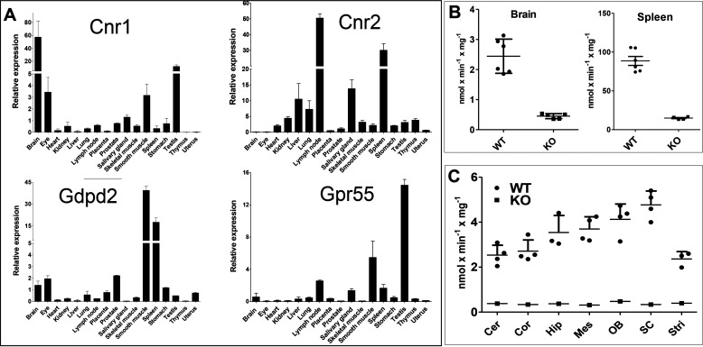
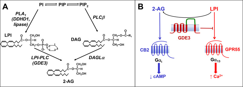
Endocannabinoid signaling plays a regulatory role in various (neuro)biological functions. 2-arachidonoylglycerol (2-AG) is the most abundant endocannabinoid, and although its canonical biosynthetic pathway involving phosphoinositide-specific phospholipase C and diacylglycerol lipase α is known, alternative pathways remain unsettled. Here, we characterize a noncanonical pathway implicating glycerophosphodiesterase 3 (GDE3, from GDPD2 gene). Human GDE3 expressed in HEK293T cell membranes catalyzed the conversion of lysophosphatidylinositol (LPI) into monoacylglycerol and inositol-1-phosphate. The enzyme was equally active against 1-acyl and 2-acyl LPI. When using 2-acyl LPI, where arachidonic acid is the predominant fatty acid, LC-MS analysis identified 2-AG as the main product of LPI hydrolysis by GDE3. Furthermore, inositol-1-phosphate release into the medium occurred upon addition of LPI to intact cells, suggesting that GDE3 is actually an ecto-lysophospholipase C. In cells expressing G-protein-coupled receptor GPR55, GDE3 abolished 1-acyl LPI-induced signaling. In contrast, upon simultaneous ex-pression of GDE3 and cannabinoid receptor CB2, 2-acyl LPI evoked the same signal as that induced by 2-AG. These data strongly suggest that, in addition to degrading the GPR55 LPI ligand, GDE3 can act as a switch between GPR55 and CB2 signaling. Coincident with a major expression of both GDE3 and CB2 in the spleen, spleens from transgenic mice lacking GDE3 displayed doubling of LPI content compared with WT mice. Decreased production of 2-AG in whole spleen was also observed, supporting the in vivo relevance of our findings. These data thus open a new research avenue in the field of endocannabinoid generation and reinforce the view of GPR55 and LPI being genuine actors of the endocannabinoid system.

Keywords: 2-arachidonoylglycerol; CB1; CB1/CB2; CB2; G-protein–coupled receptor (GPCR); GDPD2); GPR55; endocannabinoid; glycerophosphodiesterase 3 (GDE3; lysophosphatidylinositol; lysophospholipid; monoacylglycerol; phospholipase C.


Conflict of interest statement

Conflict of interest—The authors declare that they have no conflicts of interest with the contents of this article.

Figures

Figure 1.
Figure 1.
Structure of PI, LPI, GPIns, and their cleavage products. A, the predominant molecular species of PI is 1-stearoyl-2-arachidonoyl-PI. The corresponding LPI (1-stearoyl and 2-arachidonoyl) are formed upon PI hydrolysis by phospholipase A2 (PLA2) and PLA1, respectively. Further deacylation of each LPI by lysophospholipases (LysoPlases) leads to water-soluble sn-glycero-3-phosphoinositol (GPIns), which is cleaved into glycerol and inositol-1-phosphate (Ins-1P) by GDE3. B, according to our working hypothesis, hydrolysis of 2-arachidonoyl LPI by lysoPLC generates 2-AG and Ins-1P or cyclic inositol-1,2-phosphate (cIns-1,2P). The cyclization occurring during hydrolysis is not a constant event and depends on the mechanism of each enzyme.
Figure 2.
Figure 2.
Identification of GDE3 as a lysoPLC specific for LPI. A, HEK293T cells were transfected with GFP or RFP (Mock), GDE3-GFP, GDE3-R230A-GFP, GDE3-RFP, or GDE3-R230A-RFP. Membranes were isolated and used for Western blotting analysis using anti-human GDPD2 mouse polyclonal antibody (Abnova). BD, the cells were observed by confocal microscopy as described under “Experimental procedures”. E, membranes (10 µg of protein) were incubated with 50 µm of [3H]Ins-LPI (1-acyl) during 30 min in 0.1 ml of 100 mm Tris-HCl, pH 7.4, containing 2 mm CaCl2 for determination of lysoPLC activity. Data are individual values representative of five different experiments for GFP-tagged enzyme and one single experiment for RFP-tagged protein. FJ, membranes (10 µg of protein) prepared from HEK293T cells transfected with GDE3-GFP were incubated for 30 min with 50 µm [3H]Ins-LPI (1-acyl) in 0.1 ml of 100 mm Tris-HCl, pH 7.4, containing 2 mm CaCl2. After lipid extraction in the presence of 0.1 m HCl, the water-soluble extracts and standard solutions were loaded onto the HPLC column and analyzed as described. Standards loaded onto the HPLC column were pure [3H]Ins (G), GP[3H]Ins (H), cyclic [3H]Ins-1,2-phosphate (cInsP, I), or [3H]Ins-1-phosphate (Ins1P, J). The product generated by acid hydrolysis of cInsP and eluting as an early peak was not identified. Data are from one experiment representative of three experiments with very similar results. K, nonradioactive LPI (2-acyl, 50 µm) was incubated for 30 min with membranes (10 µg of protein) from HEK293T cells transfected either with GDE3-GFP or with GFP in 0.1 ml of 100 mm Tris-HCl, pH 7.4, containing 2 mm CaCl2. Lipids were extracted and analyzed by LC-MS (see “Experimental procedures”). Major and minor peaks at 3.7 and 3.8 min correspond to 2-AG and 1-AG, respectively, indicating that migration of arachidonic acid from sn-2 to sn-1 position was about 17%. The profiles of two independent experiments are shown. L, membranes (10 µg of protein) were isolated from HEK293T cells expressing GDE3-GFP, GDE3-R230A-GFP, or GFP alone. Substrate was [3H]Ins-LPI (1-acyl, 50 µm) or [3H]Ins-LPI (2-acyl, 50 µm), and lysoPLC activity was determined as described after incubation for indicated times in 0.1 ml of 100 mm Tris-HCl, pH 7.4, containing 2 mm CaCl2. Data (single determinations) are representative of two experiments with identical results. M, HEK293T cells were transfected with various vectors encoding GFP (Mock), GDE3-GFP (GDE3), GDE2-GFP (GDE2), or the various GDE2 mutants prepared as described under “Experimental procedures”. LysoPLC activity was determined using membrane fractions isolated from transfected cells (30 µg of protein) and incubated for 30 min with 50 µm [3H]Ins-LPI or 1 µm [3H]choline-LPC (1-acyl) in 0.1 ml of 100 mm Tris-HCl, pH 7.4, containing 2 mm CaCl2. Data (single values) are representative of two experiments with very similar results.
Figure 3.
Figure 3.
Conserved sequences within catalytic domains of phospholipases C (PLC) and various glycerophosphodiesterases. Alignment and definition of the consensus sequence found in PLC (Con) is identical to that provided in Fig. S6 by Rao and Sockanathan. ecGLPQ is the periplasmic glycerophosphodiesterase from Escherichia coli; h, r, m, and c refer to human, rat, mouse and chicken, respectively. The most highly conserved residues are colored in red, exceptions being highlighted in yellow.
Figure 4.
Figure 4.
Enzymatic properties of lysoPLC activity of GDE3. Membranes were isolated from HEK293T cells transfected with GDE3-GFP or GFP and incubated in 0.1-ml final volume in the presence of [3H]Ins-LPI (1-acyl). LysoPLC activity was determined as described under “Experimental procedures”. A, proteins (10 µg) were incubated for 30 min with 50 µm LPI in 0.1 m Tris-HCl buffer at the indicated pH containing 2 mm CaCl2. Data are means ± S.E. of three determinations. B, proteins (10 µg) were incubated for indicated times with 50 µm LPI in 100 mm Tris-HCl buffer, pH 7.4, containing 2 mm CaCl2. Data are individual values from one experiment representative of three experiments with very similar results. C, increasing amounts of protein incubated for 30 min with 50 µm LPI in 100 mm Tris-HCl buffer, pH 7.4, containing 2 mm CaCl2. Data are individual values from one typical experiment. D, proteins (10 µg) were incubated for 60 min with 50 µm LPI in 100 mm Tris-HCl buffer, pH 7.4, containing or absent of 2 mm CaCl2. Data are expressed as % hydrolysis and are means ± S.E. of three determinations. E, proteins (10 µg) were incubated with increasing concentrations of LPI in 0.1 ml of 100 mm Tris-HCl, pH 7.4, containing 2 mm CaCl2. Data are from one experiment representative of three experiments with very similar results. CMC, critical micelle concentration.
Figure 5.
Figure 5.
Determination of ecto-lysoPLC activity of GDE3, GDE2, and various GDE2 mutants. A, monolayers of HEK293T cells expressing GDE3 were seeded in 6-well dishes (9.5 cm2) and incubated with 11.25 µm [3H]Ins-LPI (1-acyl) added to the medium (2-ml final volume). At various times, 0.5 ml of supernatant was drawn from each well and lipids were extracted in the presence of 0.1 m HCl. The radioactivity (dpm) in upper water-methanol phase was then determined. Data are individual values from one experiment representative of three experiments with very similar results. B, HEK293T cells were transfected with GFP (Mock), GDE3-GFP (GDE3), GDE2-GFP (GDE2), or various mutants of GDE2 in fusion with GFP. Water-soluble radioactivity (cpm) was determined as in (A) on 0.5-ml aliquots submitted to the precipitation procedure using BSA and HClO4 (see “Experimental procedures”). Data are single values from one typical experiment.
Figure 6.
Figure 6.
GDE3 acts as a switch between GPR55 and CB2 signaling. A, HEK293T cells were transfected with GPR55-GFP and GDE3-RFP and examined by confocal microscopy as described under “Experimental procedures”. B, CHO-GPR55 cells transfected with GFP as a control were loaded with Fura-2-AM, stimulated by 5 µm LPI (1-acyl), and examined for fluorescence ratio 340 nm/380 nm, reflecting cytoplasmic free calcium concentration ([Ca2+]i). Arrow indicates addition of 1 µm ionomycin. C, same as in (B) with CHO-GPR55 cells transfected with GDE3-GFP. D, same as in (B) with CHO-GPR55 cells transfected with GDE3-R230A-GFP. (B), (C), and (D) show two single cell traces. E, HEK293T cells were transfected with CB2-GFP and GDE3-RFP and examined by confocal microscopy as in (A). F, CHO-CB2 cells transfected with GDE3-GFP were incubated for 20 min with 7 µm forskolin plus 200 µm IBMX and various concentrations of 2-AG or LPI (2-acyl). Cellular cAMP content was determined as described under “Experimental procedures”. Data (means ± S.E., three determinations) are expressed as % of cAMP content determined in the presence of forskolin plus IBMX only. G, Same as in (F) with CHO-CB2 cells transfected with GDE3-R230-GFP. H, Same as in (F), except that CHO-CB2 cells were preincubated or not for 15 min with 10 µm AM630 before addition of forskolin plus IBMX, followed by 5 µm LPI (2-acyl) or 5 µm 2-AG. Data are means ± S.D. of three determinations. p, probability of significance according to unpaired t independent test.
Figure 7.
Figure 7.
Expression of GDE3, CB1, CB2, and Gpr55 in various mouse tissues and lysoPLC activity in the brain and spleen. A, contents of mRNA encoding CB1 (Cnr1), CB2 (Cnr2), GDE3 (Gdpd2), and Gpr55 were determined in mouse tissues by quantitative real time PCR (see “Experimental procedures”). Data are expressed as normalized ratios relative to β-tubulin and are means ± S.E. of three determinations. B, brain and spleen membranes were prepared from WT or GDE3 KO mice as described under “Experimental procedures”. LysoPLC activity was determined in 30-min incubations using 10 µg (spleen) or 20 µg (brain) of protein and 50 µm [3H]Ins-LPI (1-acyl) as substrate in 0.1 m Tris-HCl buffer, pH 7.4, containing 2 mm CaCl2. Data are means ± S.D. (n = 5 for brain, n = 3 for spleen). C, after sacrifice, central nervous tissues were dissected to isolate cerebellum (Cer), cortex (Cor), hippocampus (Hip), mesencephalon (Mes), olfactory bulb (OB), spinal cord (SC), and striatum (Stri). LysoPLC activity was determined in crude membranes as in (B). Data (means ± S.D. (error bars)) are from four WT mice, except for hippocampus and striatum (n = 3), and from one KO mouse as a control.
Figure 8.
Figure 8.
Quantification of molecular species of LPI and monoacylglycerol in brain and spleen from WT and GDE3 KO mice. Each organ was frozen immediately after harvesting. Frozen tissues were weighed and homogenized in 2 ml of water, followed by acidification with 0.24 ml of 2 m HCl and lipid extraction in 8 ml of dichloromethane (CH2Cl2) and 4 ml of methanol. Lipids from the organic phase were submitted to solid-phase extraction using silica and eluted with a hexane-isopropanol mixture for 2-AG and methanol for LPI. Lipids were then quantified by LC-MS as described under “Experimental procedures”. Data are expressed as nmol/g wet weight tissue and are means ± S.D. (error bars) of 17 WT and 18 KO mice. 16:0, palmitoyl-; 18:0, stearoyl-; 18:1, oleoyl-; 20:4, arachidonoyl-; p, probability of significance according to unpaired t independent test (see “Experimental procedures”).
Figure 9.
Figure 9.
Role of GDE3 in 2-AG synthesis and as a switch between GPR55 and the cannabinoid receptor. A, the three major phosphoinositides, phosphatidylinositol (PI), PI 4-phosphate (PIP), and PI-bisphosphate are in permanent interconversion through four steps of phosphorylation-dephosphorylation. The canonical pathway of 2-AG synthesis involves PIP2 cleavage by PLCβ, followed by removal of stearic acid from the sn-1 position of diacylglycerol (DAG) by DAG lipase α (DAGLα). The alternative pathway requires PI hydrolysis by a phospholipase A1 (PLA1), either DDHD1 for an intracellular reaction or an extracellular lipase. GDE3 then produces 2-AG from the 2-arachidonoyl LPI species. B, as shown by data from Fig. 6, GDE3 is able to convert GPR55 signaling evoked by LPI into CB2 activation by 2-AG. The three proteins are not represented in the same membrane because the depicted exchanges might involve membranes from the same or from neighboring cells. Although not shown in this study, CB1 could also be emphasized in the switching event.

Similar articles

Cited by

References

    1. Mechoulam R., Hanuš L. O., Pertwee R., and Howlett A. C. (2014) Early phytocannabinoid chemistry to endocannabinoids and beyond. Nat. Rev. Neurosci. 15, 757–764 10.1038/nrn3811 - DOI - PubMed
    1. Maccarrone M., Bab I., Bíró T., Cabral G. A., Dey S. K., Di Marzo V., Konje J. C., Kunos G., Mechoulam R., Pacher P., Sharkey K. A., and Zimmer A. (2015) Endocannabinoid signaling at the periphery: 50 years after THC. Trends Pharmacol. Sci. 36, 277–296 10.1016/j.tips.2015.02.008 - DOI - PMC - PubMed
    1. Piomelli D. (2014) More surprises lying ahead. The endocannabinoids keep us guessing. Neuropharmacology 76, 228–234 10.1016/j.neuropharm.2013.07.026 - DOI - PMC - PubMed
    1. Hussain Z., Uyama T., Tsuboi K., and Ueda N. (2017) Mammalian enzymes responsible for the biosynthesis of N-acylethanolamines. Biochim. Biophys. Acta 1862, 1546–1561 10.1016/j.bbalip.2017.08.006 - DOI - PubMed
    1. Tsuboi K., Uyama T., Okamoto Y., and Ueda N. (2018) Endocannabinoids related N-acylethanolamines: biological activities and metabolism. Inflamm. Regen. 38, 28 10.1186/s41232-018-0086-5 - DOI - PMC - PubMed

Publication types

MeSH terms