

A New Polycaprolactone-Based Biomembrane Functionalized with BMP-2 and Stem Cells Improves Maxillary Bone Regeneration
- PMID: 32911737
- PMCID: PMC7558050
- DOI: 10.3390/nano10091774
A New Polycaprolactone-Based Biomembrane Functionalized with BMP-2 and Stem Cells Improves Maxillary Bone Regeneration
Abstract
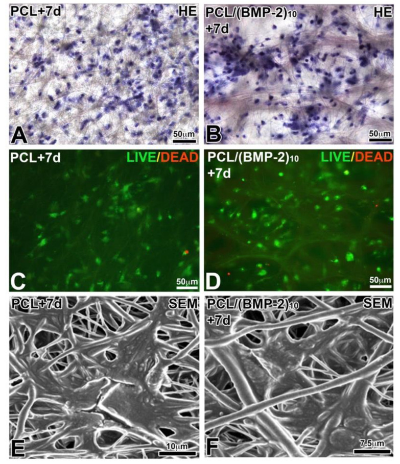
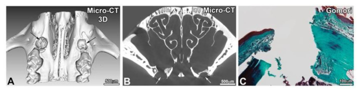
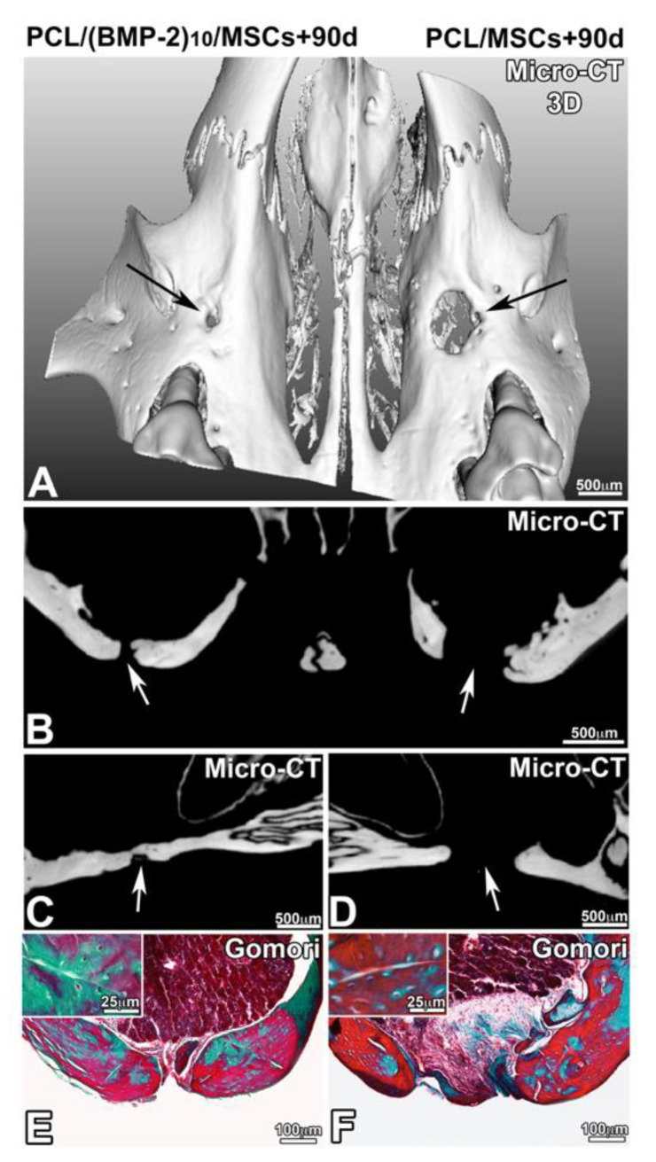
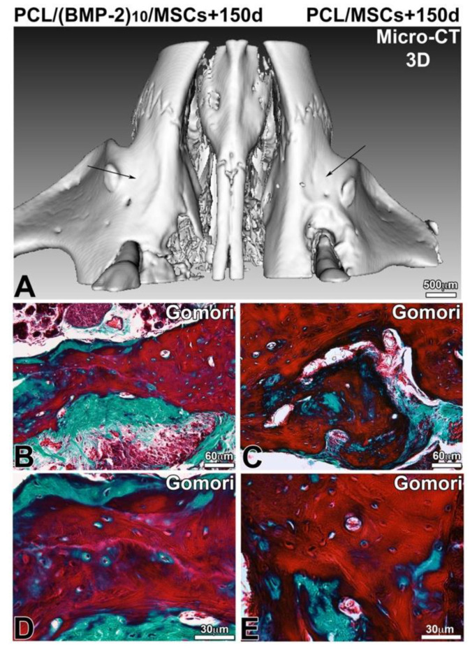
Oral diseases have an impact on the general condition and quality of life of patients. After a dento-alveolar trauma, a tooth extraction, or, in the case of some genetic skeletal diseases, a maxillary bone defect, can be observed, leading to the impossibility of placing a dental implant for the restoration of masticatory function. Recently, bone neoformation was demonstrated after in vivo implantation of polycaprolactone (PCL) biomembranes functionalized with bone morphogenic protein 2 (BMP-2) and ibuprofen in a mouse maxillary bone lesion. In the present study, human bone marrow derived mesenchymal stem cells (hBM-MSCs) were added on BMP-2 functionalized PCL biomembranes and implanted in a maxillary bone lesion. Viability of hBM-MSCs on the biomembranes has been observed using the "LIVE/DEAD" viability test and scanning electron microscopy (SEM). Maxillary bone regeneration was observed for periods ranging from 90 to 150 days after implantation. Various imaging methods (histology, micro-CT) have demonstrated bone remodeling and filling of the lesion by neoformed bone tissue. The presence of mesenchymal stem cells and BMP-2 allows the acceleration of the bone remodeling process. These results are encouraging for the effectiveness and the clinical use of this new technology combining growth factors and mesenchymal stem cells derived from bone marrow in a bioresorbable membrane.
Keywords: biomembrane; bone regeneration; nanoreservoirs; smart implant; stem cells.
Conflict of interest statement
The authors declare no conflict of interest.
Figures

Similar articles
-
Maxillary Bone Regeneration Based on Nanoreservoirs Functionalized ε-Polycaprolactone Biomembranes in a Mouse Model of Jaw Bone Lesion.Biomed Res Int. 2018 Feb 26;2018:7380389. doi: 10.1155/2018/7380389. eCollection 2018. Biomed Res Int. 2018. PMID: 29682553 Free PMC article.
-
Periodontal Tissues, Maxillary Jaw Bone, and Tooth Regeneration Approaches: From Animal Models Analyses to Clinical Applications.Nanomaterials (Basel). 2018 May 16;8(5):337. doi: 10.3390/nano8050337. Nanomaterials (Basel). 2018. PMID: 29772691 Free PMC article. Review.
-
Active implant combining human stem cell microtissues and growth factors for bone-regenerative nanomedicine.Nanomedicine (Lond). 2015;10(5):753-63. doi: 10.2217/nnm.14.228. Nanomedicine (Lond). 2015. PMID: 25816878
-
Incorporation of BMP-2 nanoparticles on the surface of a 3D-printed hydroxyapatite scaffold using an ε-polycaprolactone polymer emulsion coating method for bone tissue engineering.Colloids Surf B Biointerfaces. 2018 Oct 1;170:421-429. doi: 10.1016/j.colsurfb.2018.06.043. Epub 2018 Jun 20. Colloids Surf B Biointerfaces. 2018. PMID: 29957531
-
Bone defects and future regenerative nanomedicine approach using stem cells in the mutant Tabby mouse model.Biomed Mater Eng. 2015;25(1 Suppl):111-9. doi: 10.3233/BME-141246. Biomed Mater Eng. 2015. PMID: 25538062 Review.
Cited by
-
Modulation of immune-inflammatory responses through surface modifications of biomaterials to promote bone healing and regeneration.J Tissue Eng. 2021 Oct 26;12:20417314211041428. doi: 10.1177/20417314211041428. eCollection 2021 Jan-Dec. J Tissue Eng. 2021. PMID: 34721831 Free PMC article. Review.
-
Mesenchymal Stem Cells in Soft Tissue Regenerative Medicine: A Comprehensive Review.Medicina (Kaunas). 2023 Aug 10;59(8):1449. doi: 10.3390/medicina59081449. Medicina (Kaunas). 2023. PMID: 37629738 Free PMC article. Review.
-
Eruption of Bioengineered Teeth: A New Approach Based on a Polycaprolactone Biomembrane.Nanomaterials (Basel). 2021 May 17;11(5):1315. doi: 10.3390/nano11051315. Nanomaterials (Basel). 2021. PMID: 34067681 Free PMC article.
-
Nanomaterials for Periodontal Tissue Regeneration: Progress, Challenges and Future Perspectives.J Funct Biomater. 2023 May 24;14(6):290. doi: 10.3390/jfb14060290. J Funct Biomater. 2023. PMID: 37367254 Free PMC article. Review.
-
Oral Bone Tissue Regeneration: Mesenchymal Stem Cells, Secretome, and Biomaterials.Int J Mol Sci. 2021 May 15;22(10):5236. doi: 10.3390/ijms22105236. Int J Mol Sci. 2021. PMID: 34063438 Free PMC article. Review.
References
LinkOut - more resources
Full Text Sources
