

Anatomical Characterization of the Human Structural Connectivity between the Pedunculopontine Nucleus and Globus Pallidus via Multi-Shell Multi-Tissue Tractography
- PMID: 32906651
- PMCID: PMC7557768
- DOI: 10.3390/medicina56090452
Anatomical Characterization of the Human Structural Connectivity between the Pedunculopontine Nucleus and Globus Pallidus via Multi-Shell Multi-Tissue Tractography
Abstract
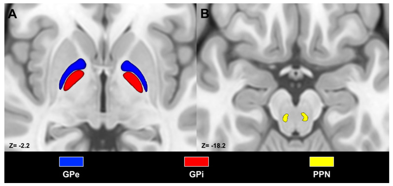
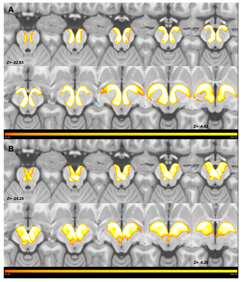
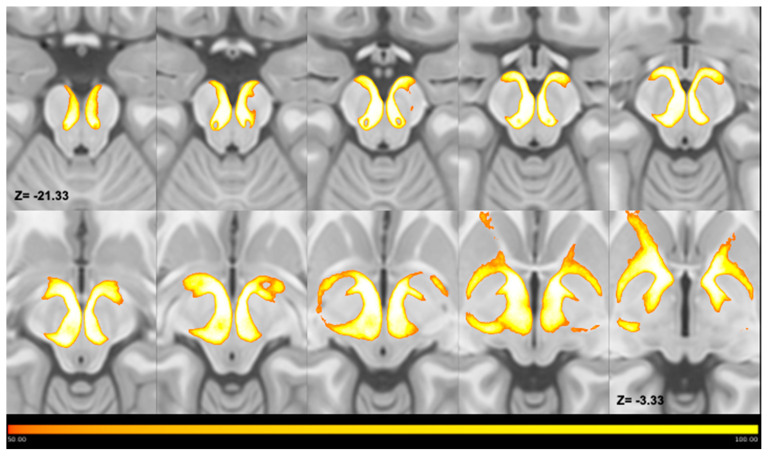
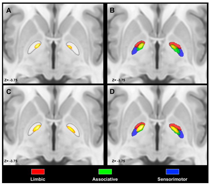
Background and objectives: The internal (GPi) and external segments (GPe) of the globus pallidus represent key nodes in the basal ganglia system. Connections to and from pallidal segments are topographically organized, delineating limbic, associative and sensorimotor territories. The topography of pallidal afferent and efferent connections with brainstem structures has been poorly investigated. In this study we sought to characterize in-vivo connections between the globus pallidus and the pedunculopontine nucleus (PPN) via diffusion tractography. Materials and Methods: We employed structural and diffusion data of 100 subjects from the Human Connectome Project repository in order to reconstruct the connections between the PPN and the globus pallidus, employing higher order tractography techniques. We assessed streamline count of the reconstructed bundles and investigated spatial relations between pallidal voxels connected to the PPN and pallidal limbic, associative and sensorimotor functional territories. Results: We successfully reconstructed pallidotegmental tracts for the GPi and GPe in all subjects. The number of streamlines connecting the PPN with the GPi was greater than the number of those joining it with the GPe. PPN maps within pallidal segments exhibited a distinctive spatial organization, being localized in the ventromedial portion of the GPi and in the ventral-anterior portion in the GPe. Regarding their spatial relations with tractography-derived maps of pallidal functional territories, the highest value of percentage overlap was noticed between PPN maps and the associative territory. Conclusions: We successfully reconstructed the anatomical course of the pallidotegmental pathways and comprehensively characterized their topographical arrangement within both pallidal segments. PPM maps were localized in the ventromedial aspect of the GPi, while they occupied the anterior pole and the most ventral portion of the GPe. A better understanding of the spatial and topographical arrangement of the pallidotegmental pathways may have pathophysiological and therapeutic implications in movement disorders.
Keywords: basal ganglia; brainstem; globus pallidus; pallidotegmental tract; pedunculopontine nucleus; structural connectivity; tractography.
Conflict of interest statement
The authors declare no conflict of interest.
Figures

Similar articles
-
Spatially coherent and topographically organized pathways of the human globus pallidus.Hum Brain Mapp. 2020 Nov;41(16):4641-4661. doi: 10.1002/hbm.25147. Epub 2020 Aug 5. Hum Brain Mapp. 2020. PMID: 32757349 Free PMC article.
-
Structural connectivity-based topography of the human globus pallidus: Implications for therapeutic targeting in movement disorders.Mov Disord. 2019 Jul;34(7):987-996. doi: 10.1002/mds.27712. Epub 2019 May 11. Mov Disord. 2019. PMID: 31077436
-
Efferent connections of the internal globus pallidus in the squirrel monkey: II. Topography and synaptic organization of pallidal efferents to the pedunculopontine nucleus.J Comp Neurol. 1997 Jun 9;382(3):348-63. J Comp Neurol. 1997. PMID: 9183698
-
Perspective on basal ganglia connections as described by Nauta and Mehler in 1966: Where we were and how this paper effected where we are now.Brain Res. 2016 Aug 15;1645:4-7. doi: 10.1016/j.brainres.2016.04.016. Epub 2016 Apr 7. Brain Res. 2016. PMID: 27064077 Review.
-
The connections of the primate subthalamic nucleus: indirect pathways and the open-interconnected scheme of basal ganglia-thalamocortical circuitry.Brain Res Brain Res Rev. 1997 Feb;23(1-2):62-78. doi: 10.1016/s0165-0173(96)00018-5. Brain Res Brain Res Rev. 1997. PMID: 9063587 Review.
Cited by
-
Red nucleus structure and function: from anatomy to clinical neurosciences.Brain Struct Funct. 2021 Jan;226(1):69-91. doi: 10.1007/s00429-020-02171-x. Epub 2020 Nov 12. Brain Struct Funct. 2021. PMID: 33180142 Free PMC article. Review.
-
Striatal topographical organization: Bridging the gap between molecules, connectivity and behavior.Eur J Histochem. 2021 Oct 13;65(s1):3284. doi: 10.4081/ejh.2021.3284. Eur J Histochem. 2021. PMID: 34643358 Free PMC article. Review.
-
Cholinergic Receptor Modulation as a Target for Preventing Dementia in Parkinson's Disease.Front Neurosci. 2021 Sep 20;15:665820. doi: 10.3389/fnins.2021.665820. eCollection 2021. Front Neurosci. 2021. PMID: 34616271 Free PMC article. Review.
-
In vivo probabilistic atlas of white matter tracts of the human subthalamic area combining track density imaging and optimized diffusion tractography.Brain Struct Funct. 2022 Nov;227(8):2647-2665. doi: 10.1007/s00429-022-02561-3. Epub 2022 Sep 17. Brain Struct Funct. 2022. PMID: 36114861 Free PMC article.
-
Structural Connectivity-Based Parcellation of the Dopaminergic Midbrain in Healthy Subjects and Schizophrenic Patients.Medicina (Kaunas). 2020 Dec 10;56(12):686. doi: 10.3390/medicina56120686. Medicina (Kaunas). 2020. PMID: 33322072 Free PMC article.
References
-
- Nieuwenhuys R. The Human Central Nervous System. Volume 53. Springer Science & Business Media; New York, NY, USA: 2008.
MeSH terms
LinkOut - more resources
Full Text Sources
