. 2020 Sep 4;21(18):6456.
doi: 10.3390/ijms21186456.

Neuroprotection by Neurotropin through Crosstalk of Neurotrophic and Innate Immune Receptors in PC12 Cells

Affiliations

Neuroprotection by Neurotropin through Crosstalk of Neurotrophic and Innate Immune Receptors in PC12 Cells

Yu Fukuda et al. Int J Mol Sci. .

Abstract

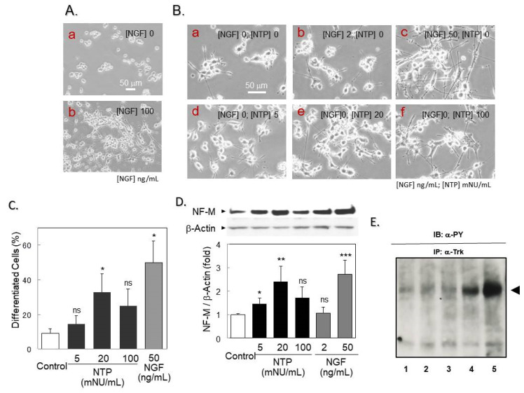
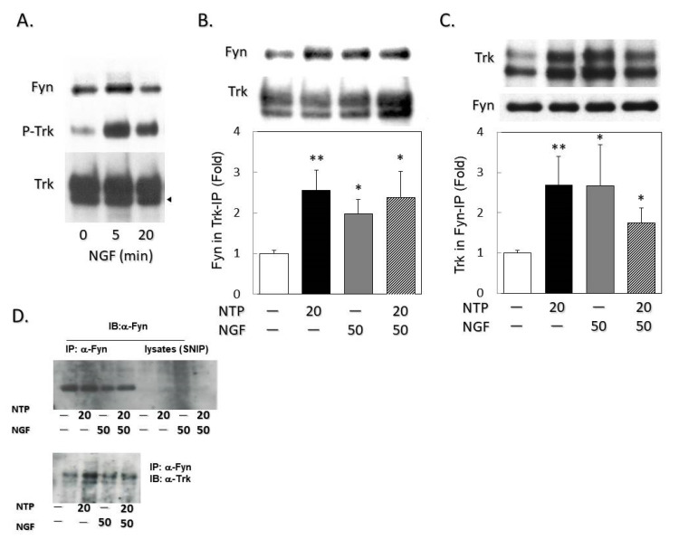
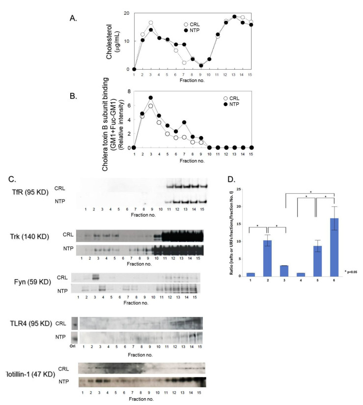
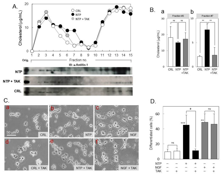
Infected or damaged tissues release multiple "alert" molecules such as alarmins and damage-associated molecular patterns (DAMPs) that are recognized by innate immune receptors, and induce tissue inflammation, regeneration, and repair. Recently, an extract from inflamed rabbit skin inoculated with vaccinia virus (Neurotropin®, NTP) was found to induce infarct tolerance in mice receiving permanent ischemic attack to the middle cerebral artery. Likewise, we report herein that NTP prevented the neurite retraction in PC12 cells by nerve growth factor (NGF) deprivation. This effect was accompanied by interaction of Fyn with high-affinity NGF receptor TrkA. Sucrose density gradient subcellular fractionation of NTP-treated cells showed heretofore unidentified membrane fractions with a high-buoyant density containing Trk, B subunit of cholera toxin-bound ganglioside, flotillin-1 and Fyn. Additionally, these new membrane fractions also contained Toll-like receptor 4 (TLR4). Inhibition of TLR4 function by TAK-242 prevented the formation of these unidentified membrane fractions and suppressed neuroprotection by NTP. These observations indicate that NTP controls TrkA-mediated signaling through the formation of clusters of new membrane microdomains, thus providing a platform for crosstalk between neurotrophic and innate immune receptors. Neuroprotective mechanisms through the interaction with innate immune systems may provide novel mechanism for neuroprotection.

Keywords: Fyn; GM1 ganglioside (GM1); Neurotropin; Toll-like receptor4 (TLR4); TrkA tyrosine kinase; lipid rafts.


Conflict of interest statement

The affiliations of each author are noted in the citation appended to the list of authors. The authors disclose the following industry relationships: Y.F. is a full-time employee of Nippon Zoki Pharmaceutical Co., Ltd. All the experimental works included in this article was performed by Y.F., K.N., T.M. All authors reported no COI to the present study.

Figures

Figure 1
Figure 1
Neurotropin (NTP) protected differentiated PC12 cells overexpressing Trk (PCtrk) cells from Nerve growth factor (NGF) deprivation. (A) NGF-mediated morphological differentiation. PCtrk cells (3000 cells in ϕ3-cm dish) were cultured for 24 h in the absence (a) or presence (b) of NGF (100 ng/mL). Phase-contrast micrographs were captured for typical areas of cultures. (Bar = 50 μm in length). (B) Effect of NTP on neurite retraction induced by NGF deprivation. Differentiated PCtrk cells were gently washed with PBS and incubated for additional 30 h in serum-free DMEM in the presence (b, 2 ng/mL; c, 50 ng/mL) or absence (a, d-f) of NGF. NTP was added to the medium at 5 mNU (d), 20 mNU (e) or 100 mNU (f) per mL. The degree of differentiation of cells in cultures supplemented with saline (Control), NTP (5, 20, or 100 mNU/mL), or NGF (50 ng/mL) was quantified as described in Materials and Methods, and summarized in (C). Data represent means and standard deviations (SD) of three independent cultures. * p < 0.05 vs. saline-treated controls. ns, not significant. (D) Neurofilament M (NF-M) expression. Cell lysates of PCtrk cells (1 × 10⁵ cells, 32 h) were subjected to Western blotting against NF-M (160 KDa) and β-actin (internal control, 45 KDa) as described in Materials and Methods (upper panel, typical blotting images). Data represent mean and SD of the intensity ratios of NF-M to β-actin from three independent cultures. * p < 0.05, ** p < 0.01 and *** p < 0.001 vs. saline-treated controls (open bar); ns, not significant (two-sided t-test). (E) Autophosphorylation status of TrkA during de-differentiation by the withdrawal of NGF. The cells were pretreated with NGF for 24 h and then further treated for 30 h either with NGF or NTP or untreated in the same way as (B). The TrkA protein was immunoprecipitated with anti-TrkA antibody and subjected to the Western blotting probed with anti-phosphotyrosine antibody. Note that there was no significant difference in the autophosphorylation status in each treatment. 1: untreated cells; 2: NGF-treated; 3: NTP-treated; 4: naïve cells; 5: NGF-treated cells (50 ng/mL, 5 min).
Figure 2
Figure 2
NTP enhanced the interaction of Trk with Fyn. (A) NGF-dependent association of Fyn with Trk. Cell lysates from PCtrk cells (5 × 10⁵ cells) treated with NGF (50 ng/mL) for indicated periods (0, 5, 20 min) were immunoprecipitated with α-Trk (clone C-14) as described in Materials and Methods. Precipitates were examined by Western blotting against Fyn (59 KDa), phosphotyrosine (P-Trk, 140 KDa), or Trk (140 KDa). α -Trk immunoblotting demonstrated split bands which represent full and truncated (arrowhead) forms of Trk in PCtrk cells. Representative images are shown. (B) Fyn was detected in α-Trk immunoprecipitates. Trk was immunoprecipitated from cell lysates of PCtrk cells (5 × 10⁵ cells) treated with NTP (20 mNU/mL, 3 h) and/or NGF (50 ng/mL, 5 min) as indicated, and subjected to Western blotting against Fyn or Trk. Data represent the band intensity of the Fyn/Trk ratio in immunoprecipitates (control = 1). (C) Trk detected in α-Fyn immunoprecipitates was similarly examined by Western blotting. Data represent the band intensity of Trk/Fyn ratio in immunoprecipitates (control = 1). All data are expressed as means and SDs of three independent cultures. * p < 0.05, and ** p < 0.01 vs. saline-treated controls (open bar); ns, not significant (two-sided t-test). Representative images are shown in upper panels. (D) To examine the enrichment of Fyn protein in respective α-Fyn immunoprecipitates, total cell-free supernatants after immunoprecipitation (SNIP) were also subjected to Western blotting. Note that almost equal Fyn protein was immunoprecipitated from lysates prepared from respective treatment. These examinations were performed four times with four different preparations with essentially identical result. Typical figure was shown.
Figure 3
Figure 3
Subcellular distribution of lipid rafts in PCtrk cells treated with NTP. PCtrk cells (5 × 10⁶ cells) were treated with 20 mNU/mL NTP (closed symbols, or NTP) or saline (open symbols, or CRL) for 3 h, lysed in 0.5% Triton X-100-containing TNE buffer and centrifuged in sucrose density gradients to isolate lipid rafts, as described in Materials and Methods. Representative data are shown. (A) Cholesterol concentrations determined by Amplex Red Cholesterol Assay Kit. (B) GM1 ± fucosyl-GM1 concentrations determined by dot blot analysis using cholera toxin subunit B subunit- conjugated with horseradish peroxidase. (C) Distribution of transferrin receptor (TfR, 95 KDa), Trk (140 KDa), Fyn (59 KDa), Toll-like receptor TLR4 (95 KDa), and flotillin-1 (47 KDa) determined by Western blotting. Detergent-insoluble typical lipid rafts were fractionated in this condition into fractions 2–5. Note that NTP treatment formed novel membrane fractions (no. 6–8) with high-buoyant density (unidentified raft-like fractions, URFs). (D) Statistical analysis of the Trk protein distribution in lipid rafts and URFs fraction. We measured the densities of each band corresponding to Trk in fractions by ImageJ software (ver. 1.51; NIH, USA; RRID: SCR_003070). Then, we calculated the ratio of densities of each fraction corresponding to rafts (fraction No. 2–5; columns 2 and 5, n = 4) and URFs (fraction No. 6–8; columns 3 and 6, n = 4) against that of fraction No. 1 (columns 1 and 4; this fraction did not contain Trk protein but exhibit background densities, n = 4) and further calculated the mean ratio ± SE of rafts fractions and of URFs. These data were subjected to the statistical analysis (Wilcoxon ranked sum test) * p < 0.05. We confirmed that Trk protein is definitely present in lipids rafts fractions in control (columns 2) and NTP-treated (column 5) cells and NTP-treatment caused a statistically significant redistribution of the Trk protein into URFs fractions (column 6) than that of control cells (column 3).
Figure 4
Figure 4
Role of Toll-like receptor 4 (TLR4) in the neuroprotection by NTP. (A) Effect of TAK-242 on NTP-induced formation of URFs. PCtrk cells (5 × 10⁶ cells) were treated in serum-free DMEM containing saline control (open symbols, or CRL), NTP (20 mNU/mL, closed symbols), or NTP and TAK-242 (10 nM, gray symbols, or TAK) for 3 h, lysed in 0.5% Triton X-100-containing TNE buffer and centrifuged in sucrose density gradients as described in Materials and Methods. Cholesterol levels in each fraction were quantified by Amplex Red Cholesterol Assay Kit to confirm peaks of the lipid rafts and URFs. Representative data are shown. Moreover, we also analyzed a rafts-marker protein, flotillin-1 distribution in sucrose density gradient fractionation of the cells treated with each treatment. NTP-treatment caused new emergence of flotillin-1 in URFs and it was abolished by TAK-242 treatment. (B) Cholesterol contents in the peak fraction of typical lipid rafts (Panel a, fraction no.3) and of URFs (Panel b, fraction no. 7) are summarized as mean and SD of three independent experiments. ns, not significant; ** p < 0.01 between groups (ANOVA). (C) Effect of TAK-242 on prevention of neurite retraction by NTP. PCtrk cells (3000 cells in ϕ3-cm dish) were differentiated by 100 ng/mL of NGF for 24 h, and treated for an additional 30 h in serum-free DMEM containing saline (a, d, CRL), NTP (b, e, 20 mNU/mL), NGF (c, f, 50 ng/mL), and/or TAK-242 (d-f, 10 nM). Representative phase-contrast micrographs were captured for typical areas of cultures. (Bar = 50 μm in length). (D) Degree of differentiated cells (%) in cultures was counted as described in Materials and Methods and is summarized as mean and SD of three independent cultures. ** p < 0.01; *** p < 0.001 vs. CRL. ns, not significant; # p < 0.05 between groups (ANOVA).

Similar articles

Cited by

References

    1. Kaplan D.R., Martin-Zenca D., Parada J.F. Tyrosine phosphorylation and tyrosine kinase activity of the trk proto-oncogene product induced by NGF. Nature. 1991;350:158–160. doi: 10.1038/350158a0. - DOI - PubMed
    1. Kaplan D.R., Hemspread B.L., Martin-Zenca D., Chao M.V., Parada L.F. The Trk proto-oncogene product: A signal transducing receptor for nerve growth factor. Science. 1991;252:554–558. doi: 10.1126/science.1850549. - DOI - PubMed
    1. Chao M.V. Neurotrophin receptors: A window into neuronal differentiation. Neuron. 1992;9:583–593. doi: 10.1016/0896-6273(92)90023-7. - DOI - PubMed
    1. Huang E.J., Reichardt L.F. Neurotrophins: Roles in neuronal development and function. Annu. Rev. Neurosci. 2001;24:677–736. doi: 10.1146/annurev.neuro.24.1.677. - DOI - PMC - PubMed
    1. Mutoh T., Tokuda A., Miyadai T., Hamagichi M., Fujiki N. Ganglioside GM1 binds to the Trk protein and regulates receptor function. Proc. Natl. Acad. Sci. USA. 1995;92:5087–5091. doi: 10.1073/pnas.92.11.5087. - DOI - PMC - PubMed

MeSH terms

LinkOut - more resources