Review
. 2020 Oct 30;48(5):2101-2115.
doi: 10.1042/BST20200261.

Understanding microcephaly through the study of centrosome regulation in Drosophila neural stem cells

Affiliations
Review

Understanding microcephaly through the study of centrosome regulation in Drosophila neural stem cells

Beverly V Robinson et al. Biochem Soc Trans. .

Abstract

Microcephaly is a rare, yet devastating, neurodevelopmental condition caused by genetic or environmental insults, such as the Zika virus infection. Microcephaly manifests with a severely reduced head circumference. Among the known heritable microcephaly genes, a significant proportion are annotated with centrosome-related ontologies. Centrosomes are microtubule-organizing centers, and they play fundamental roles in the proliferation of the neuronal progenitors, the neural stem cells (NSCs), which undergo repeated rounds of asymmetric cell division to drive neurogenesis and brain development. Many of the genes, pathways, and developmental paradigms that dictate NSC development in humans are conserved in Drosophila melanogaster. As such, studies of Drosophila NSCs lend invaluable insights into centrosome function within NSCs and help inform the pathophysiology of human microcephaly. This mini-review will briefly survey causative links between deregulated centrosome functions and microcephaly with particular emphasis on insights learned from Drosophila NSCs.

Keywords: Drosophila melanogaster; centrosomes; intellectual disability; microcephaly; neural stem cells.


Figures

Figure 1.
Figure 1.. Microcephaly associated genes are significantly enriched with centrosome genes.
(A) Venn-diagram depicts curated gene lists and overlap indicates the number of common genes present within each data set. A gene list curated by HPO indicates 1575 human genes are associated with intellectual disability (term HP:0001249; blue circle) and 965 human genes are associated with microcephaly (term HP:0000252; purple circle). Of these genes, 27 are also associated with congenital microcephaly (term: HP:0011451; purple circle inset). A gene list generated by combining all genes annotated with the following centrosome-related cell component ontology IDs curated by the Gene Ontology Resource: centrosomes (GO: 0005813), microtubule-organizing centers (GO: 0005815), and spindle pole (GO:0000922) contains 819 unique genes (yellow circle). The microcephaly phenotype and intellectual disability phenotype share 732 genes (dotted green outline). This overlap accounts for ∼75% of the microcephaly data set and ∼45% of the intellectual disability data set, indicating neurodevelopmental convergence between the neuroanatomical and behavioral phenotypes. Of the genes associated with microcephaly, 96 overlap with genes annotated with centrosome-related ontologies. Of the genes associated with intellectual disability, 152 overlap genes annotated with centrosome-related ontologies. The microcephaly and intellectual disability data sets share 78 common centrosome-related genes (red), representing ∼10% of the shared disease genes, indicating enrichment of the centrosome and centrosome-related cell components with both diseases. (B–D) Bar graphs show the most significant cellular components enriched in each data set as determined by Enrichr. P-values are displayed for centrosomes and centrosome-related cellular components (bolded text). (B) GO-cellular component analysis reveals that the centrosome and microtubule-organizing center are enriched among genes overlapping with both the microcephaly and intellectual disability phenotype data sets. (C) GO-cellular component analysis reveals that the centrosome and microtubule-organizing center are among the top five significantly enriched cellular components in the microcephaly gene data set. (C′) GO-cellular component analysis reveals that the centrosome and microtubule-organizing center are the most significantly enriched cellular components in the congenital microcephaly gene data set. (D) GO-cellular component analysis reveals that the centrosome is the most significantly enriched cellular component in the intellectual disability gene data set; **, P = 0.01.
Figure 2.
Figure 2.. Multiple centrosome-dependent cellular mechanisms are disrupted by homologous human microcephaly genes.
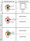
Cartoons depict the process of NSC proliferation in control (wild-type; top row) versus various mutant conditions. Asymmetric cell division defects are highlighted with gray-filled boxes. NSCs (peach circles) are oriented along the apical–basal axis with the apical polarity markers (red arc) and basal polarity determinants (gray arc) shown. Top row: During wild-type asymmetric cell division, two centrosomes (light blue cylinders) are present in late interphase. The apical centrosome is an active microtubule-organizing center with rich levels of PCM (green), while the basal centrosome is inactive (no PCM). Just prior to the onset of mitosis, cortical basal polarity (gray arc) is established. During metaphase, the spindle pole axis (dotted yellow line) aligns along the polarity axis (solid yellow line); both centrosomes are fully mature/active by this point. During anaphase, the chromosomes and polarity markers are segregated, and the cell divides along the division plane (yellow line). This asymmetric cell division generates one larger self-renewing stem cell (red outline) and one smaller differentiating cell (gray outline). 2nd row: Centrosomes and polarity. In either centrosome (e.g. cnn) or polarity (e.g. Ankle2) mutants, resultant defects include centrosome amplification with spindle morphogenesis defects or randomized spindle pole alignment, leading to failed asymmetric cell division. These errant divisions lead to cell death or symmetric cell divisions (two NSCs). 3rd row: Centrosome asymmetry. Although centrosome phenotypes are observed in interphase (note the two active centrosomes), NSCs mutant for centrosome asymmetry genes rotate misaligned spindle poles before the onset of anaphase (gray → white gradient) and then resume normal asymmetric cell division (white boxes). Not shown, some stem cells missegregate their centrosomes, resulting in too many or too few centrosomes, which may compromise NSC survival. Bottom row: Spindle assembly checkpoint. NSCs mutant for both centrosome genes and components of the SAC generate aneuploid NSCs, which undergo premature differentiation, essentially depleting the NSC pool.
Figure 3.
Figure 3.. Asymmetric protein localization directs different centrosome activity levels in interphase NSCs.
Normally, wild-type interphase NSCs exhibit asymmetric centrosome activity levels. Top row: In normal cells, the apical, daughter centrosome has high levels of PCM (green cloud) surrounding the centrioles (light blue cylinders), and it nucleates microtubules (green lines). Proteins enriched on the apical centrosome include those that promote microtubule nucleation (local protein enrichment; apical-like daughter, green font). Conversely, the mother, basal centrosome has little to no PCM. Proteins localized on the basal centrosome frequently have negative centrosome-regulating activities (basal-like mother, red font). Centrosome activity level becomes symmetrical when centrosome regulator genes are lost or overexpressed. Middle row: Loss of a positive regulator of centrosome activity (e.g. cnb) or overexpression of a negative centrosome regulator of centrosome activity (e.g. SAK) leads to two inactive, basal-like centrosomes during interphase. Bottom row: Conversely, Loss of a negative regulator of centrosome activity (e.g. plp) or overexpression of a positive regulator of centrosome activity (e.g. cnb) results in two active, apical-like centrosomes during interphase.

Similar articles

Cited by

References

    1. Allanson JE, Cunniff C, Hoyme HE, McGaughran J, Muenke M and Neri G. (2009) Elements of morphology: standard terminology for the head and face. Am. J. Med. Genet. A 149A, 6–28 10.1002/ajmg.a.32612 - DOI - PMC - PubMed
    1. Pirozzi F, Nelson B and Mirzaa G. (2018) From microcephaly to megalencephaly: determinants of brain size. Dialogues Clin. Neurosci. 20, 267–282 10.31887/DCNS.2018.20.4/gmirzaa - DOI - PMC - PubMed
    1. Passemard S, Kaindl AM and Verloes A. (2013) Microcephaly. Handb. Clin. Neurol. 111, 129–141 10.1016/B978-0-444-52891-9.00013-0 - DOI - PubMed
    1. Kohler S, Carmody L, Vasilevsky N, Jacobsen JOB, Danis D, Gourdine JP et al. (2019) Expansion of the Human Phenotype Ontology (HPO) knowledge base and resources. Nucleic Acids Res. 47, D1018–D1D27 10.1093/nar/gky1105 - DOI - PMC - PubMed
    1. Lee CE, Singleton KS, Wallin M and Faundez V. (2020) Rare genetic diseases: nature’s experiments on human development. iScience 23, 101123 10.1016/j.isci.2020.101123 - DOI - PMC - PubMed

Publication types

LinkOut - more resources