

Ketamine exhibits anti-gastric cancer activity via induction of apoptosis and attenuation of PI3K/Akt/mTOR
- PMID: 32864003
- PMCID: PMC7444715
- DOI: 10.5114/aoms.2019.85146
Ketamine exhibits anti-gastric cancer activity via induction of apoptosis and attenuation of PI3K/Akt/mTOR
Abstract
Introduction: Gastric cancer (GC) is the most widespread type of cancer after lung and liver cancer in men and after breast cancer in women. Thus, the present study was intended to evaluate the effect of ketamine (KET) on gastric cancer cells.
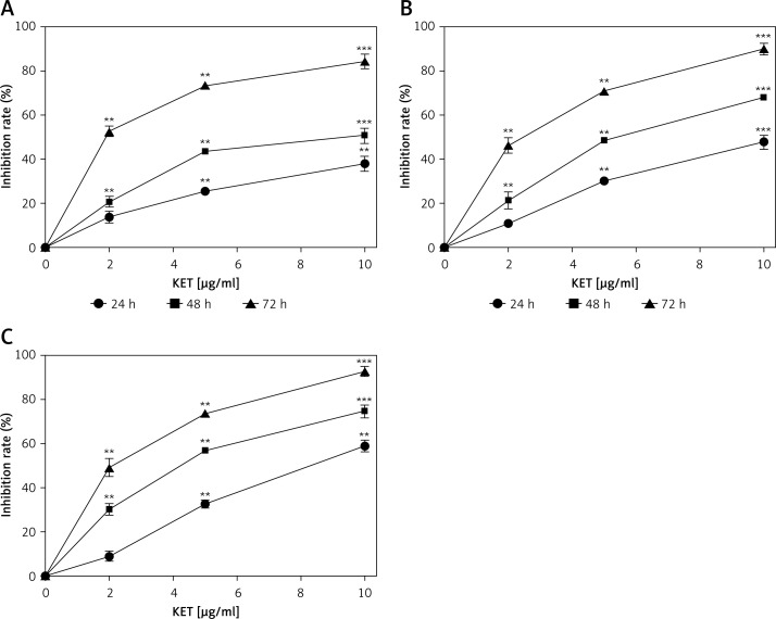
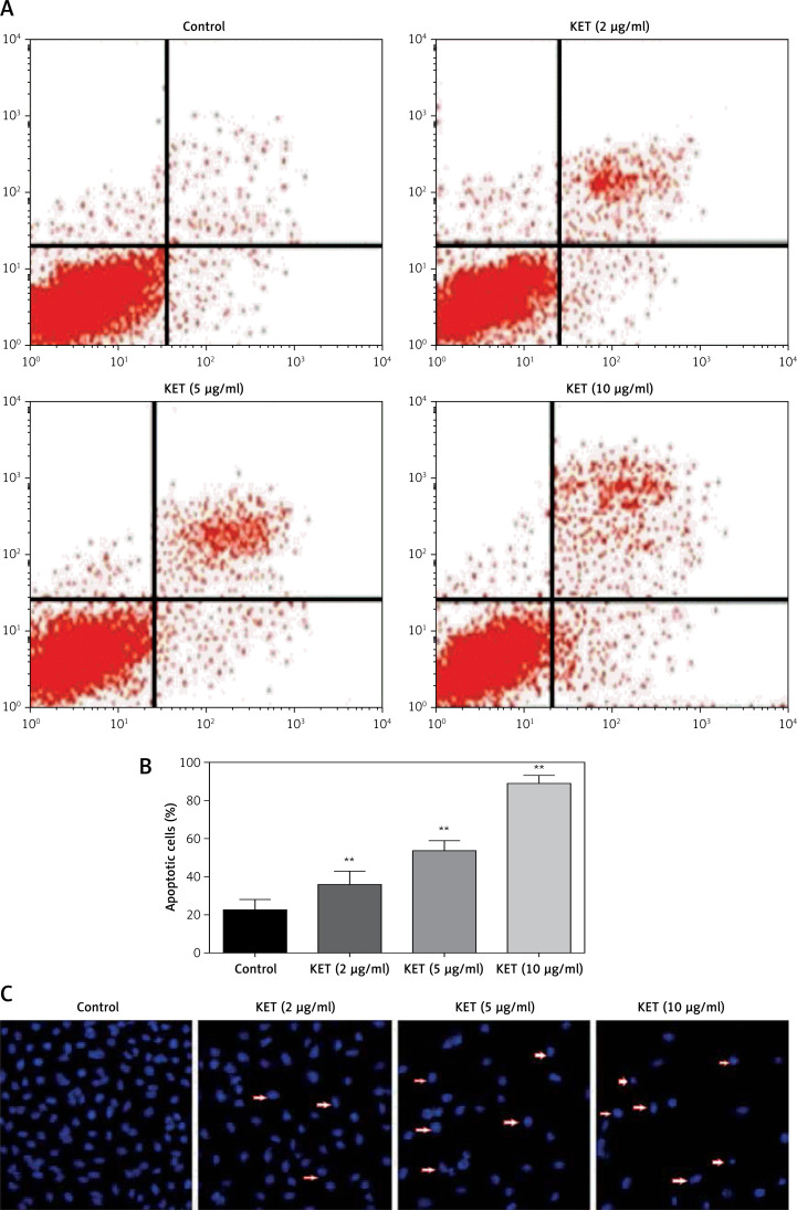
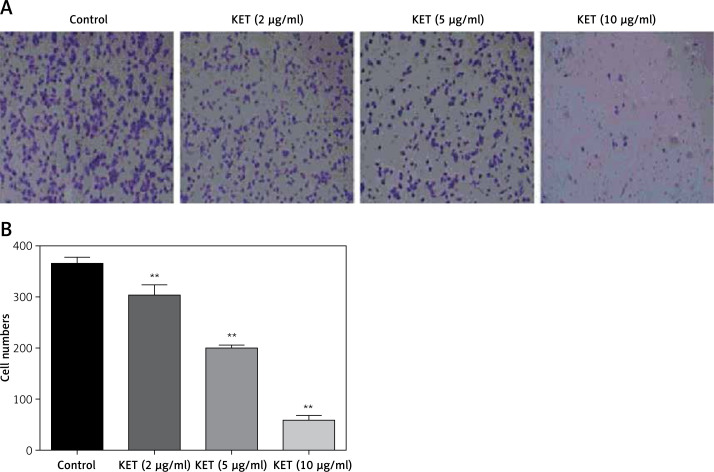
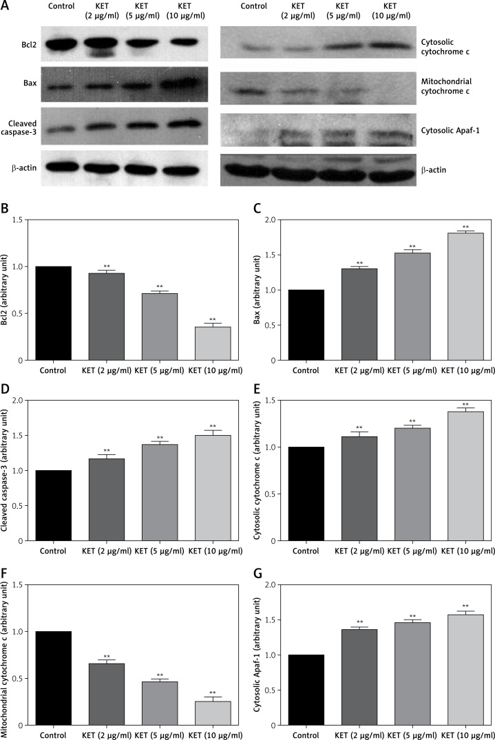
Material and methods: The effect of KET was analyzed in vitro by the MTT assay against human gastric cancer cell lines BGC-823, MKN-45 and MKN-28. The effect KET on apoptosis, cell migration and cell cycle arrest was also quantified. Western blot analysis was performed to estimate the effect of KET on apoptosis mediators and PI3K/AKT/mTOR pathway mediators. A mouse xenograft assay was also conducted to further confirm the anticancer activity.
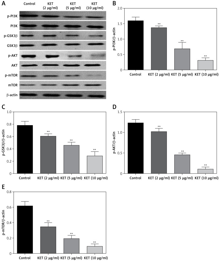
Results: KET causes reduction of cellular viability of BGC-823, MKN-45 and MKN-28, with a more significant effect against BGC-823 cells. The KET treatment showed a dose-dependent increase in apoptotic cells among BGC-823 cells. KET causes a significant dose-dependent decline in migration of treated cells. It causes induction of apoptosis mediated via the mitochondrial pathway, where it causes a decline in Bcl2 and mitochondrial cytochrome c level together with increase in expression of Bax, cytosolic cytochrome c and cytosolic apoptotic protease activating factor-1 (Apaf-1). The level of p-PI3K, p-mTOR, p-GSK3β and p-AKT was found to be downregulated in a dose-dependent manner in KET-treated cells. In a mouse xenograft model, KET causes a reduction in relative tumour volume and tumour weight.
Conclusions: Our results suggest that ketamine has the ability to inhibit progression of gastric cancer via induction of apoptosis and attenuation of PI3K/Akt/mTOR.
Keywords: Akt; PI3K; apoptosis; gastric cancer; ketamine; mTOR; viability.
Copyright: © 2019 Termedia & Banach.
Conflict of interest statement
The authors declare no conflict of interest.
Figures

Similar articles
-
The natural secolignan peperomin E induces apoptosis of human gastric carcinoma cells via the mitochondrial and PI3K/Akt signaling pathways in vitro and in vivo.Phytomedicine. 2016 Jul 15;23(8):818-27. doi: 10.1016/j.phymed.2016.04.001. Epub 2016 May 11. Phytomedicine. 2016. PMID: 27288917
-
[Curcumin induces apoptosis and protective autophagy in human gastric cancer cells with different degree of differentiation].Zhonghua Zhong Liu Za Zhi. 2017 Jul 23;39(7):490-496. doi: 10.3760/cma.j.issn.0253-3766.2017.07.003. Zhonghua Zhong Liu Za Zhi. 2017. PMID: 28728293 Chinese.
-
A new discovery: Total Bupleurum saponin extracts can inhibit the proliferation and induce apoptosis of colon cancer cells by regulating the PI3K/Akt/mTOR pathway.J Ethnopharmacol. 2022 Jan 30;283:114742. doi: 10.1016/j.jep.2021.114742. Epub 2021 Oct 13. J Ethnopharmacol. 2022. PMID: 34655668
-
Huachansu Capsule inhibits the proliferation of human gastric cancer cells via Akt/mTOR pathway.Biomed Pharmacother. 2019 Oct;118:109241. doi: 10.1016/j.biopha.2019.109241. Epub 2019 Jul 24. Biomed Pharmacother. 2019. PMID: 31351435
-
Betulin terpenoid targets OVCAR-3 human ovarian carcinoma cells by inducing mitochondrial mediated apoptosis, G2/M phase cell cycle arrest, inhibition of cell migration and invasion and modulating mTOR/PI3K/AKT signalling pathway.Cell Mol Biol (Noisy-le-grand). 2021 Sep 29;67(2):14-19. doi: 10.14715/cmb/2021.67.2.3. Cell Mol Biol (Noisy-le-grand). 2021. PMID: 34817343
Cited by
-
An Immune-Enhancing Injectable Hydrogel Loaded with Esketamine and DDP Promotes Painless Immunochemotherapy to Inhibit Breast Cancer Growth.Adv Healthc Mater. 2024 Nov;13(29):e2401373. doi: 10.1002/adhm.202401373. Epub 2024 Aug 9. Adv Healthc Mater. 2024. PMID: 39118566 Free PMC article.
-
Efficacy and safety of ketamine for the treatment of depressive symptoms in palliative care: A systematic review.Braz J Psychiatry. 2023 May 11;45(2):182-195. doi: 10.47626/1516-4446-2022-2876. Braz J Psychiatry. 2023. PMID: 36574497 Free PMC article.
-
MCRS1 Expression Regulates Tumor Activity and Affects Survival Probability of Patients with Gastric Cancer.Diagnostics (Basel). 2022 Jun 20;12(6):1502. doi: 10.3390/diagnostics12061502. Diagnostics (Basel). 2022. PMID: 35741311 Free PMC article.
-
Tumor Necrosis Factor Alpha: Implications of Anesthesia on Cancers.Cancers (Basel). 2023 Jan 25;15(3):739. doi: 10.3390/cancers15030739. Cancers (Basel). 2023. PMID: 36765695 Free PMC article. Review.
-
Anesthesia Medications and Interaction with Chemotherapeutic Agents.Oncol Ther. 2021 Jun;9(1):121-138. doi: 10.1007/s40487-021-00149-1. Epub 2021 Apr 16. Oncol Ther. 2021. PMID: 33861416 Free PMC article. Review.
References
-
- American Cancer Society, Society A, American Cancer Society I Global Cancer Facts & Figures 3rd Edition. Am Cancer Soc. 2015 doi: 10.1002/ijc.27711. - DOI
-
- National Cancer Institute . Cancer Statistics – National Cancer Institute. NCI; 2016. - DOI
-
- International Agency for Research on Cancer. World Health Organization Globocan 2012: Estimated Incidence, Mortality and Prevalence Worldwide in 2012. Cancer. 2014:1–10. doi: 10.1002/ijc.27711. - DOI
LinkOut - more resources
Full Text Sources
Research Materials
Miscellaneous