Review
. 2020 Oct;55(5):482-507.
doi: 10.1080/10409238.2020.1813070. Epub 2020 Aug 28.

Dynamic elements of replication protein A at the crossroads of DNA replication, recombination, and repair

Affiliations
Review

Dynamic elements of replication protein A at the crossroads of DNA replication, recombination, and repair

Colleen C Caldwell et al. Crit Rev Biochem Mol Biol. 2020 Oct.

Abstract

The heterotrimeric eukaryotic Replication protein A (RPA) is a master regulator of numerous DNA metabolic processes. For a long time, it has been viewed as an inert protector of ssDNA and a platform for assembly of various genome maintenance and signaling machines. Later, the modular organization of the RPA DNA binding domains suggested a possibility for dynamic interaction with ssDNA. This modular organization has inspired several models for the RPA-ssDNA interaction that aimed to explain how RPA, the high-affinity ssDNA binding protein, is replaced by the downstream players in DNA replication, recombination, and repair that bind ssDNA with much lower affinity. Recent studies, and in particular single-molecule observations of RPA-ssDNA interactions, led to the development of a new model for the ssDNA handoff from RPA to a specific downstream factor where not only stability and structural rearrangements but also RPA conformational dynamics guide the ssDNA handoff. Here we will review the current knowledge of the RPA structure, its dynamic interaction with ssDNA, and how RPA conformational dynamics may be influenced by posttranslational modification and proteins that interact with RPA, as well as how RPA dynamics may be harnessed in cellular decision making.

Keywords: Conformational protein dynamics; DNA repair; DNA replication; homologous recombination; protein-DNA interctions; replication protein A (RPA); single-molecule.


Figures
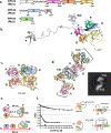
Figure 1.
Figure 1.. Structure of eukaryotic RPA-ssDNA complex.
(a) Primary structures of the three RPA subunits. The regions that make up DBDs/OB-folds A, B, C, D, E, and F, and the winged helix domain (WH) are shown as colored rectangles. The numbers below the map indicate residue numbers in the human RPA amino acid sequence where the domains start and end; in parentheses are the numbers for S. cerevisiae RPA. Areas between the domains are the flexible linkers. (b) Model of the full-length human RPA from Chen et al (36). Individual domains are shown in the same color scheme as in the primary structure. (c) Structure of the RPA-ssDNA complex from Fan and Pavletich ((98); PBD 4GOP). Ustilago maydis RPA construct consists of the DBDs A-E (depicted in the same color scheme as above); ssDNA is shown in elemental colors. TriC marks a trimerization core comprised DBDs C-D. (d) Cryo-EM reconstruction of the two S. cerevisiae RPA heterotrimers bound adjacently on ssDNA. The structure reveals an interaction between the DBDs A and E of the adjacent heterotrimers. This structure from Yates et al (50) also suggested that RPA extends ssDNA instead of folding it into a u-shaped structure as in the Ustilago maydis RPA crystal structure. Inset shows a representative 2D class average for the 2xRPA-ssDNA complex. (e) Schematic representation of the RPA-ssDNA complex in the u-shape and extended conformations. (f) FRET-based ssDNA binding experiment that suggests that in solution RPA induces ssDNA into a linear conformation (see text for details). Data are from (50).
Figure 2:
Figure 2:. Oligonucleotide/oligosaccharides binding folds (OB-folds) are conserved across all domains of life.
The cartoon at the center of the figure is the basic OB-fold motif. While there is some variation, the five beta sheets (purple) that form the mixed beta barrel and the alpha helix (teal) that caps the barrel are generally conserved. OB-folds are represented in each of the structures (bacterial SSB, archaeal SSB, archaeal RPA, and eukaryotic RPA and are also present in numerous ssDNA binding proteins represented here by the Hiran domain of HLTF and the ssDNA binding domain of human BRCA2) using the same purple beta sheet/teal alpha helix/grey loops scheme. Additional structural components are colored differently to highlight them. In the eukaryotic RPA structure the winged helix (WH) domain is depicted in red. In the BRCA2 structure the tower domain is depicted in red and the helix domain is depicted in yellow.
Figure 3:
Figure 3:. Proposed RPA-ssDNA binding modes.
Proposed structural basis for the distinct RPA-ssDNA binding modes. (a) PDB: 1JMC (76) structure of the human RPA DBD-A (pink) and DBD-B (orange) in complex with 8nt poly(dC) ssDNA represents an 8 nt binding mode schematically depicted on the right. Atoms of the four conserved aromatic residues F238 and F269 (in DBD-A) and W361 and F386 (in DBD-B) are shown as spheres. (b) PBD: 4GOP (50), which represents the high affinity (30 nt) ssDNA binding mode of Ustilago maydis RPA is shown with the same orientation of the DBDs A and B. Cartoon on the right represents the proposed transition between the 8nt and 30nt binding modes. (c) Overlap of the DBD-B from 4GOP (orange) and 1JMC (light grey) suggests different paths the ssDNA can take through this DBD likely due to the presence of the flexible linker connecting DBD-B and DBD-C. See text and (50) for extended discussion.
Figure 4:
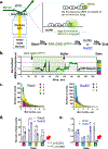
Figure 4:. Single-molecule interrogation of the RPA-ssDNA complex.
(a) Schematic representation of an ssDNA curtain experiment (see (11,39,96) for details), side view. The ssDNA molecules are stretched parallel to each other over the surface of the TIRFM slide. Binding of the RPA-GFP (green) is visualized as appearance of the fluorescence along the DNA molecules. (b) The DNA curtains experiments allowed to propose the mechanism underlying facilitated exchange of RPA on ssDNA. When no additional RPA is present in the solution, RPA molecules remain stably bound to ssDNA even though their individual binding modules may microscopically dissociate from and rebind to ssDNA (e.g. transition between 8 nt and 30 nt binding modes). In the presence of unlabeled RPA in solution, microscopic dissociation of the trimerization core (30 nt → 8 nt transition) opens a landing spot for the RPA from solution and subsequent exchange of the GFP labeled protein on the ssDNA with the unlabeled counterpart. Two color experiments with RFP-labeled RPA and GFP-labeled Rad52 showed that the recombination mediator Rad52 stabilizes some RPA molecules on ssDNA resulting in the RPA-Rad52 clusters from which the Rad51 nucleoprotein filament grows. (c) Magnetic tweezer experiment to characterize the RPA-mediated DNA duplex melting (47). (d) These experiments suggested that a microscopic association of the individual RPA DBDs creates a “toehold” that traps spontaneously melted DNA duplex at the ssDNA-dsDNA junction and promotes duplex destabilization by RPA.
Figure 5.
Figure 5.. Model of RPA dynamic binding and the role in recruitment of weaker binding downstream proteins.
(a) The dynamic binding model differentiates between two types of binding referred to as macroscopic, where the molecule as a whole binds/dissociates, and microscopic, where the molecule as a whole remains bound while individual domains bind/dissociate within the bound RPA-ssDNA complex. In contrast to the original binding modes model, which implies that DBDs A and B constitute a high affinity binding module which associates with ssDNA first and remains associated in both 8 nt and 30 nt modes, the dynamic binding model suggests that the domain binding events do not occur in a sequential manner (40). (b) With the understanding that the individual domains of RPA can dissociate, the replacement/competition model treats these microscopic dissociation events are potential windows for smaller, weaker binding proteins to access ssDNA that is otherwise saturated with RPA. In this case, Rad51, the eukaryotic recombinase, would have an opportunity to nucleate and form a filament to outcompete RPA provides that some domains dissociate from the ssDNA (87). (c) Facilitated hand-off further explains how downstream proteins may outcompete RPA. Rad52, an RPA binding protein and recombination mediator, limits the ability of the 3’ RPA DBDs to access ssDNA. Rad52 also carries Rad51, potentially loading it in the opening created by preventing these RPA domains form accessing the DNA and promoting Rad51 activity in DNA repair (40). While these models address RPA replacement in homologous recombination, other DNA replication and repair processes may exhibit similar mechanisms as many proteins interact and compete with RPA for access to ssDNA.
Figure 6.
Figure 6.. Real time single-molecule observation of the RPA conformational dynamics.
(a) smTIRFM experiment that monitors microscopic association/dissociation of the individual DBD within the macroscopically bound RPA. The ssDNA is immobilized on the surface of TIRFM flow chamber. After 30 sec of observation, RPA labeled within the DBD-D with an environmentally sensitive MB543 dye is injected in the reaction chamber. RPA binding to surface-tethered ssDNA molecules is observed as appearance of the fluorescence signal in specific spots on the flow chamber surface. After 90 sec, the unbound RPA is removed by flashing in the buffer with or without Rad52. (b) A representative trajectory (time-based change in the fluorescence in a specific spot on the slide surface) depicting conformational dynamics of an individual RPA-DBD-DMB543 molecule. The data are from (40). “ON” marks the macroscopic association of the RPA molecule with ssDNA; “OFF/bleach” indicates the moment when the signal is lost due to either RPA dissociation or MB543 dye bleaching. Raw normalized fluorescence is shown in green; the black line corresponds to the idealized trajectory after global analysis of all trajectories using ebFRET (215), which has identified four distinct states interpreted as different degrees of ssDNA engagement by DBD-D (40). (c-e) Analysis of the dwell times of the RPA conformational states. (c) Exponential fits to the dwell time distributions after buffer wash (4 states) and in the presence of Rad52 (3 states). (d) Visitation frequencies for all states in the presence and absence of Rad52 show that state 4, which is the most engaged state of the DBD-D is lost in the presence of Rad52. Visitation of the state 3 also decreases in the presence of Rad52, while states 1 and 2 are visited more often. (e) Each of the four states exists on a second time scale (τ = 1/k, where k is the decay rate constant for each exponential fit); The presence of Rad52 results in disappearance of the state 4 and longer average dwell in state 3.
Figure 7.
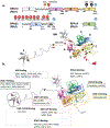
Figure 7.. Protein-protein interaction and posttranslational modifications involving RPA.
(a) Schematic representation of the RPA primary structure with sites of posttranslational modifications and the same modifications mapped on the model of human RPA. Phosphorylation sites are marked with a P in a red circle and with the amino acid noted. SUMOylation sites are marked with an S in a black circle. Ubiquitinylation (U in a blue circle) has been identified on each of the RPA subunits, though specific site are not noted. *Asterisks note phosphorylation sites that were found in yeast RPA. S187/189 have only been noted in yeast. T180 is the corresponding site in human RPA that was identified as S187 in yeast. (b) Sites of Protein-Protein interactions mapped on the structural model of human RPA. Each box is labeled with the location on the RPA molecule that the listed proteins have been found to interact with. In the case of several proteins, multiple binding sites have been identified. While some proteins have only been tested for interaction with entire subunits of RPA, others have been found to bind to specific portions of a subunit, in which case the protein in only noted in the most specific region instead of the subunit. Table 2 indicates the specific amino acid regions where it is known.

Similar articles

Cited by

References

    1. Wold MS and Kelly T (1988) Purification and characterization of replication protein A, a cellular protein required for in vitro replication of simian virus 40 DNA. Proc Natl Acad Sci U S A, 85, 2523–2527. - PMC - PubMed
    1. Wold MS (1997) Replication protein A: a heterotrimeric, single-stranded DNA-binding protein required for eukaryotic DNA metabolism. Annu Rev Biochem, 66, 61–92. - PubMed
    1. Iftode C, Daniely Y and Borowiec JA (1999) Replication protein A (RPA): the eukaryotic SSB. Crit Rev Biochem Mol Biol, 34, 141–180. - PubMed
    1. Brill SJ and Stillman B (1989) Yeast replication factor-A functions in the unwinding of the SV40 origin of DNA replication. Nature, 342, 92–95. - PubMed
    1. Nasheuer HP, von Winkler D, Schneider C, Dornreiter I, Gilbert I and Fanning E (1992) Purification and functional characterization of bovine RP-A in an in vitro SV40 DNA replication system. Chromosoma, 102, S52–59. - PubMed

Publication types

LinkOut - more resources