Inflammation, Senescence and MicroRNAs in Chronic Kidney Disease
- PMID: 32850849
- PMCID: PMC7423998
- DOI: 10.3389/fcell.2020.00739
Inflammation, Senescence and MicroRNAs in Chronic Kidney Disease
Abstract
Background: Patients with chronic kidney disease (CKD) show a chronic microinflammatory state that promotes premature aging of the vascular system. Currently, there is a growth interest in the search of novel biomarkers related to vascular aging to identify CKD patients at risk to develop cardiovascular complications.
Methods: Forty-five CKD patients were divided into three groups according to CKD-stages [predialysis (CKD4-5), hemodialysis (HD) and kidney transplantation (KT)]. In all these patients, we evaluated the quantitative changes in microRNAs (miRNAs), CD14+C16++ monocytes number, and microvesicles (MV) concentration [both total MV, and monocytes derived MV (CD14+Annexin V+CD16+)]. To understand the molecular mechanism involved in senescence and osteogenic transdifferentation of vascular smooth muscle cells (VSMC), these cells were stimulated with MV isolated from THP-1 monocytes treated with uremic toxins (txMV).
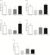
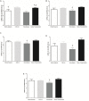
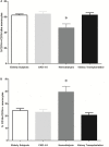
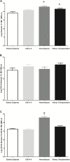
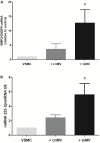
Results: A miRNA array was used to investigate serum miRNAs profile in CKD patients. Reduced expression levels of miRNAs-126-3p, -191-5p and -223-3p were observed in CKD4-5 and HD patients as compared to KT. This down-regulation disappeared after KT, even when lower glomerular filtration rates (eGFR) persisted. Moreover, HD patients had higher percentage of proinflammatory monocytes (CD14+CD16++) and MV derived of proinflammatory monocytes (CD14+Annexin V+CD16+) than the other groups. In vitro studies showed increased expression of osteogenic markers (BMP2 and miRNA-223-3p), expression of cyclin D1, β-galactosidase activity and VSMC size in those cells treated with txMV.
Conclusion: CKD patients present a specific circulating miRNAs expression profile associated with the microinflammatory state. Furthermore, microvesicles generated by monocytes treated with uremic toxins induce early senescence and osteogenic markers (BMP2 and miRNA-223-3p) in VSMC.
Keywords: chronic kidney disease; microRNAs; microvesicles; monocytes CD14+CD16++; vascular smooth muscle cells.
Copyright © 2020 Carmona, Guerrero, Jimenez, Ariza, Agüera, Obrero, Noci, Muñoz-Castañeda, Rodríguez, Soriano, Moreno, Martin-Malo and Aljama.
Figures




Similar articles
-
Proinflammatory CD14(+)CD16(+) monocytes are associated with vascular stiffness in predialysis patients with chronic kidney disease.Kidney Res Clin Pract. 2013 Dec;32(4):147-52. doi: 10.1016/j.krcp.2013.08.001. Epub 2013 Sep 26. Kidney Res Clin Pract. 2013. PMID: 26877933 Free PMC article.
-
Effect of different dialysis modalities on microinflammatory status and endothelial damage.Clin J Am Soc Nephrol. 2010 Feb;5(2):227-34. doi: 10.2215/CJN.03260509. Epub 2010 Jan 7. Clin J Am Soc Nephrol. 2010. PMID: 20056757 Free PMC article.
-
MicroRNAs 223-3p and 93-5p in patients with chronic kidney disease before and after renal transplantation.Bone. 2017 Feb;95:115-123. doi: 10.1016/j.bone.2016.11.016. Epub 2016 Nov 17. Bone. 2017. PMID: 27866993 Free PMC article.
-
CD14+CD16+ monocytes from chronic kidney disease patients exhibit increased adhesion ability to endothelial cells.Contrib Nephrol. 2011;171:57-61. doi: 10.1159/000327134. Epub 2011 May 23. Contrib Nephrol. 2011. PMID: 21625090 Review.
-
Microinflammation and endothelial damage in hemodialysis.Contrib Nephrol. 2008;161:83-88. doi: 10.1159/000130412. Contrib Nephrol. 2008. PMID: 18451662 Review.
Cited by
-
Management of Chronic Heart Failure in Dialysis Patients: A Challenging but Rewarding Path.Rev Cardiovasc Med. 2024 Jun 25;25(6):232. doi: 10.31083/j.rcm2506232. eCollection 2024 Jun. Rev Cardiovasc Med. 2024. PMID: 39076321 Free PMC article. Review.
-
Modulation of miR-29a and miR-29b Expression and Their Target Genes Related to Inflammation and Renal Fibrosis by an Oral Nutritional Supplement with Probiotics in Malnourished Hemodialysis Patients.Int J Mol Sci. 2024 Jan 17;25(2):1132. doi: 10.3390/ijms25021132. Int J Mol Sci. 2024. PMID: 38256206 Free PMC article. Clinical Trial.
-
Uremic Toxins Induce THP-1 Monocyte Endothelial Adhesion and Migration through Specific miRNA Expression.Int J Mol Sci. 2023 Aug 18;24(16):12938. doi: 10.3390/ijms241612938. Int J Mol Sci. 2023. PMID: 37629118 Free PMC article.
-
Drug repurposing screens to identify potential drugs for chronic kidney disease by targeting prostaglandin E2 receptor.Comput Struct Biotechnol J. 2023 Jul 7;21:3490-3502. doi: 10.1016/j.csbj.2023.07.007. eCollection 2023. Comput Struct Biotechnol J. 2023. PMID: 37484490 Free PMC article.
-
PTEN-induced kinase 1 is associated with renal aging, via the cGAS-STING pathway.Aging Cell. 2023 Jul;22(7):e13865. doi: 10.1111/acel.13865. Epub 2023 May 15. Aging Cell. 2023. PMID: 37183600 Free PMC article.
References
LinkOut - more resources
Full Text Sources
Other Literature Sources
Research Materials
Miscellaneous

