. 2020 Aug 4:11:1714.
doi: 10.3389/fmicb.2020.01714. eCollection 2020.

FgVps9, a Rab5 GEF, Is Critical for DON Biosynthesis and Pathogenicity in Fusarium graminearum

Affiliations

FgVps9, a Rab5 GEF, Is Critical for DON Biosynthesis and Pathogenicity in Fusarium graminearum

Chengdong Yang et al. Front Microbiol. .

Abstract

Rab GTPases play an important role in vesicle-mediated membrane trafficking in eukaryotes. Previous studies have demonstrated that deletion of RAB5/VPS21 reduces endocytosis and virulence of fungal phytopathogens in their host plants. However, Rab5 GTPase cycle regulators have not been characterized in Fusarium graminearum, the causal agent of Fusarium head blight (FHB) or head scab disease in cereal crops. In this study, we have identified and characterized a Rab5 guanine nucleotide exchange factor (GEF), the Vps9 homolog FgVps9, in F. graminearum. Yeast two hybrid (Y2H) assays have shown that FgVps9 specifically interacts with the guanosine diphosphate (GDP)-bound (inactive) forms of FgRab51 and FgRab52, the Rab5 isoforms in F. graminearum. Deletion of FgVPS9 shows impaired fungal growth and conidiation. Pathogenicity assays indicate that deletion of FgVPS9 can significantly decrease the virulence of F. graminearum in wheat. Cytological analyses have indicated that FgVps9 colocalizes with FgRab51 and FgRab52 on early endosomes and regulates endocytosis and autophagy processes. Gene expression and cytological examination have shown that FgVps9 and FgRab51 or FgRab52 function in concert to control deoxynivalenol (DON) biosynthesis by regulating the expression of trichothecene biosynthesis-related genes and toxisome biogenesis. Taken together, FgVps9 functions as a GEF for FgRab51 and FgRab52 to regulate endocytosis, which, as a basic cellular function, has significant impact on the vegetative growth, asexual development, autophagy, DON production, and plant infection in F. graminearum.

Keywords: DON; FgVps9; Fusarium graminearum; endocytosis; guanine nucleotide exchange factor; pathogenicity.


Figures

FIGURE 1
FIGURE 1
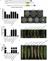
FgVps9 interacts with GDP-bound FgRab51 and FgRab52. Interactions of FgVps9 with FgRab51 and FgRab52, respectively, were examined by yeast two hybrid assays. The interaction between pGADT7-T and pGBKT7-53 was used as a positive control, and pGADT7-T and pGBKT7-Lam served as negative control. Yeast transformants express each pair of proteins were assayed for growth on SD/-Leu/-Trp/-His/-Ade and for MEL1 reporter activities.
FIGURE 2
FIGURE 2
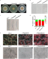
FgVps9 is involved in the vegetative growth and asexual development of F. graminearum. (A) The wild-type PH-1, ΔFgvps9 mutant, and the complementation strain ΔFgvps9com were cultured on complete media (CM) at 28°C for 3 days. (B) The wild-type PH-1, ΔFgvps9, and the complementation strain were cultured in liquid CMC media and photographed under a light microscope after 3 days. (C) Conidial morphology was observed under a light microscope after the indicated strains were cultured in liquid CMC media for 3 days. (D) A bar graph indicating the number of septa in the conidia produced by the indicated strains. (E) The perithecia produced by the wild-type PH-1, ΔFgvps9, and the complementation strain after 2 weeks of inoculation on carrot agar plates. (F) Images of the ascospores produced by the wild-type PH-1, ΔFgvps9, and the complementation strain taken from a light microscope. Bar = 20 μm.
FIGURE 3
FIGURE 3
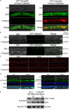
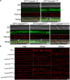
FgVps9 localizes to endosomes and participates in endocytosis and autophagic pathway. (A) FgVps9 colocalizes with both FgRab51 and FgRab52 on the endosomes in hyphae. The indicated strains were cultured in liquid complete media (CM) for 24 h. Images were captured from laser scanning confocal microscopy. White arrows indicate overlapping green fluorescent protein (GFP) and mCherry signals. (B) The number of FgRab51- and FgRab52-labeled endosomes in the tips of the hyphae in the wild-type PH-1 and ΔFgvps9 mutant. The indicated strains were cultivated in liquid CM for 24 h; the number of endosomes at the tips of the 30 hyphae (which were ∼34 μm in diameter each) was counted in each replicate. Statistical differences were calculated by multiple t-tests from three independent repeats using GraphPad Prism at p ≤ 0.05. (C) Hyphae of the wild-type PH-1, ΔFgvps9, and complement strain were incubated in liquid CM for 24 h, then stained with FM4-64 and observed under a fluorescence confocal microscope at different time points. (D) Localization of GFP-ATG8 in the wild-type PH-1 and ΔFgvps9 mutant, respectively. The indicated strains were cultured in liquid CM at 28°C for 48 h and then transferred to liquid MM-N containing 2 mM phenylmethylsulfonyl fluoride (PMSF) for 4 h. Hyphae were stained with CMAC and visualized under a laser scanning confocal microscopy. White arrows indicate autophagosomes that have not shifted to the vacuole. (E) Immunoblot analysis of GFP-FgATG8 degradation in the tested strains. The indicated strains were cultured in liquid CM at 28°C for 48 h and were transferred to liquid MM-N containing 2 mM PMSF for 4 h. Mycelia were collected at the indicated time points from which total protein was extracted and Western blot conducted to check the intensity of GFP-FgATG8 with anti-GFP antibody.
FIGURE 4
FIGURE 4
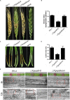
FgVps9 is required for virulence. (A) Infection of ΔFgvps9 mutant to spikelets was tremendously decreased. Flowering wheat heads were inoculated with mycelia plugs of the wild-type PH-1, ΔFgvps9, and the complementation strain. Photographs were taken at 14 days postinoculation (dpi). Inoculated spikelets were marked by black dot. (B) Graphical representation of the disease indices in panel (A). Disease index was evaluated by counting the number of symptomatic spikelets in the corresponding strains after 14 days of inoculation in field. Statistical differences was calculated by multiple t-tests from three independent repeats using GraphPad Prism at p ≤ 0.05. (C,D) Pathogenicity test of the various strains on wheat coleoptiles. The pathogenicity of the ΔFgvps9 mutant was significantly reduced. Coleoptiles were inoculated with conidia suspension from the wild-type PH-1, ΔFgvps9, and the complementation strain. Pictures were taken and lesion sizes were measured at 7 dpi. Statistical differences were calculated by multiple t-tests with three independent repeats using GraphPad Prism at p ≤ 0.05. (E) Analysis of cell-to-cell invasion in wounded wheat leaves. The lower epidermis of the detached wheat leaves were inoculated with mycelium plugs from the tested strains. Confocal images were taken at 8 and 12 h postinoculation. Red boxes indicate invasive hyphae that penetrated into surrounding cells.
FIGURE 5
FIGURE 5
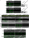
FgVps9 is pivotal for deoxynivalenol (DON) biosynthesis. (A) FgTri4- and FgTri1-labeled toxisomes formation in the indicated strains. Toxisome formation was not observed in the ΔFgvps9 mutant. Toxisome was visualized by laser scanning confocal microscopy after the indicated strains were cultivated in liquid trichothecene biosynthesis inducing (TBI) media at 28°C under dark condition for 72 h. The intensity of FgTri4-GFP and FgTri1-GFP proteins in the indicated strains were quantified by immunoblot assay using the antigreen fluorescent protein (anti-GFP) antibody (right panel). The protein Actin was used as a reference in the Western blot assay (right panel). (B) Toxisome formation in the invasive hyphae. Toxisomes in the invasive hyphae were examined 3 days after conidia from the tested strain-inoculated coleoptiles. Toxisomes were displayed by marker protein FgTri4-GFP and FgTri1-GFP. Images were captured under laser scanning confocal microscopy. White arrows indicate the sites of conidia inoculation on the coleoptiles. (C) Localization of GFP-FgSec22 [endoplasmic reticulum (ER) biomarker] in the ΔFgvps9 mutant. The tested strains were cultivated in liquid TBI media at 28°C under dark condition. Images were captured after 2 days.
FIGURE 6
FIGURE 6
FgRab5 is involved in deoxynivalenol (DON) biosynthesis. (A) The DON production of the ΔFgrab5 mutants. DON was extracted from mycelia of the indicated strains incubated in liquid trichothecene biosynthesis inducing (TBI) media under dark condition for 7 days. Asterisk represent significant differences compared to the wild-type PH-1. (B) Transcription level of TRI genes in the ΔFgrab5 mutants. Relative expression level was measured after the indicated strains incubated in liquid TBI media at 28°C under the dark condition for 3 days. (C,D) Toxisome FgTri4- and FgTri1-labeled was not observed in the ΔFgrab5 mutant. Toxisome was visualized by a laser scanning confocal microscopy after the indicated strains were cultivated in liquid TBI media at 28°C under the dark condition for 72 h. The intensity of FgTri4-GFP and FgTri1-GFP in the indicated strains were identified by immunoblot assay using the antigreen fluorescent protein (anti-GFP) antibody (right panel). The protein Actin was used as a reference in the Western blot assay (right panel).
FIGURE 7
FIGURE 7
The conserved Vps9 domain of FgVps9 is required for the biological functions of the whole protein. (A) Schematic diagram and strategy of domain deletions and point mutations of FgVps9. *525 (Asp525Ala, D525A), *562 (Glu562Ala, E562A). (B) Average colony diameters of the indicated strains incubated on complete media (CM) for 3 days. Statistical differences were calculated by multiple t-tests from three independent repeats using GraphPad Prism at p ≤ 0.05. (C) Conidiation assay. The conidia were counted after inoculation and incubation of the indicated strains in CMC media for 3 days. Statistical differences were calculated by multiple t-tests from three independent repeats using GraphPad Prism at p ≤ 0.05. (D) Deoxynivalenol (DON) production assay. DON was extracted from mycelia of the indicated strains incubated in trichothecene biosynthesis inducing (TBI) media for 7 days. Asterisks indicate significant differences at p ≤ 0.05. (E) The vegetative growth of the indicated strains cultured on CM at 28°C for 3 days. (F) Pathogenicity assay of various strains to wheat coleoptiles. Wheat coleoptiles were inoculated with conidia suspension from the indicated strains, and pictures were taken at 7 dpi. (G) Pathogenicity of the indicated strains on flowering wheat heads. The flowering wheat heads were inoculated with mycelia plugs of the indicated strains. Photographs were taken at 14 dpi. Inoculated spikelets were marked by black dots.
FIGURE 8
FIGURE 8
The Vps9 domain of FgVps9 is required for endosomal localization and endocytosis in F. graminearum. (A) Localization of the mutated GFP-FgVps9 in the indicated mutant strains. Each strain was cultivated in liquid complete media (CM) for 24 h, then stained with FM4-64 and observed under a fluorescence confocal microscope. White arrows indicate colocalization. (B) FM4-64 internalization assay in the indicated strains. Hyphae of the various strains were incubated in liquid CM at 28°C for 24 h, then stained with FM4-64 and observed under a fluorescence confocal microscope at different time points.

Similar articles

Cited by

References

    1. Abeliovich H., Klionsky D. J. (2001). Autophagy in yeast: mechanistic insights and physiological function. Microbiol. Mol. Biol. Rev. 65 463–479, table of contents. 10.1128/MMBR.65.3.463-479.2001 - DOI - PMC - PubMed
    1. Alexander N. J., Proctor R. H., McCormick S. P. (2009). Genes, gene clusters, and biosynthesis of trichothecenes and fumonisins in Fusarium. Toxin Rev. 28 198–215. 10.1080/15569540903092142 - DOI
    1. Alva A. S., Gultekin S. H., Baehrecke E. H. (2004). Autophagy in human tumors: cell survival or death? Cell Death Differ. 11 1046–1048. 10.1038/sj.cdd.4401445 - DOI - PubMed
    1. Asakura M., Ninomiya S., Sugimoto M., Oku M., Yamashita S., Okuno T., et al. (2009). Atg26-mediated pexophagy is required for host invasion by the plant pathogenic fungus Colletotrichum orbiculare. Plant Cell 21 1291–1304. 10.1105/tpc.108.060996 - DOI - PMC - PubMed
    1. Bai G., Shaner G. (1994). Scab of wheat: prospects for control. Plant Dis. 78 760–766. 10.1094/PD-78-0760 - DOI

LinkOut - more resources