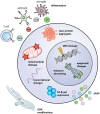
Stem Cells of the Aging Brain
- PMID: 32848716
- PMCID: PMC7426063
- DOI: 10.3389/fnagi.2020.00247
Stem Cells of the Aging Brain
Abstract
The adult central nervous system (CNS) contains resident stem cells within specific niches that maintain a self-renewal and proliferative capacity to generate new neurons, astrocytes, and oligodendrocytes throughout adulthood. Physiological aging is associated with a progressive loss of function and a decline in the self-renewal and regenerative capacities of CNS stem cells. Also, the biggest risk factor for neurodegenerative diseases is age, and current in vivo and in vitro models of neurodegenerative diseases rarely consider this. Therefore, combining both aging research and appropriate interrogation of animal disease models towards the understanding of the disease and age-related stem cell failure is imperative to the discovery of new therapies. This review article will highlight the main intrinsic and extrinsic regulators of neural stem cell (NSC) aging and discuss how these factors impact normal homeostatic functions within the adult brain. We will consider established in vivo animal and in vitro human disease model systems, and then discuss the current and future trajectories of novel senotherapeutics that target aging NSCs to ameliorate brain disease.
Keywords: aging; cell metabolism; disease modeling; mitochondria; neural stem cells; senescence; senotherapeutics.
Copyright © 2020 Nicaise, Willis, Crocker and Pluchino.
Figures


Similar articles
-
Soluble factors influencing the neural stem cell niche in brain physiology, inflammation, and aging.Exp Neurol. 2022 Sep;355:114124. doi: 10.1016/j.expneurol.2022.114124. Epub 2022 May 26. Exp Neurol. 2022. PMID: 35644426 Review.
-
Noggin rescues age-related stem cell loss in the brain of senescent mice with neurodegenerative pathology.Proc Natl Acad Sci U S A. 2018 Nov 6;115(45):11625-11630. doi: 10.1073/pnas.1813205115. Epub 2018 Oct 23. Proc Natl Acad Sci U S A. 2018. PMID: 30352848 Free PMC article.
-
Microenvironmental determinants of adult neural stem cell proliferation and lineage commitment in the healthy and injured central nervous system.Curr Stem Cell Res Ther. 2008 Sep;3(3):163-84. doi: 10.2174/157488808785740334. Curr Stem Cell Res Ther. 2008. PMID: 18782000 Review.
-
Neural stem cell niches and homing: recruitment and integration into functional tissues.ILAR J. 2009;51(1):3-23. doi: 10.1093/ilar.51.1.3. ILAR J. 2009. PMID: 20075495 Review.
-
Grafted Subventricular Zone Neural Stem Cells Display Robust Engraftment and Similar Differentiation Properties and Form New Neurogenic Niches in the Young and Aged Hippocampus.Stem Cells Transl Med. 2016 Sep;5(9):1204-15. doi: 10.5966/sctm.2015-0270. Epub 2016 May 18. Stem Cells Transl Med. 2016. PMID: 27194744 Free PMC article.
Cited by
-
Culture Protocol and Transcriptomic Analysis of Murine SVZ NPCs and OPCs.Stem Cell Rev Rep. 2023 May;19(4):983-1000. doi: 10.1007/s12015-022-10492-z. Epub 2023 Jan 9. Stem Cell Rev Rep. 2023. PMID: 36617597
-
Regulation of microglia function by neural stem cells.Front Cell Neurosci. 2023 Mar 1;17:1130205. doi: 10.3389/fncel.2023.1130205. eCollection 2023. Front Cell Neurosci. 2023. PMID: 36937181 Free PMC article. Review.
-
Modeling Neuroregeneration and Neurorepair in an Aging Context: The Power of a Teleost Model.Front Cell Dev Biol. 2021 Mar 18;9:619197. doi: 10.3389/fcell.2021.619197. eCollection 2021. Front Cell Dev Biol. 2021. PMID: 33816468 Free PMC article. Review.
-
Loss of Protein Function Causing Severe Phenotypes of Female-Restricted Wieacker Wolff Syndrome due to a Novel Nonsense Mutation in the ZC4H2 Gene.Genes (Basel). 2022 Aug 29;13(9):1558. doi: 10.3390/genes13091558. Genes (Basel). 2022. PMID: 36140726 Free PMC article.
-
Metformin's Mechanisms in Attenuating Hallmarks of Aging and Age-Related Disease.Aging Dis. 2022 Jul 11;13(4):970-986. doi: 10.14336/AD.2021.1213. eCollection 2022 Jul 11. Aging Dis. 2022. PMID: 35855344 Free PMC article.
References
Publication types
LinkOut - more resources
Full Text Sources

