. 2020 Sep;585(7824):251-255.
doi: 10.1038/s41586-020-2633-x. Epub 2020 Aug 26.

Structure of the C9orf72 ARF GAP complex that is haploinsufficient in ALS and FTD

Affiliations

Structure of the C9orf72 ARF GAP complex that is haploinsufficient in ALS and FTD

Ming-Yuan Su et al. Nature. 2020 Sep.

Abstract

Mutation of C9orf72 is the most prevalent defect associated with amyotrophic lateral sclerosis and frontotemporal degeneration1. Together with hexanucleotide-repeat expansion2,3, haploinsufficiency of C9orf72 contributes to neuronal dysfunction4-6. Here we determine the structure of the C9orf72-SMCR8-WDR41 complex by cryo-electron microscopy. C9orf72 and SMCR8 both contain longin and DENN (differentially expressed in normal and neoplastic cells) domains7, and WDR41 is a β-propeller protein that binds to SMCR8 such that the whole structure resembles an eye slip hook. Contacts between WDR41 and the DENN domain of SMCR8 drive the lysosomal localization of the complex in conditions of amino acid starvation. The structure suggested that C9orf72-SMCR8 is a GTPase-activating protein (GAP), and we found that C9orf72-SMCR8-WDR41 acts as a GAP for the ARF family of small GTPases. These data shed light on the function of C9orf72 in normal physiology, and in amyotrophic lateral sclerosis and frontotemporal degeneration.


Figures
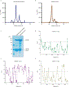
Extended Data Fig. 1:
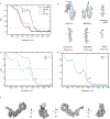
Extended Data Fig. 1:. Purification of the C9orf72-SMCR8-WDR41 and C9orf72-SMCR8 complex as well as the HDX data for trimer.
a, The superose 6 gel filtration elution profile for C9orf72-SMCR8-WDR41 complex. b, The superose 6 gel filtration elution profile for C9orf72-SMCR8 complex. mAU, milli-absorbance units. c, The purified full length C9orf72-SMCR8-WDR41 and C9orf72-SMCR8 were analyzed by SDS-PAGE. The proteins were purified at least five times with similar result (a-c). d-f, Deuterium uptake data for C9orf72-SMCR8-WDR41 complex at 0.5 sec timepoint with error bars from triplicate technical measurements. Peptides with more than 50 % deuterium uptake are the flexible regions. Y axis represents the average percent deuteration. X axis demonstrates the midpoint of a single peptic peptide.
Extended Data Fig. 2:
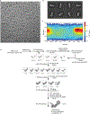
Extended Data Fig. 2:. Cryo-EM data processing.
a, A representative cryo-EM micrograph of C9orf72-SMCR8-WDR41 complex. b, Representative 2D classes. c, Orientation distribution of the aligned C9orf72-SMCR8-WDR41 particles. d, Image processing procedure.
Extended Data Fig. 3:
Extended Data Fig. 3:. Resolution estimation of the cryo-EM map as well as model building and validation.
a, Comparison between FSC curves. b, Refined coordinate model fit of the indicated region in the cryo-EM density. c, Refinement and map-vs-model FSC. d, Cross-validation test FSC curves to assess overfitting. The refinement target resolution (4.2 Å) is indicated. e. Different views of the final reconstruction.
Extended Data Fig. 4:
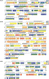
Extended Data Fig. 4:. Deuterium uptake of C9orf72-SMCR8-WDR41.
HDX- MS data are shown in heatmap format where peptides were represented using rectangular strips above the protein sequence. Absolute deuterium uptake after 0.5, 5, 50, 500 and 50,000 sec were indicated by a color gradient below the protein sequence.
Extended Data Fig. 5:
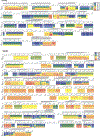
Extended Data Fig. 5:. Deuterium uptake of C9orf72-SMCR8 complex.
HDX- MS data are shown in heatmap format where peptides were represented using rectangular strips above the protein sequence. Absolute deuterium uptake after 0.5, 5, 50, 500 and 50,000 sec were indicated by a color gradient below the protein sequence.
Extended Data Fig. 6
Extended Data Fig. 6. Mapping the protected region from HDX result on the SMCR8-C9orf72-WDR41 structure.
a, The HDX uptake difference at 0.5 sec was mapped on C9orf72-SMCR8. Close view of SMCR8-WDR41 interface, highlighting the SMCR8 mutants. Close view of b, SMCR8M1, c, SMCR8M2–M5 and d, C9orf72M1 region. e, Zoom in view of WDR41 residues in SMCR8-WDR41 interface.
Extended Data Fig. 7:
Extended Data Fig. 7:. Coexpression and pull down validation of C9orf72-SMCR8 and SMCR8-WDR41 interface.
a, Close view of the residues mediating the DENN: DENN dimerization between C9orf72-SMCR8. b, Coexpression and pull down experiment of Strep-tag SMCR8 with GST-C9orf72 mutants and WDR41. c, Pull down experiment of GST-WDR41 mutants with C9orf72-SMCR8. The pull down experiments were carried out at least twice with similar results (b,c).
Extended Data Fig. 8:
Structural comparison between C9orf72-SMCR8 and FNIP2-FLCN.
Extended Data Fig. 9:
Extended Data Fig. 9:. GTPase assay for different small GTPases with C9orf72-SMCR8-WDR41.
a, SDS-PAGE of GAP protein complex (top) and GTPase proteins (bottom) used in the experiments. b, Tryptophan fluorescence GTPase signal was measured for purified RagA/C, ARL8A, ARL8B, RAB1A and RAB7A before and after addition of C9orf72-SMCR8-WDR41. The fluorescence signal upon GAP addition was normalized to 1 for each experiment. The experiments were carried out in triplicate and one representative plot was plotted. c, Tryptophan fluorescence GTPase signal was measured for purified ARF6WT or Q67L or ARF5WT and before and after addition of C9orf72-SMCR8WT-WDR41. C9orf72-SMCR8R147A- WDR41 or C9orf72-SMCR8WT. d, HPLC-based GTPase assay with ARF6, ARF5, RAB1A, RAB7A, ARL8A, ARL8B and RagA/C proteins in the absence and addition of C9orf72-SMCR8-WDR41 complex as indicated. The experiments were carried out in triplicate and one representative plot was shown. All experiments were carried out at least three times independently with similar results (a-d).
Extended Data Fig. 10:
HPLC-based GTPase assay with ARF1WT or Q71L proteins in the absence and addition of GAP complex as indicated. The experiments were carried out in triplicate and one representative plot was shown. All experiments were carried out at least three times independently with similar results.
Extended Data Fig.11:
Extended Data Fig.11:. GEF assay for different small GTPases with C9orf72-SMCR8-WDR41.
a, SDS-PAGE of C9orf72-SMCR8-WDR41 complex and GTPase proteins used in the experiments. b, GEF assay with mantGDP reloaded RAB1A, RAB5A, RAB7A, RAB8A and RAB39B proteins in the absence and addition of C9orf72-SMCR8-WDR41 complex as indicated. RAB5A treated with Rabex5 was used as a positive control reaction. Data was baseline subtracted and normalized to the signal right after GTP addition which also is the 0 s time point in the plots. Plots were the mean and standard deviation of each technical triplicate experiment. All experiments were carried out at least twice independently with similar results (a,b). c, Structural alignment of C9orf72-SMCR8-WDR41 with DENND1B-RAB35 (PDB 3TW8).
Fig. 1:
Fig. 1:. Cryo-EM structure of the C9orf72-SMCR8-WDR41 complex.
a, Schematic diagram of the domain structure of C9orf72-SMCR8-WDR41 complex. b, Cryo-EM density map (localfilter map, b-factor −50 Å2) and c, the refined coordinates of the complex shown as pipes and planks for α-helices and β-sheets, respectively. The domains color-coded as follows: SMCR8longin, cornflower blue; SMCR8DENN, dodger blue; C9orf72longin, olive; C9orf72DENN, goldenrod; WDR41, medium purple. Organizations of d, SMCR8longin: C9orf72longin and e, SMCR8DENN: C9orf72DENN arrangement.
Fig. 2:
Fig. 2:. HDX-MS of C9orf72-SMCR8 in the absence of WDR41.
a, Difference plot of percentage of deuteron incorporation of SMCR8 in heterotrimer versus dimer at 5 sec timepoint. b, Difference plot of percentage of deuteron incorporation of C9orf72 in heterotrimer versus dimer at 0.5 sec timepoint. c, Coexpression and pull down experiment of Strep-tagged SMCR8 mutants with wild type MBP-C9orf72 and GST-WDR41. d, Coexpression and pull down experiments of GST-C9orf72 mutant with wild type untagged SMCR8 and Strep-WDR41. The pull down experiments were repeated at least twice with similar results (c-d).
Fig. 3:
Fig. 3:. SMCR8 mutants fail to localize on lysosome.
a, The HDX uptake difference at 0.5 sec was mapped on C9orf72-SMCR8. Close view of SMCR8-WDR41 interface, highlighting the SMCR8 mutants. b, SMCR8-PQLC2 lysosome colocalization experiment in cells expressing the indicated SMCR8 constructs under the indicated nutrient conditions. −AA indicates cells starved for amino acids for one hr and +AA indicates cells starved and the restimulated with amino acids for 10 min. The experiment was repeated at least three times independently with similar results. c, Quantification of SMCR8 lysosomal enrichment score for immunofluorescence images in b. Plotted are mean and SD, (left to right n=11, 9, 11, 12 and 11) cells were quantified for each condition. * (p value < 0.05) and **** (p value < 0.0001) were evaluated by one-way ANOVA analysis.
Fig. 4:
Fig. 4:. C9orf72-SMCR8 is a GAP for Arf proteins.
a, Structure comparison of FNIP2-FLCN and C9orf72-SMCR8, implying a potential binding site for substrates. The conserved Arg residue was shown in spherical representation. b, Tryptophan fluorescence GTPase signal was measured for ARF1WT or Q71L before and after addition of C9orf72-SMCR8WT or C9orf72-SMCR8R147A -WDR41, C9orf72-SMCR8, FLCN-FNIP2 or GATOR1 complex. The fluorescence signal upon GAP addition was normalized to 1 for each experiment. Plots were the mean and standard deviation of triplicate technical experiments. c, Model for Arf protein family activation by C9orf72-SMCR8-WDR41.

Similar articles

Cited by

References

    1. Majounie E et al. Frequency of the C9orf72 hexanucleotide repeat expansion in patients with amyotrophic lateral sclerosis and frontotemporal dementia: a cross-sectional study. Lancet Neurol 11, 323–330 (2012). - PMC - PubMed
    1. DeJesus-Hernandez M et al. Expanded GGGGCC hexanucleotide repeat in noncoding region of C9ORF72 causes chromosome 9p-linked FTD and ALS. Neuron 72, 245–256 (2011). - PMC - PubMed
    1. Renton AE et al. A hexanucleotide repeat expansion in C9ORF72 is the cause of chromosome 9p21-linked ALS-FTD. Neuron 72, 257–268 (2011). - PMC - PubMed
    1. O’Rourke JG et al. C9orf72 is required for proper macrophage and microglial function in mice. Science (New York, N.Y.) 351, 1324–1329 (2016). - PMC - PubMed
    1. Sivadasan R et al. C9ORF72 interaction with cofilin modulates actin dynamics in motor neurons. Nature neuroscience 19, 1610–1618 (2016). - PubMed

Publication types

MeSH terms

Supplementary concepts