Review
. 2020 Oct 27;94(22):e00175-20.
doi: 10.1128/JVI.00175-20. Print 2020 Oct 27.

Structures of the Mononegavirales Polymerases

Affiliations
Review

Structures of the Mononegavirales Polymerases

Bo Liang. J Virol. .

Abstract

Mononegavirales, known as nonsegmented negative-sense (NNS) RNA viruses, are a class of pathogenic and sometimes deadly viruses that include rabies virus (RABV), human respiratory syncytial virus (HRSV), and Ebola virus (EBOV). Unfortunately, no effective vaccines and antiviral therapeutics against many Mononegavirales are currently available. Viral polymerases have been attractive and major antiviral therapeutic targets. Therefore, Mononegavirales polymerases have been extensively investigated for their structures and functions. Mononegavirales mimic RNA synthesis of their eukaryotic counterparts by utilizing multifunctional RNA polymerases to replicate entire viral genomes and transcribe viral mRNAs from individual viral genes as well as synthesize 5' methylated cap and 3' poly(A) tail of the transcribed viral mRNAs. The catalytic subunit large protein (L) and cofactor phosphoprotein (P) constitute the Mononegavirales polymerases. In this review, we discuss the shared and unique features of RNA synthesis, the monomeric multifunctional enzyme L, and the oligomeric multimodular adapter P of Mononegavirales We outline the structural analyses of the Mononegavirales polymerases since the first structure of the vesicular stomatitis virus (VSV) L protein determined in 2015 and highlight multiple high-resolution cryo-electron microscopy (cryo-EM) structures of the polymerases of Mononegavirales, namely, VSV, RABV, HRSV, human metapneumovirus (HMPV), and human parainfluenza virus (HPIV), that have been reported in recent months (2019 to 2020). We compare the structures of those polymerases grouped by virus family, illustrate the similarities and differences among those polymerases, and reveal the potential RNA synthesis mechanisms and models of highly conserved Mononegavirales We conclude by the discussion of remaining questions, evolutionary perspectives, and future directions.

Keywords: Mononegavirales polymerases; RNA-dependent RNA polymerase; cryo-EM structures; human metapneumovirus (HMPV); human respiratory syncytial virus (HRSV); rabies virus (RABV); vesicular stomatitis virus (VSV).


Figures

FIG 1
FIG 1
The genome organization and RNA synthesis of Mononegavirales. The negative-sense NNS genome is depicted from the 3′ end to the 5′ end, showing the 3′ leader (Le; cyan box), genes (gray, blue, or green box) flanking with gene start (GS; white box) and gene end (GE; black box), and 5′ trailer (Tr; yellow box). The essential genes (N, P, and L) and necessary cofactors (M2 or p30) for RNA synthesis are colored in blue and green, respectively. The RNA-dependent RNA polymerase (RdRP) sequentially produces a gradient level of Le RNA (red line) and viral mRNAs (black, blue, or green line), with the attenuation of the downstream mRNAs at each gene junction. The Le RNA (red lines inside the box) remains uncapped and nonpolyadenylated, while the viral mRNAs are 5′ capped, methylated, and 3′ polyadenylated. The lines under the Le RNA and representative viral mRNAs indicate the abundancy and gradient levels of the RNA transcripts. The promoters for transcription and replication are shown with magenta arrows.
FIG 2
FIG 2
The domain organization and architecture of L and P. (A) The domain organization and cartoon representation of the multifunctional enzyme monomeric L. The conserved regions (CRs) I to VI are labeled in gray boxes. The RNA-dependent RNA polymerization domain (RdRp), capping domain (Cap), connector domain (CD), methyltransferase domain (MT), and C-terminal domain (CTD) of L are colored in blue, green, yellow, pink, and cyan, respectively. (B) The domain organization and cartoon representation of the multimodular adapter oligomeric P. The intrinsically disordered N-terminal domain (PNTD), oligomerization domain (POD), and C-terminal domain (PCTD) are colored in magenta, red, and orange, respectively. The interaction regions with other viral proteins, including L, N, RNA-free N (N0), and accessory protein (M2-1), are labeled in gray boxes. The representative P oligomers are shown for the representative virus families Rhabdoviridae, Pneumoviridae, Paramyxoviridae, and Filoviridae.
FIG 3
FIG 3
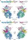
The cryo-EM structures of the Rhabdoviridae polymerases. (A) Linear domain representation of the L and P proteins of the vesicular stomatitis virus (VSV) polymerase. The cartoon view of 3.8-Å (PDB: 5A22) and 3.0-Å (PDB: 6U1X) cryo-EM structures of the VSV polymerase are shown. (B) Linear domain representation of the L and P proteins of the rabies virus (RABV) polymerase. The cartoon view of the 3.3-Å (PDB: 6UEB) cryo-EM structure of the RABV polymerase is shown. The RNA-dependent RNA polymerization domain (RdRp), capping domain (Cap), connector domain (CD), methyltransferase domain (MT), C-terminal domain (CTD) of L, and PNTD are colored in blue, green, yellow, pink, cyan, and magenta, respectively. The missing domains are colored in gray. The PNTD is highlighted as spheres, and the terminal residue numbers of the modeled P segments are indicated. The PDB accession codes are underlined.
FIG 4
FIG 4
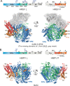
The cryo-EM structures of the Pneumoviridae polymerases. (A) Linear domain representation of the L and P proteins of the human respiratory syncytial virus (HRSV) polymerase. The cartoon view of the 3.67-Å (PDB: 6UEN) and 3.2-Å (PDB: 6PZK) cryo-EM structures of HRSV polymerase complexes. The missing domains compared with the VSV L are shown in the gray meshes. (B) Linear domain representation of the L and P proteins of the human metapneumovirus (HMPV) polymerase. The cartoon view of the 3.7-Å (PDB: 6U5O) cryo-EM structure of the HMPV polymerase is shown. The domain colorings are the same as Fig. 2. The terminal residue numbers of the modeled POD and PCTD are indicated. The PDB accession codes are underlined.
FIG 5
FIG 5
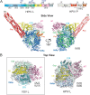
The cryo-EM structure of the Paramyxoviridae polymerase. (A) Linear domain representation of the L and P proteins of the human parainfluenza virus (HPIV) polymerase. The side view of the ribbon diagram of the 4.3-Å (PDB: 6V85) cryo-EM structure of the HPIV polymerase complex. (B) The top view of the superimposed VSV L and HPIV L shows the domain switch of the CD-MT-CTD module. The superimposition is based on the RdRp (surface view), and CD, MT, and CTD are shown as the ribbon diagram. The domain colorings are the same as Fig. 2. The VSV L is shown in the left panel (box), and the HPIV L is shown in the right panel. The HPIV L (PDB: 6V85) is colored the same as A, and another stable conformation of the HPIV L (PDB: 6V86) is colored in gray. Note the significant location switch of CTD, facing down (VSV) versus facing up (HPIV L). The PDB accession codes are underlined.
FIG 6
FIG 6
Structural comparison of the RNA-dependent RNA polymerization (RdRp) domain. (A to E) The ribbon representations of the RdRp domain of the Rhabdoviridae (VSV and RABV), Pneumoviridae (HRSV and HMPV), and Paramyxoviridae (HPIV) L in conventional orientation. The structural motifs finger, palm, thumb, and support region are in blue, red, green, and gray, respectively. The tri-residues (GDN) of the RdRp active sites at a β-hairpin tip of the palm motif are shown in magenta spheres. (F and G) Similarities of the Mononegavirales RdRp domain to other viral polymerases. Structures of the polymerases of reovirus λ3 (ReoV; PDB: 1MUK) and influenza B (FluB; PDB: 4WRT) are shown as the same orientation and coloring scheme as in A. The PDB accession codes are underlined.
FIG 7
FIG 7
Structural comparison of the Cap domain. The motifs A to E of the Cap domain of the Rhabdoviridae (VSV and RABV), Pneumoviridae (HRSV and HMPV), and Paramyxoviridae (HPIV) L are shown as ribbon diagrams in blue, yellow, red, magenta, and green, respectively. Those motifs are centered around the active site motif D (HR). The proposed priming loop (orange) is next to motif B. The PDB accession codes are underlined.
FIG 8
FIG 8
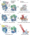
Structural comparisons of the Mononegavirales RNA polymerases. The active sites of the RdRp and Cap domains of L, GDN, and HR are shown in magenta spheres and sticks, respectively. The priming loops and supporting helix are colored in orange. (A) The structural superimposition of the Rhabdoviridae L. The VSV L is colored the same as Fig. 3A, and the RABV L is colored in gray. (B) The structural superimposition of the Rhabdoviridae P. The VSV P is colored in magenta the same as Fig. 3A, and the RABV P is colored in brown. Only the interacting domains RdRp, CD, and CTD of L are shown as surface. (C) The structural superimposition of the Pneumoviridae L. The HRSV L is colored the same as Fig. 4A, and the HMPV L is colored in gray. Note that the supporting helix is missing. (D) The superimposition of the Pneumoviridae P. The HRSV P is colored the same as Fig. 4A, and the HMPV P is colored in brown. Only the interacting domain RdRp of L is shown as surface. (E) The structural representation of the Paramyxoviridae L. The HPIV L is colored the same as Fig. 5A. (F) The location of the Paramyxoviridae P. The HPIV P is colored the same as Fig. 5A. Only the interacting domain RdRp of L is shown as surface. The PDB accession codes are underlined.
FIG 9
FIG 9
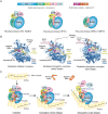
Structural models of the Mononegavirales RNA synthesis. (A) The cartoon diagrams of recently reported structures of the Rhabdoviridae (VSV and RABV), Paramyxoviridae (HPIV), and Pneumoviridae (HRSV and HMPV) polymerases. The same color scheme as Fig. 2. (B) The modeled initiation and elongation complexes. The RdRp domain of the L proteins of Rhabdoviridae (VSV), Paramyxoviridae (HPIV), and Pneumoviridae (HRSV) with modeled RNA template from reovirus λ3 polymerase (PDB: 1N1H), FluB polymerase (PDB: 6QCV), and FluB polymerase (PDB: 6QCT), respectively. The same color scheme for the RdRp domain of Mononegavirales L. The priming loop (from the Cap domain) and the support helix (from the RdRp domain) are colored in orange. The modeled RNA template and RNA transcript are shown in yellow and pink, respectively. (C) The proposed cartoon models of the initiation and elongation stages on the nucleoprotein (N) encapsidated N:RNA (NC) template. Initiation, the priming loop and support helix are at the close approximate of the GDN active site of the RdRp domain of L; elongation (early stage), the priming loop is away from but the support helix stays at the close approximate to the active site of the RdRp domain of L; elongation (late stage), the priming loop is away from the active site of the RdRp domain of L, the support helix is missing, and the CD, MT, and CTD domains of L are disordered and linked by dashed lines. The nucleoprotein (N) protein is shown as the yellow oval. The RNA template, RNA transcript, and the flexible linker are shown in the black, blue, and red lines, respectively. The priming loop and support helix are shown as the thick orange bar and cylinder, respectively. The PDB accession codes are underlined.

Similar articles

Cited by

References

    1. Lamb RA. 2013. Mononegavirales In Knipe DM, Howley PM (ed), Fields virology, 6th ed, Wolters Kluwer Health/Lippincott Williams & Wilkins, Philadelphia, PA.
    1. Whelan SP, Barr JN, Wertz GW. 2004. Transcription and replication of nonsegmented negative-strand RNA viruses. Curr Top Microbiol Immunol 283:61–119. doi:10.1007/978-3-662-06099-5_3. - DOI - PubMed
    1. Conzelmann KK. 1998. Nonsegmented negative-strand RNA viruses: genetics and manipulation of viral genomes. Annu Rev Genet 32:123–162. doi:10.1146/annurev.genet.32.1.123. - DOI - PubMed
    1. Amarasinghe GK, Ayllón MA, Bào Y, Basler CF, Bavari S, Blasdell KR, Briese T, Brown PA, Bukreyev A, Balkema-Buschmann A, Buchholz UJ, Chabi-Jesus C, Chandran K, Chiapponi C, Crozier I, de Swart RL, Dietzgen RG, Dolnik O, Drexler JF, Dürrwald R, Dundon WG, Duprex WP, Dye JM, Easton AJ, Fooks AR, Formenty PBH, Fouchier RAM, Freitas-Astúa J, Griffiths A, Hewson R, Horie M, Hyndman TH, Jiāng D, Kitajima EW, Kobinger GP, Kondō H, Kurath G, Kuzmin IV, Lamb RA, Lavazza A, Lee B, Lelli D, Leroy EM, Lǐ J, Maes P, Marzano S-YL, Moreno A, Mühlberger E, Netesov SV, Nowotny N, Nylund A, et al. . 2019. Taxonomy of the order Mononegavirales: update 2019. Arch Virol 164:1967–1980. doi:10.1007/s00705-019-04247-4. - DOI - PMC - PubMed
    1. Afonso CL, Amarasinghe GK, Bányai K, Bào Y, Basler CF, Bavari S, Bejerman N, Blasdell KR, Briand F-X, Briese T, Bukreyev A, Calisher CH, Chandran K, Chéng J, Clawson AN, Collins PL, Dietzgen RG, Dolnik O, Domier LL, Dürrwald R, Dye JM, Easton AJ, Ebihara H, Farkas SL, Freitas-Astúa J, Formenty P, Fouchier RAM, Fù Y, Ghedin E, Goodin MM, Hewson R, Horie M, Hyndman TH, Jiāng D, Kitajima EW, Kobinger GP, Kondo H, Kurath G, Lamb RA, Lenardon S, Leroy EM, Li C-X, Lin X-D, Liú L, Longdon B, Marton S, Maisner A, Mühlberger E, Netesov SV, Nowotny N, et al. . 2016. Taxonomy of the order Mononegavirales: update 2016. Arch Virol 161:2351–2360. doi:10.1007/s00705-016-2880-1. - DOI - PMC - PubMed

Publication types

MeSH terms