. 2020 Aug 25;32(8):108062.
doi: 10.1016/j.celrep.2020.108062.

Influenza-Induced Oxidative Stress Sensitizes Lung Cells to Bacterial-Toxin-Mediated Necroptosis

Affiliations

Influenza-Induced Oxidative Stress Sensitizes Lung Cells to Bacterial-Toxin-Mediated Necroptosis

Norberto Gonzalez-Juarbe et al. Cell Rep. .

Abstract

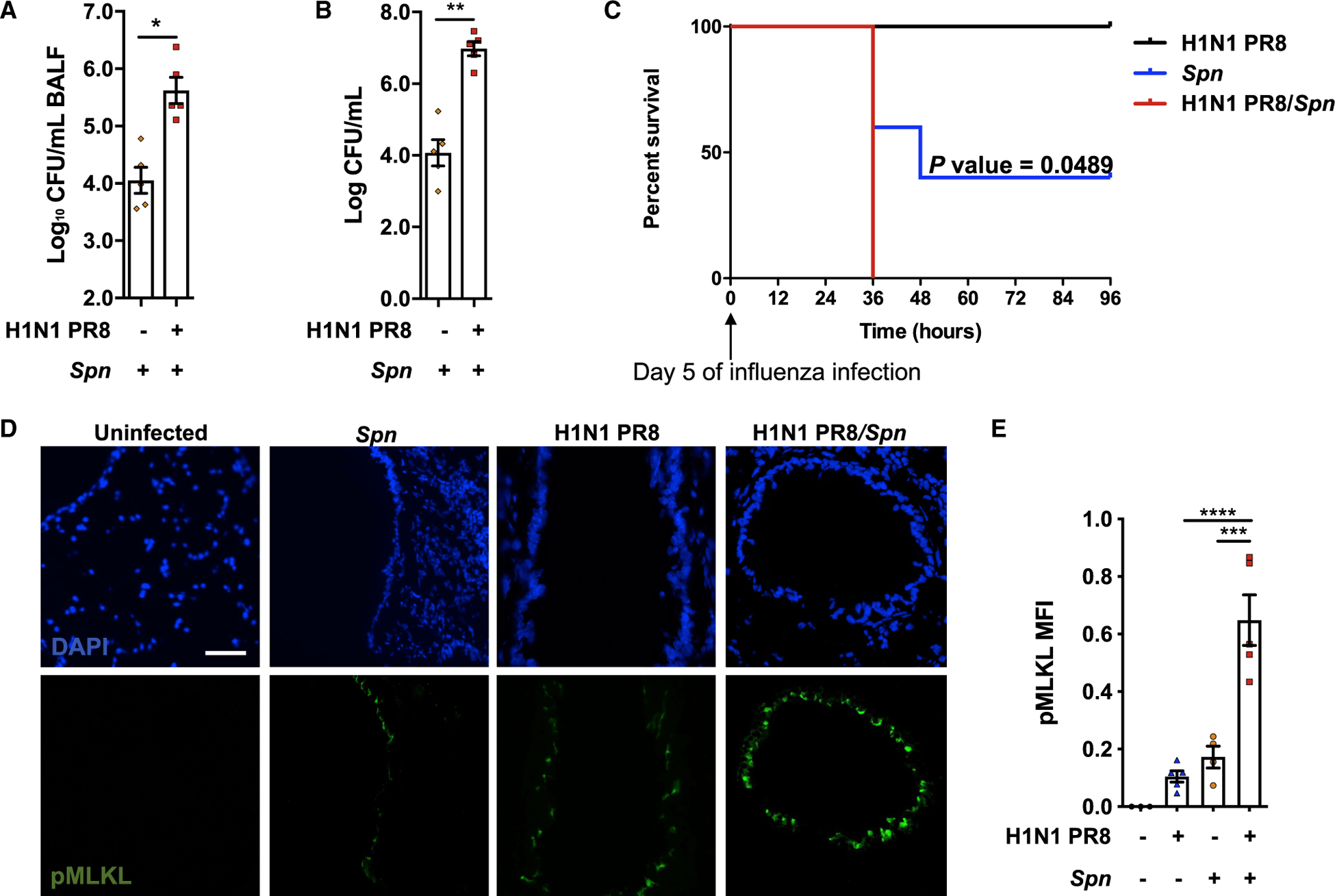
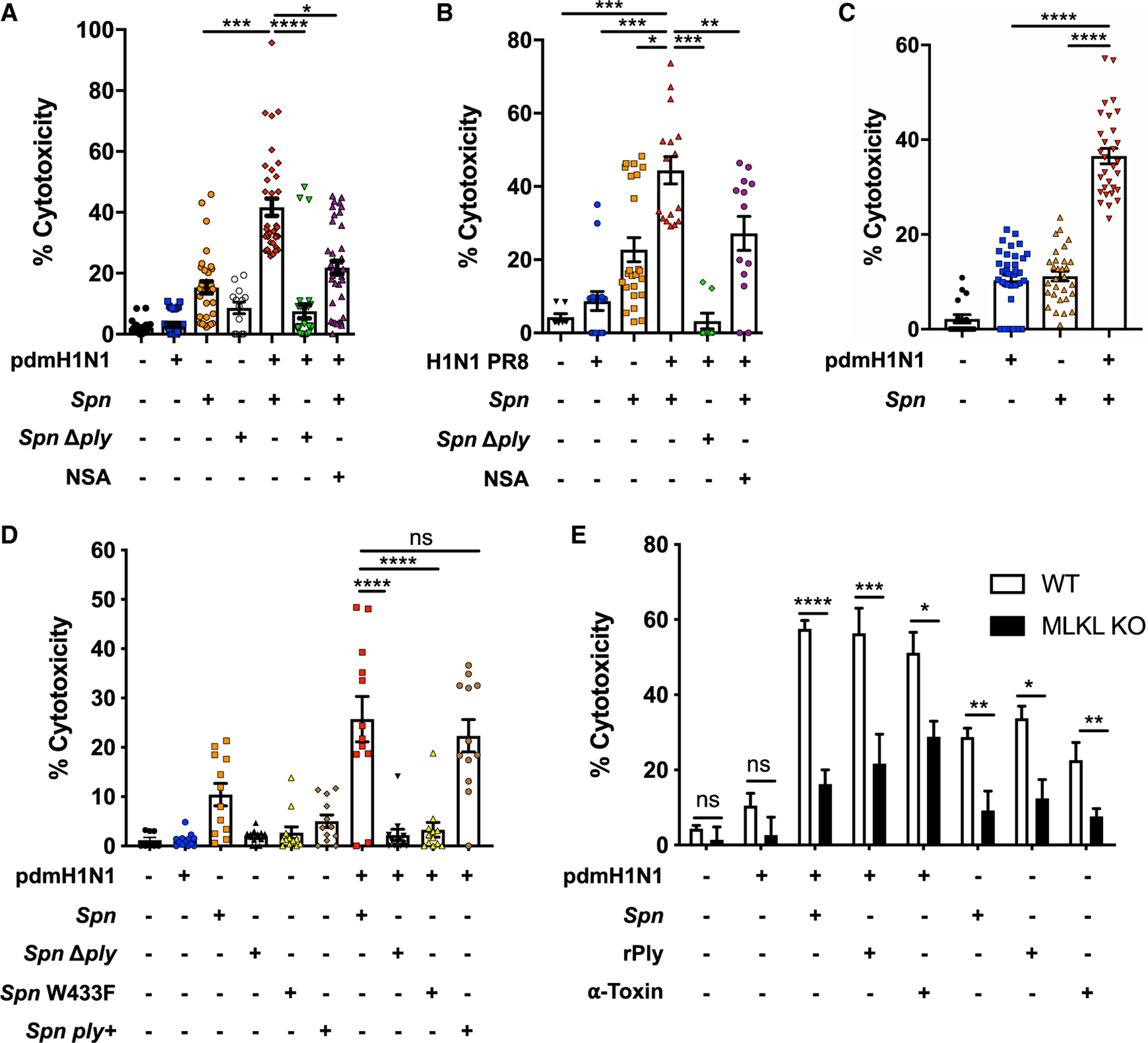
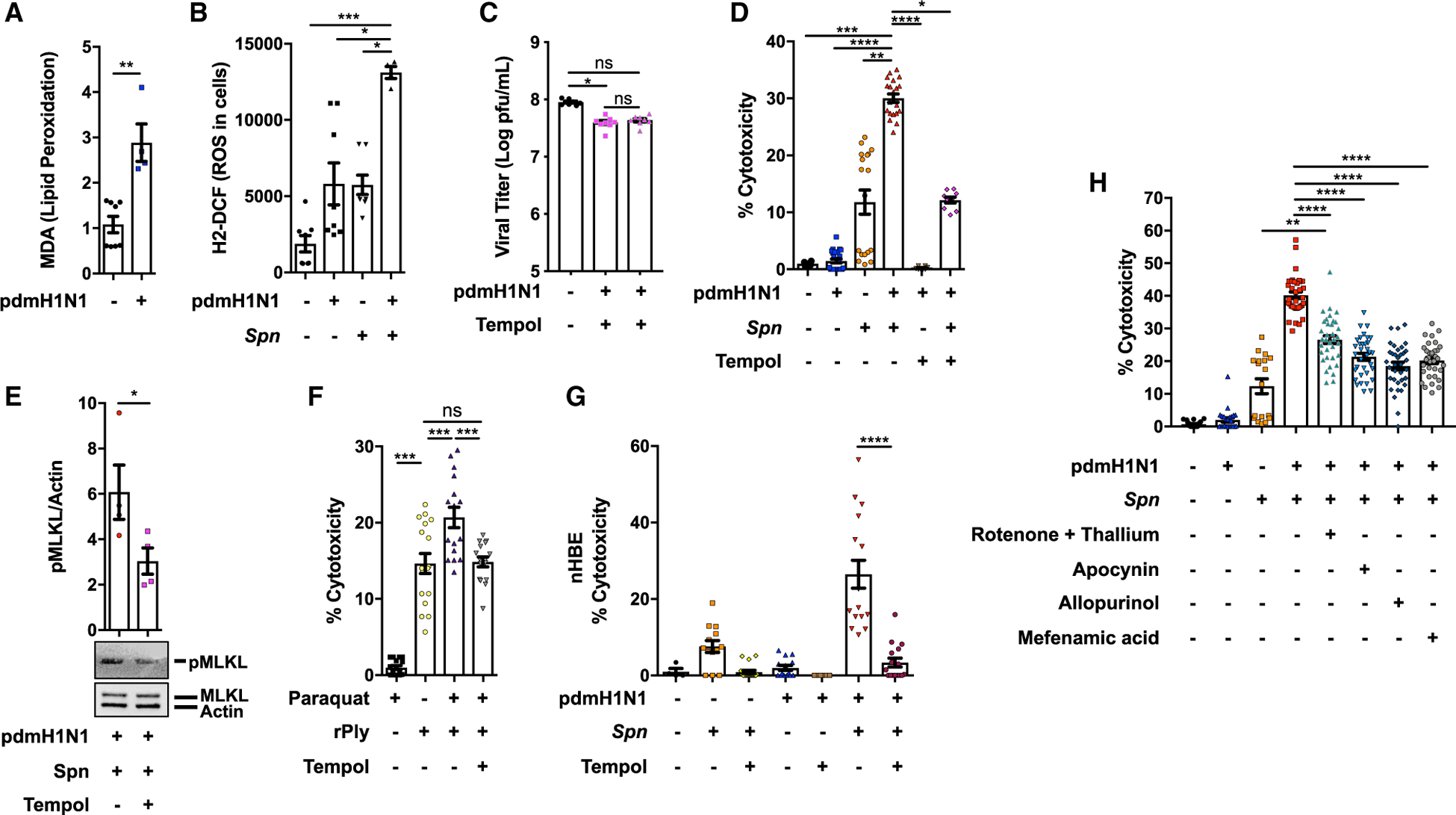
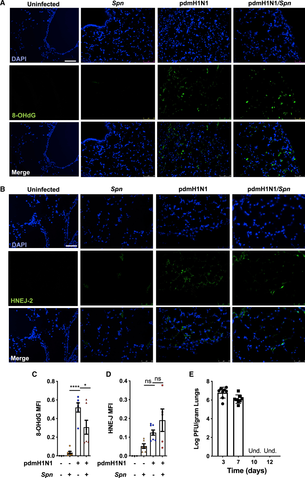
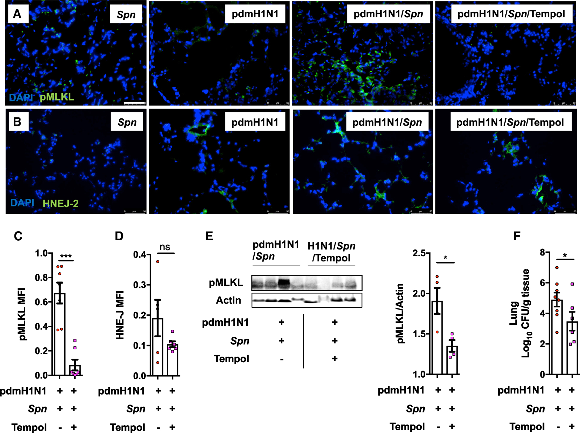
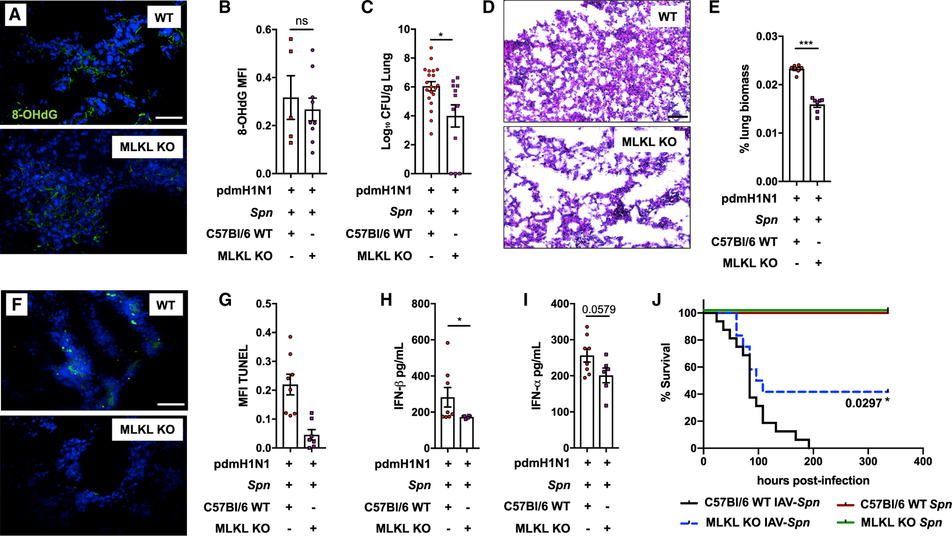
Pneumonias caused by influenza A virus (IAV) co- and secondary bacterial infections are characterized by their severity and high mortality rate. Previously, we have shown that bacterial pore-forming toxin (PFT)-mediated necroptosis is a key driver of acute lung injury during bacterial pneumonia. Here, we evaluate the impact of IAV on PFT-induced acute lung injury during co- and secondary Streptococcus pneumoniae (Spn) infection. We observe that IAV synergistically sensitizes lung epithelial cells for PFT-mediated necroptosis in vitro and in murine models of Spn co-infection and secondary infection. Pharmacoelogical induction of oxidative stress without virus sensitizes cells for PFT-mediated necroptosis. Antioxidant treatment or inhibition of necroptosis reduces disease severity during secondary bacterial infection. Our results advance our understanding on the molecular basis of co- and secondary bacterial infection to influenza and identify necroptosis inhibition and antioxidant therapy as potential intervention strategies.

Keywords: Streptococcus pneumoniae; cell death; co-infections; epithelial cells; inflammation; influenza A virus; necroptosis; oxidative stress; pneumonia; secondary bacterial infection.


Conflict of interest statement

Declaration of Interests The authors declare no competing interests.

Figures

Figure 1.
Figure 1.. IAV/Spn Co-infection Leads to Increased Mortality and Enhanced Tissue Necroptosis
(A and B) Eight-week-old C57BL/6 mice were intranasally infected with H1N1 PR8 (250 plaque-forming units [PFUs]) for 5 days and subsequently challenged intratracheally with Spn strain TIGR4 at the lethal dose (LD50) dose of 5 × 10⁵ colony-forming units [CFUs]. Mice were euthanized 24 h post-secondary infection (n = 4–5 mice/cohort). Bacterial titers in bronchoalveolar lavage fluid (BALF) (A) and blood of mice at time of sacrifice (B). (C) Survival of mice challenged with IAV, Spn, or co-infected with Spn after 5 days of IAV (n = 5). (D) Corresponding and representative images of frozen lung sections from infected mice immunofluorescently stained for p-MLKL (green) (n = 4–5/cohort). White bar denotes 50 μm. (E) Shown is the quantitation of p-MLKL levels in captured images calculated by mean fluorescent intensity (MFI). For in vivo experiments, each individual data point represents an individual animal sample. Asterisks denote the level of significance observed: = P ≤ 0.05; *** = P ≤ 0.01; *** = P ≤ 0.001; **** = P ≤ 0.0001.
Figure 2.
Figure 2.. IAV Infection Promotes PFT-Mediated Cell Death
(A and B) Lactate dehydrogenase (LDH) release was measured from A549 cells following infection with influenza A/California/7/2009 (pdmH1N1) (A) and PR8 H1N1 (B) at a MOI of 2 for 2 h and challenge with wild-type Spn (Spn, in house strain) or Ply-deficient derivative (Spn Δply) at an MOI of 10 for 4 additional h. Cells were treated with necrosulfonamide (NSA; 10 μM) when indicated. (C) LDH release was measured from MH-S alveolar macrophages following an infection workflow as in (A). (D) LDH cytotoxicity assay of supernatants from A549 cells was performed following infection with pdmH1N1 at an MOI of 2 for 2 h and challenge with Spn strains and mutants obtained from Dr. Jeffrey Weiser at an MOI of 10 for 4 h: (Zafar et al., 2017) Spn TIGR4 wild type (WT) (Spn), Ply-deficient mutant (Spn Δply), Ply point mutant deficient in pore formation (Spn W433F), and corrected mutant (Spn ply+). (E) Cytotoxicity of A549 WT (white bars) or A549 MLKL-deficient cells (black bars) was measured following the same challenge model as in (A) using Spn, recombinant pneumolysin (rPly), or alpha-toxin (α-Toxin). Individual data points in all in vitro assays represent all technical replicates collected from 3 separate experiments. Asterisks denote the level of significance observed: * = P ≤ 0.05; ** = P ≤ 0.01; *** = P ≤ 0.001; **** = P % 0.0001.
Figure 3.
Figure 3.. IAV-Mediated Oxidative Stress Potentiates Pneumolysin-Mediated Necroptosis
(A) Lipid peroxidation levels 4 h after challenge with pdmH1N1 were measured by MDA. (B) Levels of cellular ROS measured in A549 cells infected with pdmH1N1 at a MOI of 2 for 2 h and then challenged with Spn at a MOI of 10 for 2 more h. (C) Viral titers quantified (log PFU/mL) in A549 cells treated with Tempol (20 μM) for 1 h or 24 h. (D and E) Cytotoxicity (E) and corresponding p-MLKL levels (F) in A549 cells were pre-treated with Tempol for 1 h, infected with pdmH1N1 at a MOI 2 for 2 h and then challenged with Spn at an MOI of 10 for 4 additional h. (F) Cytotoxicity was measured in A549 cells pre-treated with Tempol and then treated with Paraquat (10 μM) for an additional 2 h, followed by challenge with rPly (0.1 μg) for 2 h. (G) Cytotoxicity of ex-vivo-cultured primary normal human bronchial epithelial cells pre-treated with Tempol for 1 h, infected with pdmH1N1 at a MOI of 2 for 2 h, and challenged with Spn at an MOI of 10 for 4 additional h. (H) LDH release from A549 cells pretreated with rotenone + thallium trifluoroacetate (10 nM/mL/10 nM/mL), a mitochondria-dependent ROS inhibitor; apocynin (1μM/mL), a NADPH-dependent ROS inhibitor; allopurinol (10 nM/mL), a xanthine oxidase-dependent ROS inhibitor; and mefenamic acid (20 nM/mL), a cyclooxygenase-dependent ROS inhibitor following IAV and Spn, individually and together. Individual data points in all in vitro assays represent all technical replicates collected from 3 separate experiments. Asterisks denote the level of significance observed: * = P ≤ 0.05; ** = P ≤ 0.01; *** = P ≤ 0.001; **** = P ≤ 0.0001.
Figure 4.
Figure 4.. IAV-Mediated Oxidative Stress Persists after Virus Clearance In Vivo
(A and B) Eight-week-old C57BL/6 mice were intranasally infected with A/California/7/2009 (pdmH1N1) and 10 days later challenged intratracheally with Spn. Mice were euthanized 48 h after secondary infection (n = 6–8 mice). Shown are representative immunofluorescent lung sections stained for 8-hydroxydeoxyguanosine (8-OHdG) (A) and 4-hydroxynonenal (HNE-J) (B). White bar denotes 50 μm. (C and D) Quantification of the MFI of 8-OHdG and HNE-J stainings, respectively, was performed. (E) Viral titers (log PFU/gram) in lungs at days 3, 7, 10, and 12 post-IAV infection (n = 8 mice per group) are indicated. For in vivo experiments, each individual data point represents an individual animal sample. Asterisks denote the level of significance observed: * = P ≤ 0.05; ** = P ≤ 0.01; *** = P ≤ 0.001; **** = P ≤ 0.0001.
Figure 5.
Figure 5.. Influenza Infection Potentiates Pneumolysin-Induced Necroptosis Activation during Secondary Spn Challenge
(A and B) Eight-week-old C57BL/6 mice were intranasally infected with A/California/7/2009 (pdmH1N1) and 10 days later challenged intratracheally with Spn. Mice were euthanized 48 h after secondary infection (n = 3–7 mice). Shown are bacterial titers in homogenized lung samples (A), as well as representative images of corresponding lung sections stained for p-MLKL (3 sections stained per mouse) (B). White bar denotes 50 μm. (C) MFI for p-MLKL activity was measured. (D) Densitometry and western blots for p-MLKL, MLKL, and actin from mock-, Spn-, pdmH1N1-, and pdmH1N1/Spn-infected mice (n = 3/cohort). (E–G) Eight-week-old C57BL/6 mice were intranasally infected with A/California/7/2009 (pdmH1N1) and 10 days later challenged intratracheally with Spn or Spn Δply. Mice were euthanized 48 h after secondary infection (n = 4–7 mice). Treatment with Nec1s was done intraperitoneally at 12 and 24 h following bacterial challenge. (E and F) Shown are representative images of lung tissue sections stained for p-MLKL (separate points are average of 3 pictures per mouse) (E) and MFI of pMLKL staining (F). (G) Corresponding lung bacterial titers (CFU/g tissue) calculated. (H) Survival of C57BL/6 mice following intranasal infection with pdmH1N1 for 10 days and subsequent intratracheal challenge with Spn or Spn Δply were monitored. For in vivo experiments, each individual data point represents an individual animal sample. Asterisks denote the level of significance observed: * = P ≤ 0.05; ** = P ≤ 0.01; *** = P ≤ 0.001; **** = P ≤ 0.0001.
Figure 6.
Figure 6.. Therapeutic Neutralization of ROS Reduces Necroptosis Activation during Secondary Bacterial Pneumonia
(A and B) Eight-week-old C57BL/6 mice were intranasally infected with A/California/7/2009 (pdmH1N1) and 10 days later challenged intratracheally with Spn. Mice were euthanized 48 h after secondary infection (n = 5–8 mice). Tempol treatment was done intraperitoneally at 12 and 24 h post-bacterial infection. Representative images of lung sections immunofluorescently stained for p-MLKL (A) and HNE-J (B). White bar denotes 50 μm. (C and D) Quantification of the MFI in corresponding captured images. (E) Immunoblot for pMLKL and actin of pdmH1N1-infected mice, challenged with Spn with subsequent Tempol treatment, and its densitometry quantification. (F) Bacterial titers measured in lungs at time of death. For in vivo experiments, each individual data point represents an individual animal sample. Asterisks denote the level of significance observed: * = P ≤ 0.05; ** = P ≤ 0.01; *** = P ≤ 0.001; **** = P ≤ 0.0001.
Figure 7.
Figure 7.. Inhibition of Necroptosis Reduces Disease Severity and Tissue Injury during Secondary Bacterial Pneumonia
Eight-week-old C57BL/6 mice were intranasally infected with A/California/7/2009 (pdmH1N1) and 10 days later challenged intratracheally with Spn (1,000 CFUs). Mice were euthanized 48 h after secondary infection (n > 12 mice). (A) IF of 8-OHdG. White bar denotes 50 μm. (B) Quantification of the MFI of 8-OHdG stainings. (C) Measured bacterial titers in homogenized lungs. (D) Representative H&E staining of corresponding tissue sections. Black bar denotes 100 μm. (E) Lung consolidation in tissue sections, as measured using ImageJ (white space versus lung area, separate points are the average of 3 pictures per mouse). (F and G) TUNEL stain (white bar denotes 50μm) (F) and MFI (G) of TUNEL stain quantified in lung sections. (H and I) IFN-β (H) and (I) IFN-α levels (pg/mL) (I) in lung homogenates. (J) Survival of 8-week-old WT and MLKL KO-C57BL/6 in the secondary Spn infection model (n = 8–12). For in vivo experiments, each individual data point represents an individual animal sample. Asterisks denote the level of significance observed: * = P ≤ 0.05; ** = P ≤ 0.01; *** = P ≤ 0.001; **** = P ≤ 0.0001.

Similar articles

Cited by

References

    1. Balachandran S, and Rall GF (2020). Benefits and Perils of Necroptosis in Influenza Virus Infection. J. Virol 94, e01101–19. - PMC - PubMed
    1. Brand JD, Lazrak A, Trombley JE, Shei RJ, Adewale AT, Tipper JL, Yu Z, Ashtekar AR, Rowe SM, Matalon S, and Harrod KS (2018). Influenza-mediated reduction of lung epithelial ion channel activity leads to dysregulated pulmonary fluid homeostasis. JCI Insight 3, e123467. - PMC - PubMed
    1. Brissac T, Shenoy AT, Patterson LA, and Orihuela CJ (2017). Cell invasion and pyruvate oxidase derived H2O2 are critical for Streptococcus pneumoniae mediated cardiomyocyte killing. Infect. Immun 86, e00569–17. - PMC - PubMed
    1. Brown AO, Mann B, Gao G, Hankins JS, Humann J, Giardina J, Faverio P, Restrepo MI, Halade GV, Mortensen EM, et al. (2014). Streptococcus pneumoniae translocates into the myocardium and forms unique microlesions that disrupt cardiac function. PLoS Pathog. 10, e1004383. - PMC - PubMed
    1. Byrn RA, Jones SM, Bennett HB, Bral C, Clark MP, Jacobs MD, Kwong AD, Ledeboer MW, Leeman JR, McNeil CF, et al. (2015). Pre-clinical activity of VX-787, a first-in-class, orally bioavailable inhibitor of the influenza virus polymerase PB2 subunit. Antimicrob. Agents Chemother 59, 1569–1582. - PMC - PubMed

Publication types