

Case Reports
doi: 10.1186/s41824-020-00082-y.
Epub 2020 Jul 22.
A twisted tale-radiological imaging features of COVID-19 on 18F-FDG PET/CT
Affiliations
- PMID: 32835159
- PMCID: PMC7373832
- DOI: 10.1186/s41824-020-00082-y
Item in Clipboard
Case Reports
A twisted tale-radiological imaging features of COVID-19 on 18F-FDG PET/CT
Eur J Hybrid Imaging.
2020.
Abstract
The COVID-19 pandemic has had a major impact on health care systems across the globe in a short period of time. There is a growing body of evidence surrounding the findings on hybrid imaging with FDG-PET/CT, and this case highlights the importance of molecular imaging in better understanding of the biomarkers of the disease which ultimately determine the success in building a model to predict the disease severity and monitoring the response to treatment.
Keywords: 18F-FDG PET/CT; COVID-19.
© The Author(s) 2020.
Conflict of interest statement
Competing interestsThe authors declare that they have no competing interests.
Figures
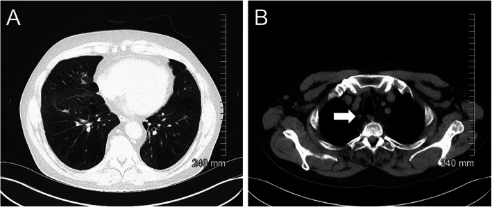
Initial CT thorax demonstrated no acute pulmonary infiltrates or lymphadenopathy (a). A morphologically normal ATS station 2R lymph node is highlighted by the white arrow (b)
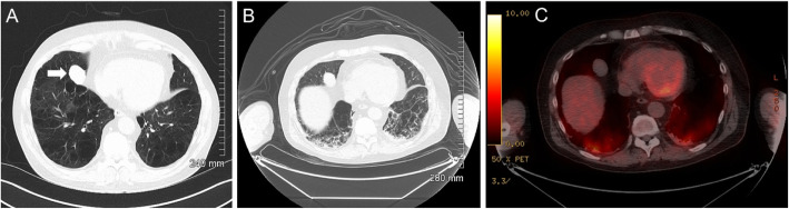
Initial CT thorax detected a right middle lobe mass (a). Subsequent PET-CT demonstrated no FDG uptake b (CT) and c (fused PET-CT)
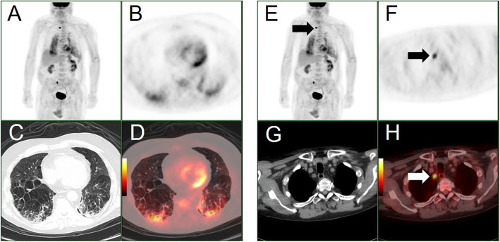
a PET maximum intensity projection (MIP), b–d: axial attenuation corrected PET, axial CT lung windows and axial fused PET/CT images through the lower thorax. e PET maximum intensity projection (MIP), f–h: axial attenuation corrected PET, axial CT mediastinal windows and axial fused PET/CT images through the superior mediastinum. These images highlight intense bilateral parenchymal metabolic activity, more prominently in the right lower lobe (images a–d) and high uptake in an ATS station 2R lymph node (e–h). Please note spurious right ureteric activity on the MIP images (images a and e)
Similar articles
-
More advantages in detecting bone and soft tissue metastases from prostate cancer using 18F-PSMA PET/CT.Hell J Nucl Med. 2019 Jan-Apr;22(1):6-9. doi: 10.1967/s002449910952. Epub 2019 Mar 7. Hell J Nucl Med. 2019. PMID: 30843003
-
Role of 2-[18F]FDG as a Radiopharmaceutical for PET/CT in Patients with COVID-19: A Systematic Review.Pharmaceuticals (Basel). 2020 Nov 10;13(11):377. doi: 10.3390/ph13110377. Pharmaceuticals (Basel). 2020. PMID: 33182811 Free PMC article. Review.
-
Multimodality approach of perioperative 18F-FDG PET/CT imaging, intraoperative 18F-FDG handheld gamma probe detection, and intraoperative ultrasound for tumor localization and verification of resection of all sites of hypermetabolic activity in a case of occult recurrent metastatic melanoma.World J Surg Oncol. 2008 Jan 10;6:1. doi: 10.1186/1477-7819-6-1. World J Surg Oncol. 2008. PMID: 18186915 Free PMC article.
-
Prognostic value of a three-scale grading system based on combining molecular imaging with 68Ga-DOTATATE and 18F-FDG PET/CT in patients with metastatic gastroenteropancreatic neuroendocrine neoplasias.Oncotarget. 2020 Feb 11;11(6):589-599. doi: 10.18632/oncotarget.27460. eCollection 2020 Feb 11. Oncotarget. 2020. PMID: 32110279 Free PMC article.
-
The role of diffusion-weighted MRI and (18)F-FDG PET/CT in the prediction of pathologic complete response after radiochemotherapy for rectal cancer: a systematic review.Radiother Oncol. 2014 Nov;113(2):158-65. doi: 10.1016/j.radonc.2014.11.026. Radiother Oncol. 2014. PMID: 25483833 Review.
Cited by
-
Not all that glitters is COVID! Differential diagnosis of FDG-avid interstitial lung disease in low-prevalence regions.Eur J Hybrid Imaging. 2020;4(1):19. doi: 10.1186/s41824-020-00088-6. Epub 2020 Oct 19. Eur J Hybrid Imaging. 2020. PMID: 33103048 Free PMC article.
-
Positron emission tomography in the COVID-19 pandemic era.Eur J Nucl Med Mol Imaging. 2021 Nov;48(12):3903-3917. doi: 10.1007/s00259-021-05347-7. Epub 2021 May 19. Eur J Nucl Med Mol Imaging. 2021. PMID: 34013405 Free PMC article. Review.
References
-
- Albano D, Bertagna F, Bertoli M, Bosio G, Lucchini S, Motta F, Panarotto MB, Peli A, Camoni L, Bengel FM, Giubbini R. Incidental findings suggestive of COVID-19 in asymptomatic patients undergoing nuclear medicine procedures in a high-prevalence region. J Nucl Med. 2020;61(5):632–636. doi: 10.2967/jnumed.120.246256. - DOI - PubMed
-
- Kirienko M, Padovano B, Serafini G, Marchianò A, Gronchi A, Seregni E, Alessi A. CT, [(18)F]FDG-PET/CT and clinical findings before and during early COVID-19 onset in a patient affected by vascular tumour. Eur J Nucl Med Mol Imaging. 2020;25:1–2. doi: 10.1007/s00259-020-04822-x. - DOI - PMC - PubMed
Publication types
LinkOut - more resources
Full Text Sources