

Quantitative Proteomic Analyses To Reveal the Key Features of Proteins in New Onset Ankylosing Spondylitis Patients
- PMID: 32832769
- PMCID: PMC7439379
- DOI: 10.1021/acsomega.0c01776
Quantitative Proteomic Analyses To Reveal the Key Features of Proteins in New Onset Ankylosing Spondylitis Patients
Abstract
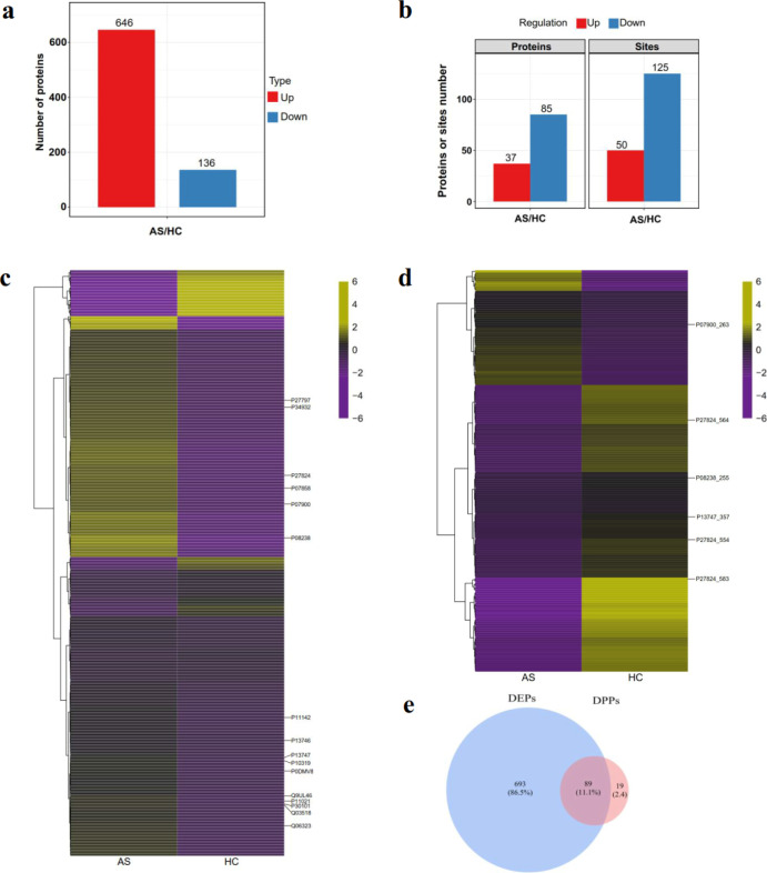
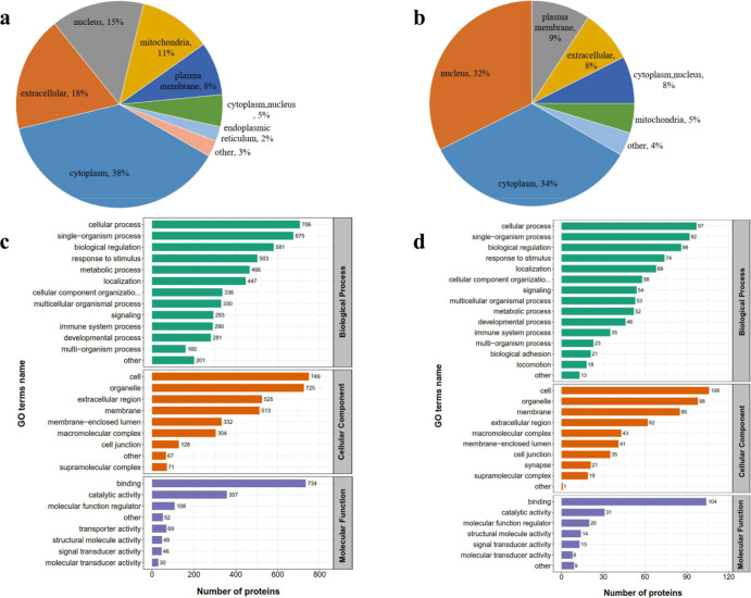
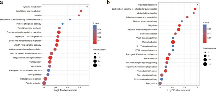
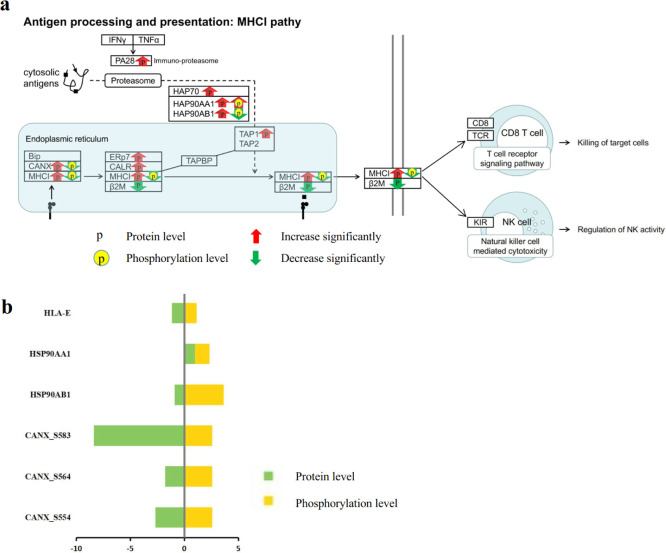
Ankylosing spondylitis (AS) is a chronic immune-mediated disease. Various immune cells play an essential role in the AS pathogenesis. However, the specific pathogenesis of AS has not been well understood. Proteomic profiles of peripheral blood mononuclear cells (PBMCs) were applied to reveal the specific pathogenesis of AS. Quantitative proteomic analyses were performed using liquid chromatography-tandem mass spectrometry (LC-MS/MS)-based methods to investigate the protein profiling of PBMCs from new-onset AS patients (n = 9) and healthy controls (n = 9). We identified 782 differentially expressed proteins (DEPs) and 527 differentially phosphorylated proteins (DPPs) between AS patients and healthy controls. The subcellular location of DEPs and DPPs showed that most of the DEPs were from the cytoplasm (n = 296, 38%), were extracellular (n = 141, 18%), and from the nucleus (n = 114, 15%); most of the DPPs were from the cytoplasm (n = 37, 34%), nucleus (n = 35, 32%), and plasma membrane (n = 10, 9%). We further identified 89 proteins with both expression and phosphorylation differences. The functional annotation of the 89 differentially expressed and phosphorylated proteins enriched in the antigen processing and presentation pathway. Four DEPs with six phosphorylated positions were found in the antigen processing and presentation pathway. The differentially expressed and phosphorylated proteins may be helpful to uncover the pathogenesis of AS. The six AS-specific proteins may serve as candidate markers for AS diagnosis and new treatment targets.
Copyright © 2020 American Chemical Society.
Conflict of interest statement
The authors declare no competing financial interest.
Figures
Similar articles
-
Global Proteomic Analyses Reveals Abnormal Immune Regulation in Patients With New Onset Ankylosing Spondylitis.Front Immunol. 2022 Mar 17;13:838891. doi: 10.3389/fimmu.2022.838891. eCollection 2022. Front Immunol. 2022. PMID: 35371008 Free PMC article.
-
Quantitative Proteomic Analysis of Peripheral Blood Mononuclear Cells in Ankylosing Spondylitis by iTRAQ.Clin Transl Sci. 2015 Oct;8(5):579-83. doi: 10.1111/cts.12265. Epub 2015 Mar 19. Clin Transl Sci. 2015. PMID: 25788137 Free PMC article.
-
Quantitative Analysis of the Global Proteome in Peripheral Blood Mononuclear Cells from Patients with New-Onset Psoriasis.Proteomics. 2018 Oct;18(19):e1800003. doi: 10.1002/pmic.201800003. Epub 2018 Sep 5. Proteomics. 2018. PMID: 30094923
-
Over-expression of talin 1 and integrin-linked kinase in PBMCs of patients with ankylosing spondylitis: a proteomic study.Clin Exp Rheumatol. 2010 Nov-Dec;28(6):828-35. Epub 2011 Jan 3. Clin Exp Rheumatol. 2010. PMID: 21122263
-
The proteomic advantage: label-free quantification of proteins expressed in bovine milk during experimentally induced coliform mastitis.Vet Immunol Immunopathol. 2010 Dec 15;138(4):252-66. doi: 10.1016/j.vetimm.2010.10.004. Epub 2010 Oct 14. Vet Immunol Immunopathol. 2010. PMID: 21067814 Review.
Cited by
-
Systematic proteomics analysis of lysine acetylation reveals critical features of placental proteins in pregnant women with preeclampsia.J Cell Mol Med. 2021 Nov;25(22):10614-10626. doi: 10.1111/jcmm.16997. Epub 2021 Oct 26. J Cell Mol Med. 2021. PMID: 34697885 Free PMC article.
-
Global Proteomic Analyses Reveals Abnormal Immune Regulation in Patients With New Onset Ankylosing Spondylitis.Front Immunol. 2022 Mar 17;13:838891. doi: 10.3389/fimmu.2022.838891. eCollection 2022. Front Immunol. 2022. PMID: 35371008 Free PMC article.
-
Systematic proteomics profiling of lysine crotonylation of the lung at Pseudoglandular and Canalicular phases in human fetus.Proteome Sci. 2023 Dec 1;21(1):22. doi: 10.1186/s12953-023-00215-8. Proteome Sci. 2023. PMID: 38041078 Free PMC article.
-
Quantitative proteomic screening uncovers candidate diagnostic and monitoring serum biomarkers of ankylosing spondylitis.Arthritis Res Ther. 2023 Apr 11;25(1):57. doi: 10.1186/s13075-023-03044-4. Arthritis Res Ther. 2023. PMID: 37041650 Free PMC article.
References
-
- Stolwijk C.; Essers I.; van Tubergen A.; Boonen A.; Bazelier M. T.; De Bruin M. L.; de Vries F. The epidemiology of extra-articular manifestations in ankylosing spondylitis: a population-based matched cohort study. Ann. Rheum. Dis. 2015, 74, 1373–1378. 10.1136/annrheumdis-2014-205253. - DOI - PubMed
-
- Gofton J. P.; Chalmers A.; Price G. E.; Reeve C. E. HL-A27 and ankylosing spondylitis in B.C. Indians. J. Rheumatol. 1975, 2, 314–318. - PubMed
-
- Londono J.; Romero-Sanchez M. C.; Torres V. G.; Bautista W. A.; Fernandez D. J.; Quiroga J. d. A.; Valle-Oñate R.; Santos A. M.; Medina J. F. The association between serum levels of potential biomarkers with the presence of factors related to the clinical activity and poor prognosis in spondyloarthritis. Rev. Bras. Reumatol. 2012, 52, 536–544. 10.1590/s0482-50042012000400006. - DOI - PubMed
LinkOut - more resources
Full Text Sources
Molecular Biology Databases
Research Materials
