

Cell intercalation in a simple epithelium
- PMID: 32829682
- PMCID: PMC7482223
- DOI: 10.1098/rstb.2019.0552
Cell intercalation in a simple epithelium
Abstract
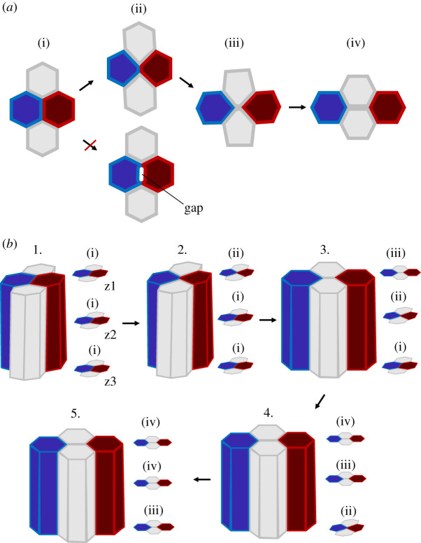
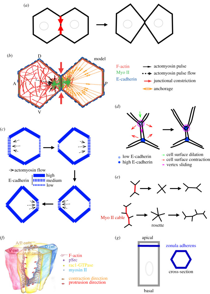
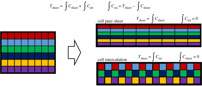
Cell intercalation is a key topological transformation driving tissue morphogenesis, homeostasis and diseases such as cancer cell invasion. In recent years, much work has been undertaken to better elucidate the fundamental mechanisms controlling intercalation. Cells often use protrusions to propel themselves in between cell neighbours, resulting in topology changes. Nevertheless, in simple epithelial tissues, formed by a single layer of densely packed prism-shaped cells, topology change takes place in an astonishing fashion: cells exchange neighbours medio-laterally by conserving their apical-basal architecture and by maintaining an intact epithelial layer. Medio-lateral cell intercalation in simple epithelia is thus an exemplary case of both robustness and plasticity. Interestingly, in simple epithelia, cells use a combinatory set of mechanisms to ensure a topological transformation at the apical and basal sides. This article is part of the discussion meeting issue 'Contemporary morphogenesis'.
Keywords: E-cadherin adhesion; actomyosin flow; actomyosin tension; junction remodelling.
Conflict of interest statement
I declare that I have no competing interests.
Figures
Similar articles
-
Neuronal immunoglobulin superfamily cell adhesion molecules in epithelial morphogenesis: insights from Drosophila.Philos Trans R Soc Lond B Biol Sci. 2020 Oct 12;375(1809):20190553. doi: 10.1098/rstb.2019.0553. Epub 2020 Aug 24. Philos Trans R Soc Lond B Biol Sci. 2020. PMID: 32829687 Free PMC article. Review.
-
The same but different: cell intercalation as a driver of tissue deformation and fluidity.Philos Trans R Soc Lond B Biol Sci. 2018 Sep 24;373(1759):20170328. doi: 10.1098/rstb.2017.0328. Philos Trans R Soc Lond B Biol Sci. 2018. PMID: 30249777 Free PMC article. Review.
-
On folding morphogenesis, a mechanical problem.Philos Trans R Soc Lond B Biol Sci. 2020 Oct 12;375(1809):20190564. doi: 10.1098/rstb.2019.0564. Epub 2020 Aug 24. Philos Trans R Soc Lond B Biol Sci. 2020. PMID: 32829686 Free PMC article. Review.
-
Dynamin-mediated endocytosis is required for tube closure, cell intercalation, and biased apical expansion during epithelial tubulogenesis in the Drosophila ovary.Dev Biol. 2016 Jan 1;409(1):39-54. doi: 10.1016/j.ydbio.2015.10.034. Epub 2015 Nov 2. Dev Biol. 2016. PMID: 26542010 Free PMC article.
-
Myosin-dependent junction remodelling controls planar cell intercalation and axis elongation.Nature. 2004 Jun 10;429(6992):667-71. doi: 10.1038/nature02590. Nature. 2004. PMID: 15190355
Cited by
-
Caenorhabditis elegans LET-413 Scribble is essential in the epidermis for growth, viability, and directional outgrowth of epithelial seam cells.PLoS Genet. 2021 Oct 21;17(10):e1009856. doi: 10.1371/journal.pgen.1009856. eCollection 2021 Oct. PLoS Genet. 2021. PMID: 34673778 Free PMC article.
-
Periderm fate and independence of tooth formation are conserved across osteichthyans.Evodevo. 2024 Oct 3;15(1):13. doi: 10.1186/s13227-024-00232-4. Evodevo. 2024. PMID: 39363199 Free PMC article.
-
Random nature of epithelial cancer cell monolayers.J R Soc Interface. 2022 May;19(190):20220026. doi: 10.1098/rsif.2022.0026. Epub 2022 May 11. J R Soc Interface. 2022. PMID: 35537474 Free PMC article.
-
Robust statistical properties of T1 transitions in a multi-phase field model of cell monolayers.Sci Rep. 2023 Jun 21;13(1):10096. doi: 10.1038/s41598-023-37064-6. Sci Rep. 2023. PMID: 37344548 Free PMC article.
-
Contemporary morphogenesis.Philos Trans R Soc Lond B Biol Sci. 2020 Oct 12;375(1809):20190549. doi: 10.1098/rstb.2019.0549. Epub 2020 Aug 24. Philos Trans R Soc Lond B Biol Sci. 2020. PMID: 32829677 Free PMC article. No abstract available.
References
Publication types
MeSH terms
LinkOut - more resources
Full Text Sources
Molecular Biology Databases
