Review
. 2020 Aug 19;21(17):5965.
doi: 10.3390/ijms21175965.

Merged Tacrine-Based, Multitarget-Directed Acetylcholinesterase Inhibitors 2015-Present: Synthesis and Biological Activity

Affiliations
Review

Merged Tacrine-Based, Multitarget-Directed Acetylcholinesterase Inhibitors 2015-Present: Synthesis and Biological Activity

Todd J Eckroat et al. Int J Mol Sci. .

Abstract

Acetylcholinesterase is an important biochemical enzyme in that it controls acetylcholine-mediated neuronal transmission in the central nervous system, contains a unique structure with two binding sites connected by a gorge region, and it has historically been the main pharmacological target for treatment of Alzheimer's disease. Given the large projected increase in Alzheimer's disease cases in the coming decades and its complex, multifactorial nature, new drugs that target multiple aspects of the disease at once are needed. Tacrine, the first acetylcholinesterase inhibitor used clinically but withdrawn due to hepatotoxicity concerns, remains an important starting point in research for the development of multitarget-directed acetylcholinesterase inhibitors. This review highlights tacrine-based, multitarget-directed acetylcholinesterase inhibitors published in the literature since 2015 with a specific focus on merged compounds (i.e., compounds where tacrine and a second pharmacophore show significant overlap in structure). The synthesis of these compounds from readily available starting materials is discussed, along with acetylcholinesterase inhibition data, relative to tacrine, and structure activity relationships. Where applicable, molecular modeling, to elucidate key enzyme-inhibitor interactions, and secondary biological activity is highlighted. Of the numerous compounds identified, there is a subset with promising preliminary screening results, which should inspire further development and future research in this field.

Keywords: Alzheimer’s disease; Friedländer reaction; acetylcholinesterase; multitarget-directed ligand; pyranopyrazole; tacrine.


Conflict of interest statement

The authors declare no conflict of interest.

Figures

Figure 1
Figure 1
(A) Hydrolysis of acetylcholine (ACh) to choline and acetate catalyzed by acetylcholinesterase (AChE). (B) View of the hAChE (PDB:4EY4) active site with key amino acid residues color coded by region (reprinted from [3], with permission from Elsevier). (C) Surface representation of the hAChE (PDB:4EY4) active site looking down the gorge with regions color coded as in (B) (reprinted from [3], with permission from Elsevier).
Figure 2
Figure 2
Structures of clinically used Alzheimer’s disease (AD) drugs. Tacrine (discontinued), donepezil, galantamine, and rivastigmine are all AChEi. Memantine is an N-methyl-d-aspartate receptor (NMDAR) antagonist. The tricyclic rings of tacrine are labeled A–C for discussion purposes.
Figure 3
Figure 3
Tacrine-based multitarget-directed ligand (MTDLs) can be classified as “linked” or “merged” based on the degree of overlap of the pharmacophores (adapted with permission from [57], copyright (2005) American Chemical Society).
Scheme 1
Scheme 1
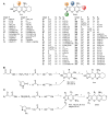
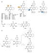
(A) Structure of pyranopyrazole tacrines 1az and 2aax [63,64,65,66,67]. (B) Synthesis of pyranopyrazole tacrines 1az. (C) Synthesis of pyranopyrazole tacrines 2aax. Reagents and conditions: (i) Ultrasonic irradiation, (S)-Pro, H2O/EtOH, rt, 10–35 min, 77–95%; (ii) cyclohexanone or cycloheptanone or tetrahydro-4H-thiopyran-4-one, AlCl3, DCE or DCM, Δ (12–24 h, 65–95%) or MWI (53–90%); (iii) DCE, Δ, 3–4 h, quant.; (iv) DABCO, EtOH, rt, 12 h, quant.
Figure 4
Figure 4
Molecular modeling of 2u in the substrate active site (CAS) of TcAChE (adapted from [66], with permission from Elsevier). The pyrazole ring forms key H-bonds with Glu199 and Tyr130 and π-π stacking with Trp84. The 4-fluorophenyl moiety forms an amide-π stacking interaction with Gly119.
Scheme 2
Scheme 2
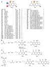
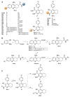
(A) Structure of pyranopyranone tacrines 5ao and 6al [68,69]. (B) Synthesis of pyranopyranone tacrines 5ao. (C) Synthesis of pyranopyranone tacrines 6al. Reagents and conditions: (i) DABCO (rt, 12 h, quant.) or Et3N (Δ, 5 min, 63–77%), EtOH, rt; (ii) cyclohexanone, AlCl3, DCE or 1,4-dioxane, Δ, 2–18 h, 50–87%.
Figure 5
Figure 5
Molecular modeling of (R)-6d in the second distinct site (PAS) of hAChE (adapted from [69]). Important interactions include π-π stacking with Trp286 and Tyr341 and H-bonds of the primary hydroxyl group with Tyr72 and the tacrine-like amino group with Tyr124.
Scheme 3
Scheme 3
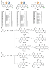
(A) Structure of pyranonaphthalene (9ac, 10a,b), pyranoquinoline (10cp), and pyranonaphthoquinone tacrines 11at [71,72,73,74]. (B) Synthesis of pyranonaphthalene (9ac, 10a,b), pyranoquinoline (10cp), and pyranonaphthoquinone tacrines 11at. Reagents and conditions: (i) 2-Naphthol, piperidine, EtOH, Δ, 12 h, 72%; (ii) cyclopentanone or cyclohexanone or cycloheptanone, AlCl3, DCE or 1,4-dioxane, Δ (2–24 h, 40–95%) or MWI (3 h, 9–70%); (iii) 1-naphthol or 8-hydroxyquinoline, piperidine, EtOH, Δ, 15 min–12 h, 47–93%; (iv) 2-hydroxy-1,4-naphthoquinone, piperidine, EtOH, Δ, 2–6 h, quant.
Scheme 4
Scheme 4
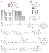
(A) Structure of other pyranotacrines 15aad and 16at [71,77,78,79]. (B) Synthesis of pyranotacrines 15aad. (C) Synthesis of pyranotacrines 16ae. (D) Synthesis of sulfamoyl-containing pyranotacrines 16ft. Reagents and conditions: (i) Ethyl benzoylacetate (R2 = CO2Et, R3 = Ph), piperidine, EtOH, Δ, 3 h, quant.; (ii) ethyl acetoacetate (R2 = CO2Et, R3 = Me) or acetylacetone (R2 = COMe, R3 = Me), piperidine, EtOH, rt, 12 h, 78–93%; (iii) cyclopentanone or cyclohexanone or cycloheptanone, AlCl3, DCE or toluene, Δ (3–24 h, 32–87%) or MWI (10 min, 70–83%); (iv) dimedone, piperidine, EtOH, rt, 12 h, 69–76%; (v) (a) SOCl2, MWI, 30 min, 88%; (b) hydroxybenzaldehyde derivative, pyridine, DCM, rt, 12 h, 85–93%; (vi) dimedone, K2CO3, EtOH, MWI, 6 min, 85–89%.
Scheme 5
Scheme 5
(A) Structure of pyridino- (19aj), indolo- (20a–c), and quinoxalinotacrines 21 [80,81,82]. (B) Synthesis of pyridinotacrines 19aj. (C) Synthesis of indolotacrines 20ac. (D) Synthesis of quinoxalinotacrine 21. Reagents and conditions: (i) Piperidine, EtOH, rt; (ii) acetone or cyclohexanone or cycloheptanone, NH4OAc, AcOH, Δ; (iii) cyclohexanone or cycloheptanone, AlCl3, DCE, Δ (18 h, 72–95%) or MWI (2 h, 16–88%); (iv) TFAA, TEA, THF, rt, 12 h, 97–99%; (v) L-Pro or picolinic acid, K2CO3, CuI, DMSO/H2O, 60 °C (12 h, 48–90%) or MWI (12 h, 26%); (vi) (a) benzaldehyde, MeOH, rt, 12 h, 97%; (b) NaBH3CN, AcOH/MeOH, rt, 12 h, 75%; (vii) (a) TEA, DMF, rt, 1.5 h, 75%; (b) Na2S2O4, H2O/MeOH, 50 °C, 3 h, 87% [83].
Scheme 6
Scheme 6
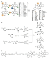
(A) Structure of pyrrolo- (22ai), pyrazolo- (22jn), and furanotacrines 22o, as well as pyrazolophthalazine tacrines 23au [84,85]. (B) Synthesis of pyrrolo- (22ai), pyrazolo- (22jn), and furanotacrine 22o. (C) Synthesis of pyrazolophthalazine tacrines 23au. Reagents and conditions: (i) EtOH, rt, 30 min, >80%; (ii) X-CH2-R2 (where X = Cl or Br, Y = CN, CO2Et, or 4-Br-Bz) TEA, Δ, 15–30 min, 63–91%; (iii) cyclopentanone or cyclohexanone, AlCl3, DCE or DCM, Δ (8–24 h, 30–95%) or MWI (30–32 min, 45–87%); (iv) malononitrile, 10% KOH, EtOH, rt, 30 min, 80%; (v) TEA, EtOH, Δ, 2 h, 67%; (vi) NiCl2·6H2O, EtOH, Δ, 4 h.
Scheme 7
Scheme 7
(A) Structure of pyrazolotacrines 24ad, 25a,b, 26ae, and 27a,b [86]. (B) Synthesis of pyrazolotacrines 24ad, 25a,b, 26ae, and 27a,b. Reagents and conditions: (i) R-CHO, piperidine, EtOH, Δ, 4 h, 80–90%; (ii) R1-NCS, EtOH, Δ, 4–6 h, 83–89%; (iii) R2-COCH2Br, NaOAc, 1,4-dioxane, Δ, 4–6 h, 60–92%; (iv) ethyl bromoacetate, NaOAc, 1,4-dioxane, Δ, 4–6 h, 68–79%.
Figure 6
Figure 6
Molecular modeling of 27a (rose) and 27b (purple) with hAChE showing the interaction with the PAS (adapted from [86], with permission from Elsevier). Key interactions noted are the tricyclic core showing favorable hydrophobic interactions with Tyr341, Tyr337, Phe338, and Val294, the 4-chlorophenyl substituent showing hydrophobic interactions with Tyr341 and Trp286, the quinolinyl nitrogen showing an H-bond with Arg296, and the thiazolidinone moiety showing favorable interactions with Glu292 and Ser293.
Scheme 8
Scheme 8
(A) Structure of thiourea tacrines 28al, 29ac, 31, and 33 and urea tacrines 30 and 32 [89,90]. (B) Synthesis of thiourea tacrines 28al. (C) Synthesis of thiourea and urea tacrines 29ac and 3033. Reagents and conditions: (i) Thiourea, NaOMe, EtOH, Δ, 12 h, 10–29%; (ii) cyclohexanone, AlCl3, DCE, MWI, 1–2 h, 46–70%; (iii) SCN-R, pyridine, Δ, 10 h, 85–86%; (iv) urea or thiourea, 300 °C, 1 h, 64–74%; (v) urea or thiourea, 200 °C, 1 h, 76–83%.
Scheme 9
Scheme 9
(A) Structure of amido- (36ap), amino- (37 and 39ae), and iminotacrines 38a,b [90,94]. (B) Synthesis of amidotacrines 36ap. (C) Synthesis of amino- (37 and 39ae) and iminotacrines 38a,b. Reagents and conditions: (i) BF3·Et2O, toluene, Δ, 4 h, 84%; (ii) 1-methylpyrazole-4-boronic acid pinacol ester or 5-pyrimidineboronic acid pinacol ester, Na2CO3, Pd(PPh3)4, 1,4-dioxane/H2O, Δ, 2 h, 52%; (iii) NH4OH or N2H4·H2O, MeOH, 60 °C, 2–3 h, 73–85%; (iv) LiOH·H2O, THF/H2O/MeOH, rt, 3 h, 79–90%; (v) benzylamine or phenylethylamine or phenylpropylamine, T3P, TEA, DMF, 60 °C, 4 h, 59–67%; (vi) 5-amino-1-methyl-1H-pyrazole, HATU, DIPEA, DMF, rt, 12 h, 63–71%; (vii) 70% H2SO4, Δ, 5 h; (viii) R-C6H4-CHO, pyridine, Δ, 6 h, 87–90%; (ix) NH2NH-Bz, Δ, 1 h, 84%; (x) X-R (where X = Cl or Br), 1,4-dioxane, Δ, 5–10 h, 69–89%.
Figure 7
Figure 7
Molecular modeling of 36c with TcAChE showing the interaction with the CAS (adapted from [94], with permission from Elsevier). Key interactions noted are the tacrine moiety π-π stacking with Trp84 and Tyr337 and H-bonding to His447 and the hydrazide H-bonding with Tyr337.
Scheme 10
Scheme 10
(A) Structure of naphthalene pyranopyrimidinones 42ai and 43ai, naphthoquinone pyranopyrimidinones 44al, and naphthalene pyrimidinimines 42jo and 43jo [95,96,97]. (B) Synthesis of naphthalene pyranopyrimidinones 42ai and 43ai and naphthoquinone pyranopyrimidinones 44al. (C) Synthesis of naphthalene pyranopyrimidinimines 42jo and 43jo. Reagents and conditions: (i) 2-Naphthol or 1-naphthol, piperidine, EtOH, MWI, 10 min, 68–95%; (ii) γ-butyrolactam or δ-valerolactam or ε-caprolactam, POCl3, DCE or PhBr, MWI, 15 min, 70–96%; (iii) potassium phthalimide-N-oxyl, H2O, Δ, 30–60 min, >90%.
Figure 8
Figure 8
Molecular modeling of (S)-42n with hAChE showing the interaction with the mid-gorge region (adapted from [96], © 2016 WILEY-VCH Verlag GmbH & Co. KGaA, Weinheim). Key interactions noted are the π-π stacking between Trp86 and the benzochromeno moiety and an H-bond between Asp74 and the imino moiety.
Scheme 11
Scheme 11
(A) Structure of 2-methoxyhuprine (50) ((−)-huperine A shown for reference) and propargyl tacrines 51ac and propargyl ethylenediamine tacrines 52a,b [106,107]. (B) Synthesis of 2-methoxyhuprine (50). (C) Synthesis of propargyl tacrines 51ac. (D) Synthesis of propargyl ethylenediamine tacrines 52a,b. Reagents and conditions: (i) 1 M NaOH, 1,4-dioxane, Δ, 18 h, 82%; (ii) 5% Pd/C, H2, EtOH, rt, 2 h, quant.; (iii) AlCl3, DCE, MWI, 2 h, 80%; (iv) propargyl bromide (1.1 eq), KOH or K2CO3, MeCN, rt, 12–36 h, 35–82%; (v) propargyl bromide (2.1 eq), KOH or K2CO3, MeCN, rt, 12–36 h, 35–82%.
Scheme 12
Scheme 12
(A) Structure of semicarbazone tacrines 53a,b and indenoquinolines 54ao [108,111]. (B) Synthesis of semicarbazone tacrines 53a,b. (C) Synthesis of indenoquinolines 54ao. Reagents and conditions: (i) 1,4-Doxane, Δ, 24 h, 80%; (ii) thiosemicarbazide hydrochloride or semicarbazide hydrochloride, EtOH, Δ, 3 h, 75–79%; (iii) (a) InCl3, toluene, Δ, 24 h; (b) 2 M NaOH, Δ, 24 h, 13–50%; (iv) PhB(OH)2 (1.3 or 2.6 eq.), Pd(PPh3)4, K2CO3, 1,4-dioxane, Δ, 4 h, 80–96%.

Similar articles

Cited by

References

    1. Silman I., Sussman J.L. Acetylcholinesterase: ‘classical’ and ‘non-classical’ functions and pharmacology. Curr. Opin. Pharmacol. 2005;5:293–302. doi: 10.1016/j.coph.2005.01.014. - DOI - PubMed
    1. Quinn D.M. Acetylcholinesterase: Enzyme structure, reaction dynamics, and virtual transition states. Chem. Rev. 1987;87:955–979. doi: 10.1021/cr00081a005. - DOI
    1. Ochoa R., Rodriguez C.A., Zuluaga A.F. Perspectives for the structure-based design of acetylcholinesterase reactivators. J. Mol. Graph. Model. 2016;68:176–183. doi: 10.1016/j.jmgm.2016.07.002. - DOI - PubMed
    1. Bajda M., Więckowska A., Hebda M., Guzior N., Sotriffer C.A., Malawska B. Structure-based search for new inhibitors of cholinesterases. Int. J. Mol. Sci. 2013;14:5608–5632. doi: 10.3390/ijms14035608. - DOI - PMC - PubMed
    1. Mercey G., Verdelet T., Renou J., Kliachyna M., Baati R., Nachon F., Jean L., Renard P.-Y. Reactivators of acetylcholinesterase inhibited by organophosphorus nerve agents. Acc. Chem. Res. 2012;45:756–766. doi: 10.1021/ar2002864. - DOI - PubMed

MeSH terms

LinkOut - more resources