. 2020 Aug 10;18(1):310.
doi: 10.1186/s12967-020-02478-7.

Impact of HIV-1 Vpr manipulation of the DNA repair enzyme UNG2 on B lymphocyte class switch recombination

Affiliations

Impact of HIV-1 Vpr manipulation of the DNA repair enzyme UNG2 on B lymphocyte class switch recombination

Patrick Eldin et al. J Transl Med. .

Abstract

Background: HIV-1 Vpr encodes a 14 kDa protein that has been implicated in viral pathogenesis through modulation of several host cell functions. In addition to pro-apoptotic and cytostatic properties, Vpr can redirect cellular E3 ubiquitin ligases (such as DCAF1-Cul4A E3 ligase complex) to target many host proteins and interfere with their functions. Among them, Vpr binds the uracil DNA glycosylase UNG2, which controls genome uracilation, and induces its specific degradation leading to loss of uracil removal activity in infected cells. Considering the essential role of UNG2 in antibody diversification in B-cells, we evaluated the impact of Vpr on UNG2 fate in B lymphocytes and examined the functional consequences of UNG2 modulations on class switch recombination (CSR).

Methods: The impact of Vpr-induced UNG2 deregulation on CSR proficiency was evaluated by using virus-like particles able to deliver Vpr protein to target cells including the murine model CSR B cell line CH12F3 and mouse primary B-cells. Co-culture experiments were used to re-examine the ability of Vpr to be released by HIV-1 infected cells and to effectively accumulate in bystander B-cells. Vpr-mediated UNG2 modulations were monitored by following UNG2 protein abundance and uracil removal enzymatic activity.

Results: In this study we report the ability of Vpr to reduce immunoglobulin class switch recombination (CSR) in immortalized and primary mouse B-cells through the degradation of UNG2. We also emphasize that Vpr is released by producing cells and penetrates bystander B lymphocytes.

Conclusions: This work therefore opens up new perspectives to study alterations of the B-cell response by using Vpr as a specific CSR blocking tool. Moreover, our results raise the question of whether extracellular HIV-1 Vpr detected in some patients may manipulate the antibody diversification process that engineers an adapted response against pathogenic intruders and thereby contribute to the intrinsic B-cell humoral defect reported in infected patients.

Keywords: Class switch recombination; Human immunodeficiency virus; Uracil DNA glycosylase 2; Uracilation; Vpr.


Conflict of interest statement

The authors declare no competing interests.

Figures
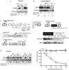
Fig. 1
Fig. 1
HA-Vpr-delivering VLPs decrease UNG2 expression in human B lymphocytes. a Daudi cells were infected with VSV-G pseudotyped wt HIV-1 at MOI of 25. 24 and 72 h after infection UNG2 levels were examined by immunoblot with anti-UNG2 antibody or anti-α-tubulin as loading control. b Daudi-CD4 + cells were infected with either HIV-1 wt, HIV-1ΔVpr or VSV-G pseudotyped wt HIV-1 at the indicated MOI. 72 h after infection UNG2 levels were examined by immunoblot with anti-UNG2 antibody or anti-α-tubulin as loading control (Ni: non-infected). c HA-Vpr-delivering VLPs were generated by cotransfecting HEK 293T cells with pCMV HA-Vpr along with a packaging vector pCMVΔR8.2ΔVpr, an Envelope vector pCMV VSV-G and the defective lentiviral vector pHR-GFP. VLPs produced in the presence (VLP HA-Vpr) or absence of pCMV HA-Vpr (VLP ΔVpr) were purified on sucrose gradients, and titrated by p24 ELISA kit. VLP titers were also determined by transducing 293T cells with serial dilutions of virus suspensions. Virus extracts corresponding to the indicated p24 amount were resolved on PAGE-SDS gels, and immunoblotted with specific antibodies for their content in p24 and HA-Vpr. d Daudi B-cells were transduced with VLP HA-Vpr or ΔVpr at M.O.I of 10. Cells were lysed at different time points and examined by immunoblot for UNG2 content with anti-UNG2 antibody or anti-α-tubulin as loading control. Lanes were regrouped from two different gels, see Supplementary dataset for detail. UNG2 levels at the various time points were quantified with ImageJ and normalized with their corresponding α-tubulin levels and plotted as a UNG2% of untransduced (Ut) cells. Lanes were cropped from blot membranes serially probed with anti-UNG2 then with anti-αtubulin antibodies (see Additional file 2: Supplementary dataset 2)
Fig. 2
Fig. 2
VLP-delivered HA-Vpr induces a proteasome-dependent decrease in human B lymphocyte uracil removal capacity. a Daudi B-cells were transduced with VLP HA-Vpr wt or mutant (R90K, Q65R or W54R) or ΔVpr at M.O.I of 10. 72 h later cells were lysed and examined by immunoblot for UNG2 content (with anti-UNG2 antibody or anti-α tubulin as control). Lanes were cropped from different parts of a unique gel (see Additional file 2: Supplementary dataset 2). Corresponding UNG activity was measured in the presence or absence of the UNG inhibitor UGI. b Daudi cells were treated for 1 h with 50 µM MG132 and then transduced with the indicated VLPs at MOI of 10. 24 h later, UNG2 content was analyzed with anti-UNG2 by western blotting (lanes were cropped from different parts of a unique gel (see Additional file 2: Supplementary dataset 2)) and UNG activity as in (a). Values are the means of triplicate measurements ± SD. Statistical significance was determined using the ANOVA test. Ut: untransduced
Fig. 3
Fig. 3
Transduction of Daudi B-cells by HA-Vpr-delivering VLPs increases genome uracilation. Daudi B-cells were transduced with VLP HA-Vpr at a MOI of 5. a UNG2 levels were analyzed by western blot with anti-UNG2 antibodies at 24 and 120 h post infection. Lanes were cropped from blot membranes serially probed with anti-UNG2 then with anti-αtubulin antibodies (see Additional file 2: Supplementary dataset 2). UNG activity was measured in the corresponding whole cell extracts. b DNA was extracted from cells 120 h post-transduction and treated with Methoxyamine (Mx) to protect pre-existing AP sites. Recombinant E. coli UDG was then used to excise uracil and generate AP sites which were quantified by ELISA. Numbers of AP sites per 105 bp were measured in triplicate with the error bars representing the standard error of the mean. c DNA of ethanol-fixed cells was labeled with propidium iodide. Cell cycle progression was followed by flow cytometry. G2/M:G0/G1 ratios are indicated in the upper right corner of each histogram. d Apoptosis was followed by flow cytometry after labeling with Annexin V-FITC in the presence of CaCl2. Ut: untransduced
Fig. 4
Fig. 4
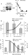
Impact of Vpr on CSR in immortalized B-cells. a Left panel, CH12F3 cells were transduced with VLP HA-Vpr at a M.O.I. of 10. 48 h later, UNG2 content was assessed by western blotting with anti-UNG2 antibodies as described in Materials and methods. Lanes were cropped from blot membranes serially probed with anti-UNG2 then with anti-αtubulin antibodies (see Additional file 2: Supplementary dataset 2). Right panel, CH12F3 cells transduced with VLP HA-Vpr (M.O.I. of 5) were examined for UNG activity at days 0, 1, 2, 3, 4 and 7. b After 24 h in culture CSR was induced by IL-4/α-CD40/TGF-β stimulation for 3 days. Class switch efficiency from IgM-to-IgA isotype was evaluated by flow cytometry by measuring the  % IgGA+ B220+ cells (left panel). DNA from corresponding cells was extracted and uracil content (dUrd per 105 bp) was determined by LC/MS/MS. Cells transduced with VLP ΔVpr and mock-transduced cells are shown as controls. c CH12F3 B-cells were transduced with increasing MOI of VLP HA-Vpr. 24 h later, corresponding isotype switching was induced by cytokine stimulation for 3 days and evaluated by flow cytometry. Values are the mean of duplicate experiments ± SD. Corresponding UNG activities were measured in the presence or absence of the UNG inhibitor UGI. Stim: stimulated; Unstim: unstimulated
Fig. 5
Fig. 5
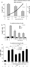
Impact of VLP-delivered Vpr on CSR efficiency in primary B-cells. a Primary B-cells were transduced with VLPs HA-Vpr or ΔVpr at a M.O.I. of 10. 24 h later CSR was induced by IL-4/α-CD40/TGF-β stimulation for 3 days. IgM-to-IgG1 isotype class switch efficiency was evaluated by flow cytometry by measuring the % IgG1+ B220+. DNA from corresponding cells was extracted and uracil content (dUrd per 105 bp) was determined by LC/MS/MS. b Primary B-cells isolated from either wt C57BL/6 or AID−/− mice, were transduced with increasing MOI of VLP HA-Vpr. 24 h later, isotype switching was induced by cytokine stimulation for 3 days and evaluated by flow cytometry. Values are the mean of duplicate experiments ± SD. Ut, untransduced, non stim: non stimulated. c Primary B-cells were transduced with VLP HA-Vpr (wt or mutants R90K, Q65R, W54R) or ΔVpr. 24 h later CSR was induced as stated above. Class switch efficiency was evaluated by flow cytometry by measuring % IgG1+ B220+ cells. For VLP-transduced cells, % switched cells was determined from GFP+-gated populations. Values are derived from three independent experiments with n corresponding to the number of replicates
Fig. 6
Fig. 6
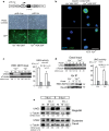
Vpr released from HEK 293T producing cells and Magic5B HIV-1 infected cells can be taken up by Daudi B-cells. a VLPs able to drive the constitutive expression of Vpr by delivery of the pHR-Vpr (Vpr+) lentiviral expression cassette or control VLPs containing a pHR-ΔVpr cassette were used to transduce HEK 293T. After 72 h, transduction efficiency was confirmed by the expression of GFP. b Cells were then washed and co-cultured with Daudi B-cells suspended in culture hanging inserts. After an additional 72 h, Daudi cells were collected, immobilized on poly(l-lysine) coated cover slips, formalin-fixed and labeled with DAPI and anti-Vpr antibodies to visualize nuclei and Vpr content, respectively. For each condition, whole cell extracts of producer HEK 293T cells (c) and of target Daudi cells (d) were analyzed for UNG2 content by immunoblot using αtubulin as a loading control. Lanes were cropped from blot membranes serially probed with anti-UNG2 then with anti-αTubulin antibodies (see Supplementary dataset). Uracil DNA glycosylase activity in the lysate was determined in the absence (NT) or presence of the UNG inhibitor UGI (0.2 U). Values are the means of triplicate experiments ± SD. Statistical significance was determined using the ANOVA test. Vpr content was determined by immunoprecipitation using anti-Vpr antibodies. Vpr was visualized by extending exposition time as specified in Additional file 2: Supplementary dataset 2. e Daudi cells maintained in hanging cell inserts were left in contact for the indicated time with MAGIC5B cells previously infected with HIV-1 wt or ΔVpr at MOI of 10. Target Daudi and producer MAGICB cell populations were harvested and whole cell extracts were prepared for immunoblot analysis of their UNG2 levels using αtubulin as a loading control. Lanes were cropped from blot membranes serially probed with anti-UNG2 then with anti-αTubulin antibodies (see Additional file 2: Supplementary dataset 2)

Similar articles

Cited by

References

    1. Guenzel CA, Herate C, Benichou S. HIV-1 Vpr-a still ‘enigmatic multitasker’. Front Microbiol. 2014;5:127. - PMC - PubMed
    1. Goh WC, et al. HIV-1 Vpr increases viral expression by manipulation of the cell cycle: a mechanism for selection of Vpr in vivo. Nat Med. 1998;4:65–71. - PubMed
    1. Connor RI, Chen BK, Choe S, Landau NR. Vpr is required for efficient replication of human immunodeficiency virus type-1 in mononuclear phagocytes. Virology. 1995;206:935–944. - PubMed
    1. Popov S, Rexach M, Ratner L, Blobel G, Bukrinsky M. Viral protein R regulates docking of the HIV-1 preintegration complex to the nuclear pore complex. J Biol Chem. 1998;273:13347–13352. - PubMed
    1. Agostini I, et al. The human immunodeficiency virus type 1 Vpr transactivator: cooperation with promoter-bound activator domains and binding to TFIIB. J Mol Biol. 1996;261:599–606. - PubMed

Publication types

Substances