Establishment of international autoantibody reference standards for the detection of autoantibodies directed against PML bodies, GW bodies, and NuMA protein
- PMID: 32776893
- PMCID: PMC7855248
- DOI: 10.1515/cclm-2020-0981
Establishment of international autoantibody reference standards for the detection of autoantibodies directed against PML bodies, GW bodies, and NuMA protein
Abstract
Objectives: Reference materials are important in the standardization of autoantibody testing and only a few are freely available for many known autoantibodies. Our goal was to develop three reference materials for antibodies to PML bodies/multiple nuclear dots (MND), antibodies to GW bodies (GWB), and antibodies to the nuclear mitotic apparatus (NuMA).
Methods: Reference materials for identifying autoantibodies to MND (MND-REF), GWB (GWB-REF), and NuMA (NuMA-REF) were obtained from three donors and validated independently by seven laboratories. The sera were characterized using indirect immunofluorescence assay (IFA) on HEp-2 cell substrates including two-color immunofluorescence using antigen-specific markers, western blot (WB), immunoprecipitation (IP), line immunoassay (LIA), addressable laser bead immunoassay (ALBIA), enzyme-linked immunosorbent assay (ELISA), and immunoprecipitation-mass spectrometry (IP-MS).
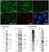
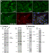
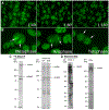
Results: MND-REF stained 6-20 discrete nuclear dots that colocalized with PML bodies. Antibodies to Sp100 and PML were detected by LIA and antibodies to Sp100 were also detected by ELISA. GWB-REF stained discrete cytoplasmic dots in interphase cells, which were confirmed to be GWB using two-color immunofluorescence. Anti-Ge-1 antibodies were identified in GWB-REF by ALBIA, IP, and IP-MS. All reference materials produced patterns at dilutions of 1:160 or greater. NuMA-REF produced fine speckled nuclear staining in interphase cells and staining of spindle fibers and spindle poles. The presence of antibodies to NuMA was verified by IP, WB, ALBIA, and IP-MS.
Conclusions: MND-REF, GWB-REF, and NuMA-REF are suitable reference materials for the corresponding antinuclear antibodies staining patterns and will be accessible to qualified laboratories.
Keywords: GW body; NuMA; autoimmunity; multiple nuclear dots; reference materials.
Conflict of interest statement
Figures


Similar articles
-
Autoantibodies to GW bodies and other autoantigens in primary biliary cirrhosis.Clin Exp Immunol. 2011 Feb;163(2):147-56. doi: 10.1111/j.1365-2249.2010.04288.x. Epub 2010 Nov 22. Clin Exp Immunol. 2011. PMID: 21091667 Free PMC article.
-
Two major autoantigen-antibody systems of the mitotic spindle apparatus.Arthritis Rheum. 1996 Oct;39(10):1643-53. doi: 10.1002/art.1780391006. Arthritis Rheum. 1996. PMID: 8843854
-
Autoantibodies to mRNA processing pathways (glycine and tryptophan-rich bodies antibodies): prevalence and clinical utility in a South Australian cohort.Pathology. 2019 Dec;51(7):723-726. doi: 10.1016/j.pathol.2019.07.008. Epub 2019 Oct 17. Pathology. 2019. PMID: 31630877
-
Antinuclear antibodies as ancillary markers in primary biliary cirrhosis.Expert Rev Mol Diagn. 2012 Jan;12(1):65-74. doi: 10.1586/erm.11.82. Expert Rev Mol Diagn. 2012. PMID: 22133120 Review.
-
Autoantibodies against "nuclear dots" in primary biliary cirrhosis.Semin Liver Dis. 1997 Feb;17(1):71-8. doi: 10.1055/s-2007-1007184. Semin Liver Dis. 1997. PMID: 9089912 Review.
Cited by
-
Anti-dense fine speckled 70 (DFS70) autoantibodies: correlates and increasing prevalence in the United States.Front Immunol. 2023 Jun 23;14:1186439. doi: 10.3389/fimmu.2023.1186439. eCollection 2023. Front Immunol. 2023. PMID: 37426660 Free PMC article.
-
Application of collagen triple helix repeat containing-1 and mitotic spindle apparatus antibody in small cell lung cancer diagnosis.J Clin Lab Anal. 2022 May;36(5):e24412. doi: 10.1002/jcla.24412. Epub 2022 Apr 6. J Clin Lab Anal. 2022. PMID: 35385156 Free PMC article.
-
Strong Association of the Myriad Discrete Speckled Nuclear Pattern With Anti-SS-A/Ro60 Antibodies: Consensus Experience of Four International Expert Centers.Front Immunol. 2021 Oct 5;12:730102. doi: 10.3389/fimmu.2021.730102. eCollection 2021. Front Immunol. 2021. PMID: 34675922 Free PMC article.
References
-
- Meroni PL, Schur PH. ANA screening: an old test with new recommendations. Ann Rheum Dis 2010. August;69:1420–2. - PubMed
-
- Chan EKL, Fritzler MJ, Wiik A, Andrade LE, Reeves WH, Tincani A, et al. Autoantibody standardization committee in 2006. Autoimmun Rev 2007. September;6:577–80. - PubMed
-
- Ching RW, Dellaire G, Eskiw CH, Bazett-Jones DP JJocs. PML bodies: a meeting place for genomic loci?. J Cell Sci 2005;118: 847–54. - PubMed
Publication types
MeSH terms
Substances
Grants and funding
LinkOut - more resources
Full Text Sources
Research Materials
Miscellaneous
