. 2020 Jul 15:14:214.
doi: 10.3389/fncel.2020.00214. eCollection 2020.

Spinal Inhibition of GABAB Receptors by the Extracellular Matrix Protein Fibulin-2 in Neuropathic Rats

Affiliations

Spinal Inhibition of GABAB Receptors by the Extracellular Matrix Protein Fibulin-2 in Neuropathic Rats

Marie-Amélie Papon et al. Front Cell Neurosci. .

Abstract

In the central nervous system, the inhibitory GABAB receptor is the archetype of heterodimeric G protein-coupled receptors (GPCRs). Receptor interaction with partner proteins has emerged as a novel mechanism to alter GPCR signaling in pathophysiological conditions. We propose here that GABAB activity is inhibited through the specific binding of fibulin-2, an extracellular matrix protein, to the B1a subunit in a rat model of neuropathic pain. We demonstrate that fibulin-2 hampers GABAB activation, presumably through decreasing agonist-induced conformational changes. Fibulin-2 regulates the GABAB-mediated presynaptic inhibition of neurotransmitter release and weakens the GABAB-mediated inhibitory effect in neuronal cell culture. In the dorsal spinal cord of neuropathic rats, fibulin-2 is overexpressed and colocalized with B1a. Fibulin-2 may thus interact with presynaptic GABAB receptors, including those on nociceptive afferents. By applying anti-fibulin-2 siRNA in vivo, we enhanced the antinociceptive effect of intrathecal baclofen in neuropathic rats, thus demonstrating that fibulin-2 limits the action of GABAB agonists in vivo. Taken together, our data provide an example of an endogenous regulation of GABAB receptor by extracellular matrix proteins and demonstrate its functional impact on pathophysiological processes of pain sensitization.

Keywords: GABAB receptor; disinhibition; fibulin-2; neuropathic pain; spinal cord.


Figures

FIGURE 1
FIGURE 1
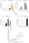
Fibulin-2 regulates baclofen-induced GABAB conformational changes in vitro. Fibulin-2 dose-dependently prevents agonist-induced conformational changes. FRET efficiency is measured between GFP-GABAB1a and DsRed-GABAB2 with different concentrations of baclofen (1 nM to 1 mM) (black circles, control). When fibulin-2 expression is inhibited with anti-fibulin-2 siRNA (blue triangles), efficacy of baclofen is increased (maximal effect of blue plot vs. black plot, p < 0.05). In contrast, in cells treated with exogenous fibulin-2 protein at 25 nM (green plot), maximal effect of baclofen is decreased as compared to control (maximal effect of fibulin-2 at 25 nM vs. control, ∗∗p < 0.01). With 50 nM of fibulin-2 (red plot), the FRET efficiency between the two subunits is abolished. 20 < n < 30. Two Way ANOVA, Tukey post hoc test: p < 0.05; ∗∗p < 0.01.
FIGURE 2
FIGURE 2
Fibulin-2 regulates agonist-induced GABAB activation in vitro. (A) FRET efficiency between GFP-GABAB1a and DsRed-GABAB2 ± baclofen at 10–6M. FRET efficiency is decreased after overexpression of fibulin-2 and is restored by anti-fibulin-2 siRNA application in a dose-dependent manner (20 or 40 pmol siRNA). ∗∗∗p < 0.001 vs. “B1a/B2 + baclofen” unless indicated; ∗∗p < 0.01; (n = 21 cells). (B) FRET efficiency between GFP-GABAB1a and DsRed-GABAB2 ± baclofen at 10–6M. FRET efficiency is increased after anti-fibulin-2 siRNA application in basal conditions without baclofen (∗∗∗p < 0.001 “B1a/B2” vs. “B1a/B2 + siRNA”). Agonist-induced receptor activation is increased after transfection with anti-fibulin-2 siRNA but not after transfection with mmRNA (n = 21 cells). p < 0.05; ∗∗∗p < 0.001; n.s.: p > 0.05 vs. “B1a/B2 + baclofen” unless indicated. (C) FRET efficiency between GFP-GABAB1a and DsRed-GABAB2 ± GABA at 10–4M. FRET efficiency is decreased after overexpression of fibulin-2. GABA effect is increased after anti-fibulin-2 siRNA application but not after mmRNA transfection (25 < n < 35). p < 0.05; ∗∗p < 0.01; n.s.: p > 0.05 vs. “B1a/B2 + GABA” unless indicated. (D) FRET efficiency between GFP-GABAB1b and DsRed-GABAB2 ± baclofen at 10–6M. Fibulin-2 overexpression has no effect on the agonist-induced conformational changes of GABAB1b/GABAB2 (16 < n < 31). p < 0.05; ∗∗∗p < 0.001; n.s.: p > 0.05 vs. “B1b/B2 + baclofen” unless indicated. (E) Fibulin-2 regulates GABAB signaling in vitro. (A) Stimulation (% of the basal activity) of [35S]GTPγS binding observed from 1 nM of baclofen to 100 μM. Primary cell cultures of rat cortex treated with anti- fibulin-2 siRNA show higher maximal effect than control cultures (257% ± 29 vs. 155% ± 23) but similar EC50. However, fibulin-2 treated cultures show higher EC50 than control cultures (20 vs. 0.32 μM). Data are expressed as percentage of stimulation.
FIGURE 3
FIGURE 3
Fibulin-2 interacts with GABAB and alters antagonist-induced effects in vitro. (A) Fibulin-2-Flag co-immunoprecipitates with myc-GABAB1a (lane 1, left panel) in COS-7 cells. In the absence of HA-GABAB2, the co-immunoprecipitation is largely reduced (lane 2, left panel). Fibulin-2-Flag does not co-immunoprecipitate with the GABAB1b subunit (lane 3, left panel). No signal is observed in the absence of myc-GABAB1a or fibulin-2-Flag (lane 3 and 4, left panel). Conversely, myc-GABAB1a co-immunoprecipitates with fibulin-2-Flag (right panel). IP, immunoprecipitation; WB, Western-Blot. (B) GABAB1a-GFP subunit co-immunoprecipitates with HA-GABAB2 in COS-7 cells in the presence (lane 1, left panel) or in the absence (lane 2, left panel) of fibulin-2-Flag. Fibulin-2-flag transfection actually results in the expression of the protein (right panel). IP: immunoprecipitation; WB: Western-Blot. (C) One site fitting of the [3H]-CGP54626 binding sites (GABAB receptor) inhibited by baclofen in rat primary cell cultures (expressed in % of inhibition). The membrane preparations from cell cultures were incubated with 2 nM [3H]-CGP54626 and increasing concentrations of baclofen. The affinity of the GABAB receptor to the specific agonist baclofen was similar with fibulin-2 (formula image); anti-fibulin-2 siRNA (formula image) controls (■). pIC50control: –5.0 ± 0.1; pIC50siRNA: –4.6 ± 0.1; pIC50fibulin: –4.7 ± 0.1. (D) Effects of saclofen on FRET efficiency between GFP-GABAB1a and DsRed-GABAB2 ± baclofen at 10–6M. Baclofen effect is reversed by saclofen at 100 μM and 1 mM. Application of anti-fibulin-2 siRNA (20 or 40 pmol) suppresses this reversion at 100 μM dose (***p < 0.001 “B1a/B2 + saclo100 μM” vs. “B1a/B2 + saclo100 μM + siRNA40”). At high dose (1 mM), saclofen remains efficient even in the presence of anti-fibulin-2 siRNA (NS: p > 0.05 “B1a/B2 + saclo1 mM” vs. “B1a/B2 + saclo1 mM + siRNA40”) (n = 21 cells). *p < 0.05; **p < 0.01; ***p < 0.001; n.s.: p > 0.05 vs. “B1a/B2 + baclofen” unless indicated. (E) Effects of CGP55845 on FRET efficiency between GFP-GABAB1a and DsRed-GABAB2 ± baclofen at 10–6M. Baclofen effect is reversed by CGP55845 at 0.5 and 50 μM. Application of anti-fibulin-2 siRNA (40 pmol) suppresses this reversion at 0.5 μM dose (*p < 0.05 “B1a/B2 + CGP 0.5 μM” vs. “B1a/B2 + CGP 0.5 μM + siRNA40”). At high dose (50 μM), CGP remains efficient even in the presence of anti-fibulin-2 siRNA (NS: p > 0.05 “B1a/B2 + CGP50 μM” vs. “B1a/B2 + CGP50 μM + siRNA40”) (n = 21 cells). *p < 0.05; **p < 0.01; ***p < 0.001; n.s.: p > 0.05 vs. “B1a/B2 + baclofen” unless indicated.
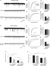
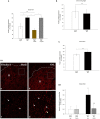
FIGURE 4
FIGURE 4
Fibulin-2 controls the sensitivity to baclofen and saclofen of GABAB-mediated presynaptic inhibition. (A1) Spontaneous miniature EPSCs (mEPSCs) recorded from cultured spinal neurons transfected with mismatch siRNA before and after application of baclofen (10 μM) and baclofen + saclofen (100 μM). (A2) Amplitude cumulative probability for the experiment shown in (A1). Inset trace shows superimposed average of 25 mEPSCs obtained in control and in presence of baclofen. (A3) Inter-event interval cumulative probability for the experiment shown in (A1). The distribution is significantly shifted toward the right during baclofen application. (A4,A5) average variation in mEPSC amplitude (A4) and frequency (A5), expressed as percent of control (n = 9). (B1) mEPSCs recorded from cultured spinal neurons transfected with fibulin-2 siRNA before and after application of baclofen (10 μM), saclofen (100 μM), and CGP (50 μM). (B2) Amplitude cumulative probability for the experiment shown in (B1). Inset trace shows superimposed average of 25 mEPSCs obtained in control and in presence of baclofen. (B3) Inter-event interval cumulative probability for the experiment shown in (B1). The distribution is shifted toward the right during baclofen and baclofen + saclofen application. (B4,B5) Average variation in mEPSC amplitude (B4) and frequency (B5), expressed as percent of control (n = 7). (C1,C2) Average frequencies (C1) and amplitudes (C2) of mEPSCs recorded in preparations transfected with mismatch (n = 9) or anti fibulin-2 siRNA (n = 7), in control conditions and in the presence of baclofen (10 μM).
FIGURE 5
FIGURE 5
Fibulin-2 is upregulated in the spinal cord of neuropathic rats. (A) qRT-PCR analysis of fibulin-2 mRNA in the ipsilateral spinal dorsal horn of sham rats, neuropathic rats (SNL), and SNL rats injected with anti-fibulin-2 siRNA (SNL + siRNA) or with mismatch RNA (SNL + mmRNA). Data are expressed as percentage of sham ± SEM. Fibulin-2 expression increased in ipsilateral dorsal horn of neuropathic rats. This increase was efficiently prevented by siRNA injection. The injection of mmRNA had no effect on fibulin-2 expression. *p < 0.05, **p < 0.01 “sham” vs. “SNL”; n.s.: p > 0.05. (B) qRT-PCR analysis of mRNA of fibulin-2 in the ipsilateral dorsal root ganglia of sham and neuropathic (SNL) rats. Data are expressed as percentage of sham ± SEM. No significant changes are seen (n.s.: p > 0.05 “sham” vs. “SNL). (C) qRT-PCR analysis of mRNA of fibulin-1 in the ipsilateral spinal dorsal horn of sham and neuropathic (SNL) rats. Data are expressed as percentage of sham ± SEM. No significant changes are observed (n.s.: p > 0.05 “sham” vs. “SNL). (D1) Immunohistochemistry for endogenous fibulin-2 in the ipsilateral dorsal horn of sham (a), and neuropathic (SNL) (b) rats. The dotted line indicates the dorsal limit of the spinal cord section. Framed areas of the lamina II (II) in (a,b) are displayed at higher magnification in (c,d), respectively. Fibulin-2 expression was very low in sham animals (arrowheads in c) whereas an intense staining could be seen in the SNL group (arrowheads in d). (D2) The quantification of the signal intensity confirmed the significant difference in the ipsilateral dorsal horn between sham and SNL groups. In contrast, no changes is seen in the contralateral dorsal horn (n = 3 sections from 7 animals in sham and SNL groups). **p < 0.01; n.s.: p > 0.05.
FIGURE 6
FIGURE 6
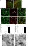
Endogenous co-expression of B1a and fibulin-2 in sham and neuropathic rats. (A1) Immunohistochemistry for endogenous B1a (green) and fibulin-2 (red) in lamina II (II) of the dorsal horn of sham (a), and neuropathic (SNL) (b) rats. The dotted line indicates the dorsal limit of the spinal cord section. (A2) Micrographs are higher magnification of the framed areas in (A1) and correspond to sham (a–c), and SNL (d–f) rats. In sham animals, B1a subunit (green, arrows) and fibulin-2 (red, arrowheads) are distributed throughout the dorsal horn but show very little colocalization. In contrast, colocalization was more frequent in SNL rats (double arrowheads). However, some single labeling was also found for B1a (arrow) and fibulin-2. Bar = 15 μm (same for all). (B) Quantification of colocalization in the ipsilateral dorsal horn, expressed as the percentage of B1a labeling colocalized with fibulin-2 (n = 8 sections from 3 animals in the sham group; n = 7 sections from 3 animals in the SNL group). ∗∗p < 0.01. (C) Double immunogold labeling for B1a (15 nm colloidal gold diameter) and fibulin-2 (5 nm colloidal gold diameter) in the ipsilateral dorsal horn of SNL rats. Both proteins colocalized at the plasma membrane of spinal neurons (double arrowheads in a,b). Gold particles association was found essentially at extra-synaptic sites (b, the synapse is indicated with large arrows). Single fibulin-2 labeling was also found in the vicinity of the plasma membrane (a, arrow). A, axonal nerve ending; bar = 100 nm.
FIGURE 7
FIGURE 7
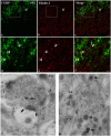
Endogenous co-expression of fibulin-2 and CGRP in neuropathic rats. (a–f) Immunohistochemistry for endogenous fibulin-2 and CGRP in lamina II of the dorsal horn of neuropathic (a–f) rats. Framed areas of the lamina II (II) in a to c are displayed at higher magnification in (d–f), respectively. CGRP (green) and fibulin-2 (red) staining was frequently seen overlapping or in close apposition (double arrowheads). Bar = 15 μm (same for all). (g,h) Double immunogold labeling for fibulin-2 (15 nm colloidal gold diameter) and CGRP (5 nm colloidal gold diameter) in the ipsilateral dorsal horn of neuropathic rats. CGRP was found in secretory granules (d,e, arrows). Secretory granules-containing processes were surrounded by fibulin-2 labeling, at the plasma membrane (d,e, arrowheads), mostly at extra-synaptic sites (large arrows in d). Bar = 100 nm in (d); 50 nm in (e).
FIGURE 8
FIGURE 8
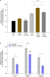
Intrathecal injection of anti-fibulin-2 siRNA potentiates the antinociceptive effect of baclofen in neuropathic rats. (A) Effects of anti-fibulin-2 siRNA and baclofen injections in SNL rats. The effects are quantified with or without baclofen, and before (SNL, SNL + baclo) or after (SNL + siRNA, SNL + baclo + siRNA) intrathecal injections of anti-fibulin-2 siRNA or mismatch RNA (SNL + mmRNA, SNL + baclo + mmRNA). Threshold to mechanical stimulation was measured with the von Frey test. In the absence of baclofen, the injection of anti-fibulin-2 siRNA, but not mmRNA, induces an analgesic effect per se (*p < 0.05 “SNL” vs. “SNL + siRNA”). Baclofen displayed an antinociceptive effect in SNL rats (**p < 0.01 “SNL” vs. “SNL + baclo”). Anti-fibulin-2 siRNA potentiated the antinociceptive effect of baclofen in SNL rats (**p < 0.01 “SNL + Baclo” vs. “SNL + baclo + siRNA”). The withdrawal threshold remained unchanged after injection of mismatch RNA. The withdrawal threshold before nerve ligation was set to 100%. *p < 0.05; **p < 0.01; n.s.: p > 0.05. (B) Effects of baclofen and saclofen co-application after injections of iFect (control condition), anti-fibulin-2 siRNA (siRNA) or mismatch RNA (mmRNA) in neuropathic rats. Saclofen inhibited the baclofen effect before anti-fibulin-2 siRNA injection (**p < 0.01 “baclofen” vs. “baclofen + saclofen”). This effect of saclofen is abolished after anti-fibulin-2 siRNA injections (n.s.: p > 0.05, siRNA, “baclofen” vs. “baclofen + saclofen”) but not after mmRNA injection (*p < 0.05, mmRNA, “baclofen” vs. “baclofen + saclofen”). After anti-fibulin-2 siRNA injection, the withdrawal threshold was significantly higher than after iFect injection (##p < 0.01 siRNA “baclofen + saclofen” vs. iFect “baclofen + saclofen”). The withdrawal threshold in neuropathic rats before injection was set to 100%.

Similar articles

Cited by

References

    1. Attal N., Cruccu G., Haanpaa M., Hansson P., Jensen T. S., Nurmikko T., et al. (2006). EFNS guidelines on pharmacological treatment of neuropathic pain. Eur. J. Neurol. 13 1153–1169. 10.1111/j.1468-1331.2006.01511.x - DOI - PubMed
    1. Balasubramanian S., Fam S. R., Hall R. A. (2007). GABAB receptor association with the PDZ scaffold Mupp1 alters receptor stability and function. J. Biol. Chem. 282 4162–4171. 10.1074/jbc.M607695200 - DOI - PubMed
    1. Bettler B., Kaupmann K., Mosbacher J., Gassmann M. (2004). Molecular structure and physiological functions of GABA(B) receptors. Physiol. Rev. 84 835–867. 10.1152/physrev.00036.2003 - DOI - PubMed
    1. Biermann B., Ivankova-Susankova K., Bradaia A., Abdel Aziz S., Besseyrias V., Kapfhammer J. P., et al. (2010). The Sushi domains of GABAB receptors function as axonal targeting signals. J. Neurosci. 30 1385–1394. 10.1523/JNEUROSCI.3172-09.2010 - DOI - PMC - PubMed
    1. Blein S., Ginham R., Uhrin D., Smith B. O., Soares D. C., Veltel S., et al. (2004). Structural analysis of the complement control protein (CCP) modules of GABA(B) receptor 1a: only one of the two CCP modules is compactly folded. J. Biol. Chem. 279 48292–48306. 10.1074/jbc.m406540200 - DOI - PubMed

LinkOut - more resources