Ethanolic Extract of the Fungus Trichoderma asperelloides Induces Ultrastructural Effects and Death on Leishmania amazonensis
- PMID: 32760675
- PMCID: PMC7373754
- DOI: 10.3389/fcimb.2020.00306
Ethanolic Extract of the Fungus Trichoderma asperelloides Induces Ultrastructural Effects and Death on Leishmania amazonensis
Abstract
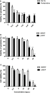
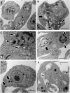
The Trichoderma genus comprises several species of fungi whose diversity of secondary metabolites represents a source of potential molecules with medical application. Because of increased pathogen resistance and demand for lower production costs, the search for new pharmacologically active molecules effective against pathogens has become more intense. This is particularly evident in the case of American cutaneous leishmaniasis due to the high toxicity of current treatments, parenteral administration, and increasing rate of refractory cases. We have previously shown that a fungus from genus Trichoderma can be used for treating cerebral malaria in mouse models and inhibit biofilm formation. Here, we evaluated the effect of the ethanolic extract of Trichoderma asperelloides (Ext-Ta) and its fractions on promastigotes and amastigotes of Leishmania amazonensis, a major causative agent of cutaneous leishmaniasis in the New World. Ext-Ta displayed leishmanicidal action on L. amazonensis parasites, and its pharmacological activity was associated with the low-molecular-weight fraction (LMWF) of Ext-Ta. Ultrastructural analysis demonstrated morphological alterations in the mitochondria and the flagellar pocket of promastigotes, with increased lipid body and acidocalcisome formation, microtubule disorganization of the cytoplasm, and intense vacuolization of the cytoplasm when amastigotes were present. We suggest the antiparasitic activity of Trichoderma fungi as a promising tool for developing chemotherapeutic leishmanicidal agents.
Keywords: Leishmania amazonensis; Trichoderma; chemotherapy; leishmaniasis; leishmanicidal.
Copyright © 2020 Lopes, Santos, Dos Anjos, Silva Júnior, Paula, Vannier-Santos, Silva-Jardim, Castro-Gomes, Pirovani and Lima-Santos.
Figures


Similar articles
-
Leishmanicidal activity of Piper marginatum Jacq. from Santarém-PA against Leishmania amazonensis.Exp Parasitol. 2020 Mar;210:107847. doi: 10.1016/j.exppara.2020.107847. Epub 2020 Jan 28. Exp Parasitol. 2020. PMID: 32004535
-
Efficacy of lapachol on treatment of cutaneous and visceral leishmaniasis.Exp Parasitol. 2019 Apr;199:67-73. doi: 10.1016/j.exppara.2019.02.013. Epub 2019 Feb 21. Exp Parasitol. 2019. PMID: 30797783
-
A lupane-triterpene isolated from Combretum leprosum Mart. fruit extracts that interferes with the intracellular development of Leishmania (L.) amazonensis in vitro.BMC Complement Altern Med. 2015 Jun 6;15:165. doi: 10.1186/s12906-015-0681-9. BMC Complement Altern Med. 2015. PMID: 26048712 Free PMC article.
-
Selective effects of Euterpe oleracea (açai) on Leishmania (Leishmania) amazonensis and Leishmania infantum.Biomed Pharmacother. 2018 Jan;97:1613-1621. doi: 10.1016/j.biopha.2017.11.089. Epub 2017 Nov 28. Biomed Pharmacother. 2018. PMID: 29793323
-
Ethanolic extract of Croton blanchetianus Ball induces mitochondrial defects in Leishmania amazonensis promastigotes.An Acad Bras Cienc. 2020 Oct 28;92(suppl 2):e20180968. doi: 10.1590/0001-3765202020180968. eCollection 2020. An Acad Bras Cienc. 2020. PMID: 33146273
Cited by
-
Unveiling the anticancer potential of the ethanolic extract from Trichoderma asperelloides.Front Pharmacol. 2024 May 1;15:1398135. doi: 10.3389/fphar.2024.1398135. eCollection 2024. Front Pharmacol. 2024. PMID: 38751785 Free PMC article.
-
Trichoderma harzianum as fungicide and symbiont: is it safe for human and animals?Vet Res Forum. 2023;14(11):604-614. doi: 10.30466/vrf.2023.561862.3618. Epub 2023 Nov 15. Vet Res Forum. 2023. PMID: 38169556 Free PMC article.
-
Anti-leishmanial compounds from microbial metabolites: a promising source.Appl Microbiol Biotechnol. 2021 Nov;105(21-22):8227-8240. doi: 10.1007/s00253-021-11610-6. Epub 2021 Oct 9. Appl Microbiol Biotechnol. 2021. PMID: 34625819 Review.
References
-
- Adade C. M., Souto-Padrón T. (2010). Contributions of ultrastructural studies to the cell biology of trypanosmatids: targets for anti-parasitic drugs. Open Parasitol. J. 4, 178–187. 10.2174/1874421401004010178 - DOI
-
- Atanasova L., Druzhinina I., Jaklitsch W. (2013). “Two hundred Trichoderma species recognized on the basis of molecular phylogeny,” in Trichoderma: Biology and Applications, eds Mukherjee P. K., Horwitz B. A., Singh U. S., Mukherjee M., Schmoll M. (London: CAB International; ), 10–42. 10.1079/9781780642475.0010 - DOI
Publication types
MeSH terms
Substances
Supplementary concepts
LinkOut - more resources
Full Text Sources

