. 2020 Aug 6;16(8):e1008696.
doi: 10.1371/journal.ppat.1008696. eCollection 2020 Aug.

Selective Upregulation of CTLA-4 on CD8+ T Cells Restricted by HLA-B*35Px Renders them to an Exhausted Phenotype in HIV-1 infection

Affiliations

Selective Upregulation of CTLA-4 on CD8+ T Cells Restricted by HLA-B*35Px Renders them to an Exhausted Phenotype in HIV-1 infection

Shokrollah Elahi et al. PLoS Pathog. .

Abstract

HLA-B*35Px is associated with HIV-1 disease rapid progression to AIDS. However, the mechanism(s) underlying this deleterious effect of this HLA allele on HIV-1 infection outcome has not fully understood. CD8+ T cells play a crucial role to control the viral replication but impaired CD8+ T cells represent a major hallmark of HIV-1 infection. Here, we examined the effector functions of CD8+ T cells restricted by HLA-B*35Px (HLA-B*35:03 and HLA-B*35:02), HLA-B*27/B57 and non-HLA-B*27/B57 (e.g. HLA-A*01, A*02, A*03, A*11, A*24, A*26, B*40, B*08, B*38, B*44). CD8+ T cells restricted by HLA-B*35Px exhibited an impaired phenotype compared with those restricted by HLA-B*27/B57 and even non-HLA-B*27/B57. CD8+ T cells restricted by non-HLA-B*27/B57 when encountered their cognate epitopes upregulated TIM-3 and thus became suppressed by regulatory T cells (Tregs) via TIM-3: Galectin-9 (Gal-9). Strikingly, CD8+ T cells restricted by HLA-B*35Px expressed fewer TIM-3 and therefore did not get suppressed by Tregs, which was similar to CD8+ T cells restricted by HLA-B*27/B57. Instead, CD8+ T cells restricted by HLA-B*35Px upon recognition of their cognate epitopes upregulated CTLA-4. The transcriptional and impaired phenotype (e.g. poor effector functions) of HIV-specific CD8+ T cells restricted by HLA-B*35 was related to persistent CTLA-4, elevated Eomes and blimp-1 but poor T-bet expression. As such, anti-CTLA-4 antibody, Ipilimumab, reversed the impaired proliferative capacity of antigen-specific CD8+ T cells restricted by HLA-B*35Px but not others. This study supports the concept that CD8+ T resistance to Tregs-mediated suppression is related to allele restriction rather than the epitope specificity. Our results aid to explain a novel mechanism for the inability of HIV-specific CD8+ T cells restricted by HLA-B*35Px to control viral replication.


Conflict of interest statement

The authors have declared that no competing interests exist.

Figures
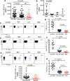
Fig 1
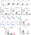
Fig 1. Diminished cytokine production by HLA-B*35Px-restricted CD8+ T cells.
(A) Cumulative data showing quantification of IFN-γ secreting cells (spot count)/ 106 PBMCs following stimulation with different 15 mer peptides restricted by HLA-B*27/B57, non-HLA-B*27/B57 (HLA-A*03, A*02, A*24, A*26 and B*40) and HLA-B*35Px/B*53 using ELISpot assay. (B) Cumulative data showing the breadth of IFN-γ response to different HIV-proteins in PBMCs of HLA-B*35Px individuals using ELISpot assay. (C) Representative flow cytometry plots, and (D) cumulative data showing percentages of IL-2 secreting CD8+ T cells restricted by HLA-B*27/B57, non-HLA-B*27/B57 and HLA-B*35 in HIV-infected individuals following stimulation with their cognate epitopes (2 μg/ml) for 72 hrs as measured by ICS. (E) Representative flow cytometry plots, and (F) cumulative data showing percentages of TNF-α secreting CD8+ T cells restricted by HLA-B*27/B57, non-HLA-B*27/B57 and HLA-B*35 in HIV-infected individuals following stimulation of PBMCs with their cognate epitopes (2 μg/ml) for 72 hrs using ICS. (G) Representative flow cytometry plots, and (H) cumulative data showing percentages of IFN-γ secreting CD8+ T cells restricted by HLA-B*27/B57, non-HLA-B*27/B57 and HLA-B*35 in HIV-infected individuals following stimulation with their cognate epitopes (2 μg/ml) for 72 hrs as measured by ICS. (I) Cumulative data showing quantification of IFN-γ secreting cells (spot count)/ 106 PBMCs following stimulation with different epitopes restricted by non-HLA-B*27/B57 (HLA-A*03, A*01, A*02, A*24, A*11, B*38, B*08 and B*44) and HLA-B*35Px/B*53 from 5 HLA-B35/B53 individuals using ELISpot assay. Each point represents data from an epitope. Bar, mean ± one standard error.
Fig 2
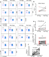
Fig 2. HLA-B*35/53-restricted CD8+ T cells have impaired expression of perforin, GzmB with low proliferative capacity.
(A-D) Representative plots and cumulative data showing expression of GzmB, perforin, and their co-expression respectively, in CD8+ T cells restricted by HLA-B*27/B57, non-HLA-B*27/B57 and HLA-B*35Px/B*53 in HIV-infected individuals following stimulation of PBMCs with their cognate epitopes (2 μg/ml) for 72 hrs using ICS. (E) Cumulative data showing co-expression of GzmB/perforin in CD8+ T cells restricted by HLA-B*27/B*57 versus those restricted by HLA-B*35/B*53 within individuals having HLA-B35Px. (F) Representative plots, and (G) cumulative data indicating percentages of proliferated CD8+ T cells restricted by different HLA-alleles following stimulation with their cognate epitopes (2 μg/ml) for 96 hrs measured by CFSE titration assay. (H) Cumulative data showing percentages of proliferated CD8+ T cells restricted by HLA-B*27/B*57 versus those restricted by HLA-B*35/B*53 within individuals having HLA-B35Px. Each point represents data from an epitope. Bar, mean ± one standard error.
Fig 3
Fig 3. The proliferation of HLA-B*35-restricted CD8+ T cells is not suppressed by Tregs.
(A-B) Representative plots showing proliferation of CD8+ T cells restricted by different HLA-alleles within an HLA-B*35Px patient (PTID:PR-03) following stimulation with their cognate epitopes in the presence or absence of Tregs for 96 hrs. Red symbols representing CD8+ T cells restricted by HLA-B*35/B*53 and black symbols those restricted by HLA-A*03/A*11. (C-D) Representative plots showing proliferation of CD8+ T cells restricted by different HLA-alleles within an HLA-B*35Px patient (PTID:PR-05) following stimulation with their cognate epitopes (2 μg/ml for 96 hrs) in the presence or absence of Tregs. (E) Cumulative data showing percentages of proliferated antigen-specific CD8+ T cells restricted by HLA-B*35Px following stimulation with their cognate epitopes. Red symbols representing CD8+ T cells restricted by HLA-B*35 and black symbols those restricted by HLA-A*03/A*11, HLA-B*44 and HLA-A*301. (F) Cumulative data showing percentages of proliferated antigen-specific CD8+ T cells restricted by HLA-A*01, A*02, A*03, A*08, A*11, A*24, B*38 and B*44 following stimulation with their cognate epitopes. Each point represents data from an epitope.
Fig 4
Fig 4. Differential suppression of TNF-α by Tregs depending on the HLA-allele restriction of CTLs.
A) Representative flow cytometry plots of % TNF-α production by CD8+ T cells restricted by different HLA-alleles, as shown, following stimulation with their cognate epitopes (2 μg/ml for 72 hrs) measured by ICS in the presence or absence of autologous Tregs. B-D) Cumulative data showing %TNF-α secreting CD8+ T cells restricted by either HLA-B*27/B57, HLA-B*35Px or non-HLA-B*27/57 following stimulation with their cognate epitopes (2 μg/ml) for 72 hrs as measured by ICS. Each point represents data from an epitope.
Fig 5
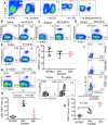
Fig 5. Differential expression of co-inhibitory receptors by CD8+ T cells upon recognition of their cognate epitopes in vitro.
(A) Representative plots showing gating strategy for identification of antigen-specific T cells using tetramer staining. (B, C) Representative flow cytometry plot showing % co-expression of Tetramer+PD-1+ CD8+ T cells following stimulation of PBMCs with B27-KK10 (KRWIILGLNK) epitope (2 μg/ml) for 72 hrs in patient #LNP01 and patients #LNP03 respectively. (D, E) Representative flow cytometry plot showing % co-expression of Tetramer+PD-1+ CD8+ T cells following stimulation of PBMCs with A03-RK9 (RLRPGGKKK) and B35-FL9 (FPVKPQVPL) epitopes (2 μg/ml) for 72 hrs in patient #PR03 and patients #PR01 respectively. (F) Cumulative data showing % tetramer+PD-1+ in CD8+ T cells restricted by different HLA-alleles, as shown, following encounter with their cognate epitopes for 72 hrs. (G) Representative flow cytometry plot showing % co-expression of Tetramer+TIM-3+ CD8+ T cells following stimulation of PBMCs with A02-SL9 (SLYNTVATL) and B35-FL9 (FPVKPQVPL) epitopes (2 μg/ml) for 72 hrs in patients #PR04 and #PR01, respectively. (H) Cumulative data showing % TIM-3 expressing antigen-specific CD8+ T cells following stimulation with their cognate epitopes for 72 hrs. (I, J) Representative flow cytometry plot showing % co-expression of Tetramer+CTLA-4+ CD8+ T cells following stimulation of PBMCs with B35-FL9 (FPVKPQVPL), A03-RK9 (RLRPGGKKK), and B57-KF-11 (KAFSPEVIPMF) epitopes (2 μg/ml) for 72 hrs in patients #PR01 and #PR03 and #LNP02, respectively. (K) Cumulative data showing % TIM-3 expressing antigen-specific CD8+ T cells following stimulation with their cognate epitopes for 72 hrs. Each point represents data from an epitope. Bar, mean ± one standard error.
Fig 6
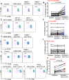
Fig 6. Differential effects of anti-TIM-3, anti-PD-1 and anti-CTLA-4 blocking antibodies on rescuing of the proliferative capacity of CD8+ T cells restricted by different HLA-alleles.
(A) Representative flow cytometry plots, and (B) cumulative data showing proliferation of antigen-specific CD8+ T cells restricted by non-HLA-B*27/B57 following stimulation with their cognate-epitopes in the absence or presence of anti-PD-1 (Pembrolizumab 5 μg/ml), anti-TIM-3 (10 μg/ml LEAF) antibodies for 96 hours as measured by CFSE dilution assay, QK9 (QVPLRPMTYK), patient #LNP04. (C) Representative flow cytometry plots, and (D) cumulative data showing proliferation of antigen-specific CD8+ T cells restricted by HLA-B*27/B57 following stimulation with their cognate-epitopes in the absence or presence of anti-PD-1 (Pembrolizumab 5 μg/ml), anti-TIM-3 (10 μg/ml LEAF) antibodies for 96 hours as measured by CFSE dilution assay, KF11 (KAFSPEVIPMF), patient #LNP04. (E) Representative flow cytometry plots, and (F) cumulative data showing proliferation of antigen-specific CD8+ T cells restricted by HLA-B*35Px following stimulation with their cognate-epitopes in the absence or presence of anti-PD-1 (Pembrolizumab 5 μg/ml), anti-TIM-3 (10 μg/ml LEAF) antibodies for 96 hours as measured by CFSE dilution assay, QM9 (QVTNSATIM), patient #PR05. (G) Representative flow cytometry plots, and (H) cumulative data showing proliferation of antigen-specific CD8+ T cells restricted by HLA-B*35Px following stimulation with their cognate-epitopes in the absence or presence of anti-CTLA-4 (Ipilimumab 5 μg/ml) antibody for 96 hours as measured by CFSE dilution assay, QM9 (QVTNSATIM), patient #PR05. (I) Representative flow cytometry plots showing proliferation of antigen-specific CD8+ T cells restricted by HLA-B*27 following stimulation with GY9 (GLNKIVRMY) epitope, and (J) CD8+ T cells restricted by HLA-A24 following stimulation with epitope RL9 (RYLKDQQLL) in the absence or presence of anti-CTLA-4 (Ipilimumab 5 μg/ml) antibody for 96 hours as measured by CFSE dilution assay, patient #LNP01. Each point represents data from an epitope.
Fig 7
Fig 7. HLA-B*35Px restricted CTLs exhibit an exhausted phenotype at the gene level.
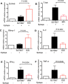
(A) Relative mRNA expression of Eomes, (B) T-bet, (C) blimp-1, (D) IL-2, E) IFN-γ and F) TNF-α in CD8+ T cells restricted by HLA-B*35Px versus those restricted by HLA-B*27/B57 as quantified by qPCR. Data are from a minimum of five antigen-specific CD8+ T cells restricted by each HLA-type and isolated by sorting for RNA isolation and qPCR. Bar, mean ± one standard error.

Similar articles

Cited by

References

    1. Williams DW, Elahi S. Profound immune consequences for young adults infected with HIV perinatally or during childhood: a cautionary tale regarding adherence to antiretroviral therapy. Aids. 2019;33(14):2251–2. 10.1097/QAD.0000000000002328 - DOI - PMC - PubMed
    1. Altfeld M, Addo MM, Rosenberg ES, Hecht FM, Lee PK, Vogel M, et al. Influence of HLA-B57 on clinical presentation and viral control during acute HIV-1 infection. Aids. 2003;17(18):2581–91. 10.1097/00002030-200312050-00005 - DOI - PubMed
    1. Elahi S, Dinges WL, Lejarcegui N, Laing KJ, Collier AC, Koelle DM, et al. Protective HIV-specific CD8+ T cells evade Treg cell suppression. Nature medicine. 2011;17(8):989–95. 10.1038/nm.2422 - DOI - PMC - PubMed
    1. Elahi S, Horton H. Association of HLA-alleles with the immune regulation of chronic viral infections. The international journal of biochemistry & cell biology. 2012;44(8):1361–5. - PMC - PubMed
    1. Carrington M, Nelson GW, Martin MP, Kissner T, Vlahov D, Goedert JJ, et al. HLA and HIV-1: heterozygote advantage and B*35-Cw*04 disadvantage. Science. 1999;283(5408):1748–52. 10.1126/science.283.5408.1748 - DOI - PubMed

Publication types

MeSH terms